

The DOI matches its Crossref record; 167 of the 167 references it cites were found in a registry or an index.
A new look at cerebrospinal fluid circulation
Open accessAbstract
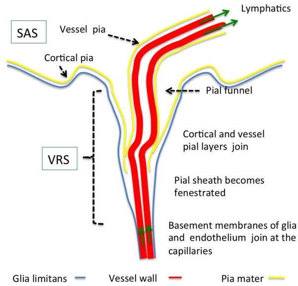
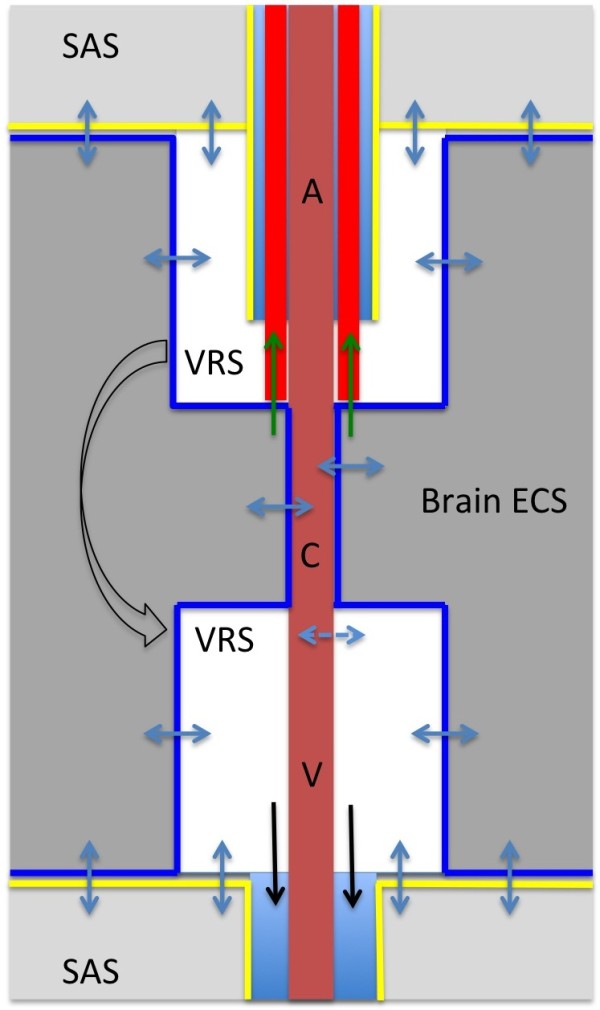
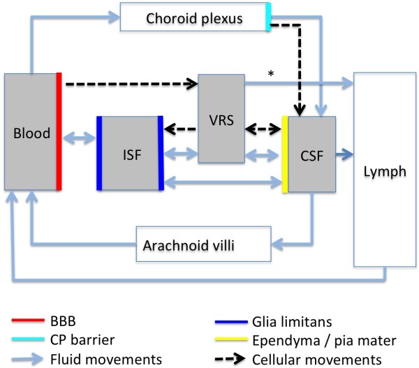
According to the traditional understanding of cerebrospinal fluid (CSF) physiology, the majority of CSF is produced by the choroid plexus, circulates through the ventricles, the cisterns, and the subarachnoid space to be absorbed into the blood by the arachnoid villi. This review surveys key developments leading to the traditional concept. Challenging this concept are novel insights utilizing molecular and cellular biology as well as neuroimaging, which indicate that CSF physiology may be much more complex than previously believed. The CSF circulation comprises not only a directed flow of CSF, but in addition a pulsatile to and fro movement throughout the entire brain with local fluid exchange between blood, interstitial fluid, and CSF. Astrocytes, aquaporins, and other membrane transporters are key elements in brain water and CSF homeostasis. A continuous bidirectional fluid exchange at the blood brain barrier produces flow rates, which exceed the choroidal CSF production rate by far. The CSF circulation around blood vessels penetrating from the subarachnoid space into the Virchow Robin spaces provides both a drainage pathway for the clearance of waste molecules from the brain and a site for the interaction of the systemic immune system with that of the brain. Important physiological functions, for example the regeneration of the brain during sleep, may depend on CSF circulation.
Overview
The claims and statistics of this paper haven’t been checked yet.
Run the check — free, no account needed. It reads the open-access text and checks the sources it cites.
Integrity signals
Registry
Cites
Statistics
Figures
Verification
Claims the paper makes
The claims and statistics of this paper haven’t been checked yet.
Run the check — free, no account needed. It reads the open-access text and checks the sources it cites.
Claims other papers make about this one
Each is checked against this paper’s own text: does it say what the citing paper says it does?
The cited source says what the claim attributes to it.
In the citing paper
“Uptake into arachnoid villi, as proposed more than one hundred years ago, seems to be of minor importance in humans, as reviewed by Brinker et al. (50) and as indicated in our recent study (13).”
From Brain-wide glymphatic enhancement and clearance in humans assessed with MRI. (2018) · cites this paper as [50]
Self-check
Percentages, sums and reported differences re-computed from the paper’s own numbers.
The self-check needs the paper’s full text.
Citations
Cites
Iliff, J. J., Wang, M., Liao, Y., Plogg, B. A., Peng, W., Gundersen, G. A., Benveniste, H., Vates, G. E., Deane, R., Goldman, S. A., et al. (2012) · Science Translational Medicine · doi:10.1126/scitranslmed.3003748 · Cited by 5,824
Sleep Drives Metabolite Clearance from the Adult Brain
Xie, L., Kang, H., Xu, Q., Chen, M. J., Liao, Y., Thiyagarajan, M., O’Donnell, J., Christensen, D. J., Nicholson, C., Iliff, J. J., et al. (2013) · Science · doi:10.1126/science.1241224 · Cited by 5,411
Structure and function of the blood–brain barrier
Abbott, N. J., Patabendige, A. A. K., Dolman, D. E. M., Yusof, S. R., & Begley, D. J. (2010) · Neurobiology of Disease · doi:10.1016/j.nbd.2009.07.030 · Cited by 5,185
Daneman, R., & Prat, A. (2015) · Cold Spring Harbor Perspectives in Biology · doi:10.1101/cshperspect.a020412 · Cited by 3,437
Aquaporin-4 deletion in mice reduces brain edema after acute water intoxication and ischemic stroke
Manley, G. T., Fujimura, M., Ma, T., Noshita, N., Filiz, F., Bollen, A. W., Chan, P. H., & Verkman, A. S. (2000) · Nature Medicine · doi:10.1038/72256 · Cited by 1,501
Diffusion in Brain Extracellular Space
Syková, E., & Nicholson, C. (2008) · Physiological Reviews · doi:10.1152/physrev.00027.2007 · Cited by 1,481
Nielsen, S. M., Nagelhus, E. A., Amiry-Moghaddam, M. R., Bourque, C. W., Agre, P. C., & Ottersen, O. P. (1997) · Journal of Neuroscience · doi:10.1523/jneurosci.17-01-00171.1997 · Cited by 1,440
Brain-wide pathway for waste clearance captured by contrast-enhanced MRI
Iliff, J. J., Lee, H., Yu, M., Feng, T., Logan, J., Nedergaard, M., & Benveniste, H. (2013) · Journal of Clinical Investigation · doi:10.1172/jci67677 · Cited by 1,148
Blood–brain barrier structure and function and the challenges for CNS drug delivery
Abbott, N. J. (2013) · Journal of Inherited Metabolic Disease · doi:10.1007/s10545-013-9608-0 · Cited by 951
Mathiisen, T. M., Lehre, K. P., Danbolt, N. C., & Ottersen, O. (2010) · Glia · doi:10.1002/glia.20990 · Cited by 893
Research works printed but not linked: 1
- 10.1186/1743-8454-5-10 · DOI · Paper page
Cited by
Sorted by citations
Items 501–600 of 821
Brain lymphatic drainage system in fetus and newborn: Birth of a new era of exploration.
Angelis, L. C. D., Witte, M., Bellini, T., Bernas, M. J., Boccardo, F. M., Ramenghi, L. A., & Bellini, C. (2018) · PubMed · Cited by 6
Kratz, S. V. (2021) · SAGE Open Medical Case Reports · doi:10.1177/2050313x20952224 · Cited by 7
Páez-Nova, M., Andaur, K., Campos, G., García-Ballestas, E., Moscote‐Salazar, L. R., Osvaldo Koller, C., & Valenzuela, S. (2021) · Child s Nervous System · doi:10.1007/s00381-021-05325-2 · Cited by 6
Lyons, H., Ramalingam, S., Mitchell, J., Yiangou, A., Thaller, M., Sinclair, A. J., & Mollan, S. P. (2022) · Journal of Medical Case Reports · doi:10.1186/s13256-022-03687-y · Cited by 6
Hyphenated Mass Spectrometry Techniques in the Diagnosis of Amyloidosis
Spodzieja, M., Rodziewicz‐Motowidło, S., & Szymańska, A. (2017) · Current Medicinal Chemistry · doi:10.2174/0929867324666171003113019 · Cited by 6
Future Directions in Sports-Related Concussion Management
Kerr, H. A., Bakken, B. ̧., & House, G. (2020) · Clinics in Sports Medicine · doi:10.1016/j.csm.2020.08.009 · Cited by 6
Deng, P., Halmai, J. A. N. M., Beitnere, U., Cameron, D. L., Martinez, M. L., Lee, C. C., Waldo, J. J., Thongphanh, K., Adhikari, A., Copping, N. A., et al. (2022) · Frontiers in Molecular Neuroscience · doi:10.3389/fnmol.2021.789913 · Cited by 6
Bücker, P., Buzzi, R. M., Akeret, K., Mosberger, L., Richter, H., Sperling, M., Hugelshofer, M., Schaer, D. J., & Kärst, U. (2022) · Metallomics · doi:10.1093/mtomcs/mfac057 · Cited by 6
Body Fluids Modulate Propagation of Tumor Treating Fields
Wong, E. T., & Lok, E. (2023) · Advances in Radiation Oncology · doi:10.1016/j.adro.2023.101316 · Cited by 6
Toma, A. K. (2018) · Elsevier eBooks · doi:10.1016/b978-0-323-43140-8.00060-3 · Cited by 6
Muñoz‐Montecinos, C., Romero, A. D. B., Sepúlveda, V., Vira, M. Á., Fehrmann-Cartes, K., Marcellini, S., Aguilera, F., Caprile, T., & Fuentes, R. (2022) · Frontiers in Cell and Developmental Biology · doi:10.3389/fcell.2021.801652 · Cited by 8
Cerebrospinal Fluid Pressure Dynamics
Czosnyka, M., Schuhmann, M. U., Czosnyka, Z. H., Pickard, J. D., & Whitfield, P. C. (2019) · Springer eBooks · doi:10.1007/978-3-319-27250-4_3 · Cited by 6
Tang, K., Zhou, J., Yang, H., Tan, X., Xie, M., Tao, X., Ge, X., & Xu, J. (2010) · PubMed · Cited by 6
Integrative Medicine for PTSD and TBI: Two Innovative Approaches
Leskowitz, E. (2016) · Medical Acupuncture · doi:10.1089/acu.2016.1168 · Cited by 6
de Rezende, V. L., da Costa, M. D. A., Martins, C. D., Mathias, K., Gonçalves, C. L., Barichello, T., & Petronilho, F. C. (2025) · Neurochemical Research · doi:10.1007/s11064-025-04361-7 · Cited by 5
Lateral ventricular choroid plexus enlargement in patients with spontaneous intracranial hypotension
Yoon, S., Ha, W., Jeong, J., Song, S.-W., Yum, J., Cho, S., & Chu, M. K. (2025) · Headache The Journal of Head and Face Pain · doi:10.1111/head.15073 · Cited by 5
Vakhtin, A. A., Lin, H. C., Richardson, S. P., Shaff, N. A., & Ryman, S. G. (2025) · Journal of Cerebral Blood Flow & Metabolism · doi:10.1177/0271678x251399120 · Cited by 5
Fabi, A., Alves, A. S., Neutzner, A., Frank, S., Lariu, A., Muller, L., Ismail, T., Gkotsoulias, D. G., Guzman, R. G., Schaefer, D. J., et al. (2025) · JPRAS Open · doi:10.1016/j.jpra.2025.12.013 · Cited by 6
Water exchange across the blood–CSF barrier: A systematic review
Hjørnevik, T., & Eide, P. K. (2026) · Journal of Cerebral Blood Flow & Metabolism · doi:10.1177/0271678x251413926 · Cited by 5
Peeler, D. J., Luera, N., Horner, P. J., Pun, S. H., & Sellers, D. L. (2020) · Journal of Controlled Release · doi:10.1016/j.jconrel.2020.03.025 · Cited by 5
Lis-Bartos, A., Szarek, D., Krok−Borkowicz, M., Marycz, K., Jarmundowicz, W., & Laska, J. (2020) · Polymers · doi:10.3390/polym12112693 · Cited by 5
Intrathecal catheter implantation decreases cerebrospinal fluid dynamics in cynomolgus monkeys
Khani, M., Fu, A. Q., Pluid, J., Gibbs, C. P., Oshinski, J. N., Xing, T., Stewart, G. R., Zeller, J. R., & Martin, B. A. (2020) · PLoS ONE · doi:10.1371/journal.pone.0244090 · Cited by 5
Outcomes of Cranioplasty: A Single-Center Experience
Fallatah, M. A., Aldahlawi, A. K., Babateen, E. M., Saif, S., Alnejadi, W., Bamsallm, M., & Lary, A. (2023) · Cureus · doi:10.7759/cureus.35213 · Cited by 5
The Emerging Role of Water Loss in Dog Aging
Guelfi, G., Capaccia, C., Ratto, V. F., Bufalari, A., Leonardi, L., Mechelli, L., Cenci, S., & Maranesi, M. (2025) · Cells · doi:10.3390/cells14070545 · Cited by 5
Low Reynolds number pumping near an elastic half space
Trevino, A., Powers, T. R., Zenit, R., & Rodriguez, M. (2025) · Physical Review Fluids · doi:10.1103/physrevfluids.10.054003 · Cited by 5
Cavaglià, M., & Tuszyński, J. A. (2025) · Biosystems · doi:10.1016/j.biosystems.2025.105669 · Cited by 5
Temporal changes of spinal subarachnoid space patency after graded spinal cord injury in rats
Franco-Bourland, R. E., Reyes‐Alva, H. J., Quintana-Armenta, A., Martínez‐Cruz, A., Madrazo, I., & Guízar‐Sahagún, G. (2015) · Injury · doi:10.1016/j.injury.2015.01.007 · Cited by 5
The modelling error in multi-dimensional time-dependent solute transport models
Masri, R., Zeinhofer, M., Kuchta, M., & Rognes, M. E. (2024) · ESAIM Mathematical Modelling and Numerical Analysis · doi:10.1051/m2an/2024060 · Cited by 5
Eide, P. K., Undseth, R., Gjertsen, Ø., Valnes, L. M., Ringstad, G. A., & Lindstrøm, E. K. (2024) · Fluids and Barriers of the CNS · doi:10.1186/s12987-024-00587-9 · Cited by 5
An introduction to hydrocephalus: types, treatments and management
Thompson, S. D. (2017) · British Journal of Neuroscience Nursing · doi:10.12968/bjnn.2017.13.1.36 · Cited by 5
Suzuki, J.-I., Dezawa, M., & Kitada, M. (2017) · Brain Research · doi:10.1016/j.brainres.2017.08.033 · Cited by 5
Cerebrospinal Fluid Dynamics and Intrathecal Delivery
Tangen, K. M., Mehta, A. I., & Linninger, A. A. (2018) · Elsevier eBooks · doi:10.1016/b978-0-12-805353-9.00067-x · Cited by 6
Lee, J., Lee, J. H., & Lee, J. M. (2021) · Children · doi:10.3390/children8010027 · Cited by 5
A review of fluid flow in and around the brain, modeling, and abnormalities
Prichard, R., Gibson, M., Joseph, C. R., & Strasser, W. S. (2021) · Elsevier eBooks · doi:10.1016/b978-0-12-818144-7.00015-3 · Cited by 6
Saeed, S., Haroon, H. B., Jhanjhi, N. Z., Naqvi, M., & Ahmad, M. (2022) · Advances in healthcare information systems and administration book series · doi:10.4018/978-1-7998-8929-8.ch007 · Cited by 5
Plata, K. S., Cruz, G., & Lezcano, H. (2022) · Radiologia Brasileira · doi:10.1590/0100-3984.2021.0120 · Cited by 5
Head Phantom for the Acquisition of Pulsatile Optical Signals for Traumatic Brain Injury Monitoring
Roldán, M., & Kyriacou, P. A. (2023) · Photonics · doi:10.3390/photonics10050504 · Cited by 5
Cerebrospinal Fluid Flow Parameters in Normal Subjects above 40 Years of Age
Rohilla, S., Kumar, P., & Singh, I. (2023) · Indian journal of radiology and imaging - new series/Indian journal of radiology and imaging/Indian Journal of Radiology & Imaging · doi:10.1055/s-0043-1776413 · Cited by 5
Cerebrospinal fluid flow dynamics in Huntington's disease evaluated by phase contrast MRI
Rodrigues, F. B., Byrne, L. M., Vita, E. D., Johnson, E. B., Hobbs, N. Z., Thornton, J. S., Scahill, R. I., & Wild, E. J. (2019) · European Journal of Neuroscience · doi:10.1111/ejn.14356 · Cited by 5
Post-mortem biochemistry differences between vitreous humour and cerebrospinal fluid
Garland, J., Philcox, W., McCarthy, S., Kesha, K., Lam, L., Spark, A., Palmière, C., Elstub, H., Cala, A. D., Stables, S. R., et al. (2019) · Australian Journal of Forensic Sciences · doi:10.1080/00450618.2019.1597920 · Cited by 5
Subarachnomegaly—venous congestion of infancy
Sainz, L. V., & Schuhmann, M. U. (2021) · Child s Nervous System · doi:10.1007/s00381-021-05328-z · Cited by 5
Development of the Cerebrospinal Fluid Pathways During Embryonic and Fetal Life in Humans
Catala, M. (2019) · Springer eBooks · doi:10.1007/978-3-319-27250-4_2 · Cited by 4
Kudelić, N., Koprek, I., Radoš, M., Orešković, D., Jurjević, I., & Klarica, M. (2023) · Frontiers in Neurology · doi:10.3389/fneur.2023.1234396 · Cited by 4
Disruption of the Blood-Brain Barrier During Neuroinflammatory and Neuroinfectious Diseases
Salimi, H., & Klein, R. S. (2024) · Springer eBooks · doi:10.1007/978-3-031-60006-7_7 · Cited by 4
Anatomiye Klinik ve Morfolojik Yaklaşımlar
Direk, F. K., Direk, F. K., Durmaz, S. C., Karabaş, S. A., Filik, R., Ceranoğlu, F. G., YILMAZ, M. T., Saygın, D. A., & Türkmen, B. (2024) · Özgür Yayınları eBooks · doi:10.58830/ozgur.pub626 · Cited by 4
Aquino, G., Palagini, L., Alfì, G., Feige, B., Spiegelhalder, K., Piarulli, A., & Gemignani, A. (2025) · Journal of Sleep Research · doi:10.1111/jsr.70069 · Cited by 5
On reduced-order modeling of drug dispersion in the spinal canal
Parras-Martos, F. J., Sánchez, A. L., Martı́nez-Bazán, C., Coenen, W., & Gutiérrez-Montes, C. (2025) · Fluids and Barriers of the CNS · doi:10.1186/s12987-025-00657-6 · Cited by 4
Buongiorno, M., Tur, C., Giraldo, D. M., Cullell, N., Krupiński, J., Lanzillo, R., & Sánchez‐Benavides, G. (2025) · Brain Sciences · doi:10.3390/brainsci15070766 · Cited by 5
Huang, M., Zhang, P., Chen, Z., Wang, X., Li, H., Xu, S., Li, L., Zheng, W., & Xiao, H. (2025) · European journal of medical research · doi:10.1186/s40001-025-03048-7 · Cited by 4
Kollarik, S., Humer, S. K., Zurfluh, C. E., Gousopoulos, E., & Lindenblatt, N. (2025) · Cellular and Molecular Neurobiology · doi:10.1007/s10571-025-01611-8 · Cited by 4
Cranial nerves as pathways for human cerebrospinal fluid efflux: In vivo evidence
Falkenberg‐Jensen, B., Pripp, A. H., Ringstad, G. A., & Eide, P. K. (2025) · Journal of Cerebral Blood Flow & Metabolism · doi:10.1177/0271678x251386232 · Cited by 4
Wong, A. W., Sim, N. H. S., Thng, C. B., Sim, D. P.-Y., Low, Z. L., & Foo, A. S. C. (2025) · Plastic & Reconstructive Surgery · doi:10.1097/prs.0000000000012627 · Cited by 4
Quantitative and Qualitative Analysis of CSF Flow Dynamics
Токарев, А. С., Talypova, D. A., Terekhin, I. A., & Grin, A. A. (2022) · Russian Sklifosovsky Journal Emergency Medical Care · doi:10.23934/2223-9022-2022-11-1-86-95 · Cited by 4
Kajiwara, S., Nakamura, H., Sakata, K., Komaki, S., Negoto, T., & Morioka, M. (2022) · BMC Pediatrics · doi:10.1186/s12887-022-03743-y · Cited by 4
Tertiary Lymphoid Structures in Central Nervous System Disorders
Vaccaro, A., Pereira, B. D. A., van de Walle, T., & Dimberg, A. (2024) · Methods in molecular biology · doi:10.1007/978-1-0716-4184-2_2 · Cited by 4
Reduced subarachnoid fluid diffusion in enlarged subarachnoid spaces of infancy
Whitehead, M. T., Lee, B., McCarron, A., Fricke, S. T., & Vézina, G. (2017) · The Neuroradiology Journal · doi:10.1177/1971400916689803 · Cited by 4
Trumbore, C. N. (2018) · Journal of Alzheimer s Disease · doi:10.3233/jad-171080 · Cited by 4
Application of an Aptamer-Based Proteomics Assay (SOMAscan™) in Rat Cerebrospinal Fluid
Simats, A., Ramiro, L., Montaner, J., & García‐Berrocoso, T. (2019) · Methods in molecular biology · doi:10.1007/978-1-4939-9706-0_13 · Cited by 4
Corbo, C. P., & Fulop, Z. L. (2020) · Microscopy Research and Technique · doi:10.1002/jemt.23457 · Cited by 4
In vivo uptake of a fluorescent conjugate of melanin-concentrating hormone in the rat brain
Ruiz-Viroga, V., Urbanavicius, J., Torterolo, P., & Lagos, P. (2021) · Journal of Chemical Neuroanatomy · doi:10.1016/j.jchemneu.2021.101959 · Cited by 5
Neuroimaging in Pediatric Hydrocephalus
Raybaud, C. (2020) · Springer eBooks · doi:10.1007/978-3-319-72168-2_15 · Cited by 4
Choroid Plexus: Source of Cerebrospinal Fluid and Regulator of Brain Development and Function
Snodgrass, R., & Johanson, C. E. (2019) · Springer eBooks · doi:10.1007/978-3-319-27250-4_38 · Cited by 4
Glymphatics, Cranium, and the Brain Homeostasis
Zibitt, M., Makhoul, V., Yu, J. C.-K., Nguyen, K. D., & Williams, J. K. (2023) · FACE · doi:10.1177/27325016231160291 · Cited by 6
Idiopathic Normal Pressure Hydrocephalus
Bech-Azeddine, R., & Gjerris, F. (2013) · CRC Press eBooks · doi:10.3109/9781420016284-19 · Cited by 4
Ventricular Catheters for Hydrocephalus
Galarza, M., Giménez, Á., Pellicer, O., Valero, J., & Amigó, J. M. (2018) · Springer eBooks · doi:10.1007/978-3-319-31889-9_72-1 · Cited by 4
Cliver, R. N., Ayers, B., Brady, A., Firestein, B. L., & Vázquez, M. (2020) · Journal of Tissue Engineering and Regenerative Medicine · doi:10.1002/term.3164 · Cited by 4
Hydrocephalus and Its Diagnosis - A Review
Surti, A., & Usmani, A. (2019) · Journal of Bahria University Medical and Dental College · doi:10.51985/jbumdc2019139 · Cited by 4
Hydrocephalus with lateral ventricular lesions: case series and review of literature
Alselisly, A. M., Ashry, A. H., & Mahmoud, A. T. (2021) · The Egyptian Journal of Neurology Psychiatry and Neurosurgery · doi:10.1186/s41983-021-00283-7 · Cited by 4
Bilgili, A., Tuna, İ. S., Rahman, M., Naney, T. D., & Albayram, S. (2022) · Magnetic Resonance Imaging · doi:10.1016/j.mri.2022.06.013 · Cited by 4
Zakaria, F. H., Ismail, S., & Khadijah, N. M. J. (2022) · Research Journal of Pharmacy and Technology · doi:10.52711/0974-360x.2022.00637 · Cited by 4
Cerebrospinal Fluid (CSF) Directed Antineoplastic Agents: A Review of Current Knowledge
Laber, D. A. (2015) · Journal of Cancer Prevention & Current Research · doi:10.15406/jcpcr.2015.02.00026 · Cited by 4
Amyloid Deposits and Idiopathic Normal-Pressure Hydrocephalus: An 18F-Florbetaben Study
Kang, K., Yoon, U., Hong, J., Jeong, S. Y., Ko, P., Lee, S., & Lee, H. (2018) · European Neurology · doi:10.1159/000487133 · Cited by 4
Good barriers make good neighbors
Ransohoff, R. M. (2014) · Science · doi:10.1126/science.1260705 · Cited by 4
Nørager, N. H., Riedel, C. S., Munch, T. N., Kelsen, J., & Juhler, M. (2025) · Journal of Neurosurgery Case Lessons · doi:10.3171/case25226 · Cited by 3
Hegen, H., Andréasson, U., Archelos-Garcia, J.-J., Bachmaier, G., Blennow, K., Bsteh, G., Hubeek, I., Khalil, M., Kühle, J., Kuiperij, H. B., et al. (2025) · Fluids and Barriers of the CNS · doi:10.1186/s12987-025-00747-5 · Cited by 3
An Overview of Central Nervous System Tumours
Flepisi, B. T., & Balmith, M. (2021) · SciMedicine Journal · doi:10.28991/scimedj-2021-0304-8 · Cited by 3
Zhen-zhu, H., Li, Z., Shi, Y., Liu, S., Shen, Y., Hu, F., Li, Q., Liu, X., Gou, X., Chen, Z., et al. (2023) · Neurological Sciences · doi:10.1007/s10072-023-07208-7 · Cited by 3
CSF Flow Dynamics in Relation to Intrathecal Drug Transport
Linninger, A. A., Tangen, K. M., Ayansiji, A. O., Gehrke, D. S., Venugopal, I., Yaksh, T. L., Mehta, A. I., & Singh, M. R. (2023) · Springer eBooks · doi:10.1007/978-3-031-39558-1_9 · Cited by 3
Combined Predictive Model for Endoscopic Third Ventriculostomy Success in Adults and Children
Sistiaga, I. L., Catalán-Uribarrena, G., Pérez-Fernández, S., Carrasco, A., Iglesias, J., de Gopegui, E. R., & Pomposo, I. (2024) · World Neurosurgery · doi:10.1016/j.wneu.2024.02.119 · Cited by 3
Rewiring Neuroimmunity: Nanoplatform Innovations for CNS Disease Therapy
Akbar, M. U., Guo, W., Shen, X. M., Fang, Y., Liu, Y., Wang, P., Elnathan, R., & Chen, Y. (2025) · Advanced Therapeutics · doi:10.1002/adtp.202500124 · Cited by 3
Ma, R., Hao, J., Ding, J., Yu, H., & Xia, S. (2025) · Quantitative Imaging in Medicine and Surgery · doi:10.21037/qims-2024-2571 · Cited by 3
Circulation of Cerebrospinal Fluid (CSF)
Soytürk, H., Yılmaz, M., Önal, C., Suveren, E., & Kılıç, Ü. (2021) · IntechOpen eBooks · doi:10.5772/intechopen.99621 · Cited by 3
Syringomyelia; an update on clinicopathological studies, diagnosis, and management
Rizk, E. (2022) · Elsevier eBooks · doi:10.1016/b978-0-12-819507-9.00006-5 · Cited by 3
Schmid, S., Bevot, A., Neunhoeffer, F., Michel, J., Kumpf, M., Reimold, M., Hofbeck, M., & Schuhmann, M. U. (2023) · Pediatric Neurosurgery · doi:10.1159/000530387 · Cited by 3
Khan, D. Z., Tariq, K., Lee, K. S., Dyson, E., Russo, V., Watkins, L. D., & Russo, A. (2024) · British Journal of Neurosurgery · doi:10.1080/02688697.2023.2290101 · Cited by 3
Nassir, A., Rosenthal, G., Zadka, Y., Houri, S., Doron, O., & Barnea, O. (2024) · Computer Methods in Biomechanics & Biomedical Engineering · doi:10.1080/10255842.2024.2308695 · Cited by 3
Visualization of Cerebrospinal Fluid Outflow and Egress along the Nerve Roots of the Lumbar Spine
Vucevic, D., Malis, V., Bae, W. C., Ota, H., Oshio, K., McDonald, M. A., & Miyazaki, M. (2024) · Bioengineering · doi:10.3390/bioengineering11070708 · Cited by 3
Velocity of cerebrospinal fluid in the aqueduct measured by phase‐contrast MRI in rat
Ding, G., Li, L., Chopp, M., Zhang, L., Li, Q., Luo, H., Wei, M., Zhang, J., Boyd, E., Zhang, Z., et al. (2024) · NMR in Biomedicine · doi:10.1002/nbm.5233 · Cited by 5
Ha, J., Lee, S., Kim, S., Lee, J. S., Ahn, J. H., Cho, J. W., Fasano, A. A., & Youn, J. (2024) · Movement Disorders Clinical Practice · doi:10.1002/mdc3.14255 · Cited by 3
Madhavan, R. K., Faryami, A., Tappen, N., Gopalakrishnan, P., Ajaz, S. H., Roberts, C., & Harris, C. (2025) · Frontiers in Bioengineering and Biotechnology · doi:10.3389/fbioe.2024.1519499 · Cited by 3
Dezena, R. A. (2017) · Springer eBooks · doi:10.1007/978-3-319-50068-3_1 · Cited by 3
Ko, P.-W., Lee, H., Lee, M., Youn, Y. C., Kim, S., Kim, J.-H., Kang, K., & Suk, K. (2021) · Journal of the Neurological Sciences · doi:10.1016/j.jns.2021.117353 · Cited by 3
Венозна дисциркуляція та когнітивні порушення
Oros, M. M., & Oros, M. M. (2021) · INTERNATIONAL NEUROLOGICAL JOURNAL · doi:10.22141/2224-0713.17.6.2021.242235 · Cited by 3
An in vitro experimental investigation of oscillatory flow in the cerebral aqueduct
Sincomb, S., Moral-Pulido, F., Campos, O., Martı́nez-Bazán, C., Haughton, V. M., & Sánchez, A. L. (2024) · European Journal of Mechanics - B/Fluids · doi:10.1016/j.euromechflu.2024.01.010 · Cited by 3
Tokuda, T. (2014) · Rinsho Shinkeigaku · doi:10.5692/clinicalneurol.54.1193 · Cited by 3
Yamada, S., Ishikawa, M., Iwamuro, Y., & Yamamoto, K. (2017) · Surgery for Cerebral Stroke · doi:10.2335/scs.45.189 · Cited by 3
Blending Established and New Perspectives on Choroid Plexus-CSF Dynamics
Johanson, C. E., & Keep, R. F. (2020) · Physiology in health and disease · doi:10.1007/978-1-0716-0536-3_2 · Cited by 3
The flow limiting operator: a new approach to environmental control in flow bioreactors
Horbatiuk, J., Alazzawi, L., & Harris, C. A. (2020) · RSC Advances · doi:10.1039/d0ra05128d · Cited by 3
Cerebrospinal Fluid Circulation
Longatti, P., Basaldella, L., Feletti, A., & Fiorindi, A. (2019) · Springer eBooks · doi:10.1007/978-3-319-27250-4_39 · Cited by 3
A comparison of two mathematical models of the cerebrospinal fluid dynamics
Donatelli, D., Marcati, P., & Romagnoli, L. (2019) · Mathematical Biosciences & Engineering · doi:10.3934/mbe.2019140 · Cited by 3
Metadata
- Authors
- Brinker, T., Stopa, E., Morrison, J., & Klinge, P.
- Affiliations
- Brown University (US)
- Corresponding author
- Brinker, Thomas
- ORCID
- Klinge, Petra
- Venue
- Fluids and Barriers of the CNS
- Published
- 2014
- Volume
- 11
- Issue
- 1
- Pages
- 10
- ISSN
- 2045-8118 (ISSN-L)
- PMC
- PMC4016637
- OpenAlex
- W2157256907
- Cited by
- 819
- References
- 167
- Citations by year
- 2026: 32 · 2025: 63 · 2024: 66 · 2023: 72 · 2022: 105 · 2021: 72 · 2020: 70 · 2019: 91 · 2018: 74 · 2017: 68 · 2016: 48 · 2015: 42 · 2014: 13
- FWCI
- 6.51
- Type
- journal article
- Language
- en
- Open access
- gold
- doi.org
- Licence
- cc-by
- Locations
- Fluids and Barriers of the CNS (publishedVersion, cc-by)
- Europe PMC (PubMed Central) (publishedVersion, cc-by)
- PubMed Central (publishedVersion, cc-by)
- PubMed (publishedVersion)
- Topics
- Health Sciences › Medicine › Clinical Neurology › Cerebrospinal fluid and hydrocephalus
- Life Sciences › Biochemistry, Genetics and Molecular Biology › Molecular Biology › Ion Transport and Channel Regulation
- Life Sciences › Neuroscience › Neurology › Barrier Structure and Function Studies
- Keywords
- cerebrospinal fluid flow, choroid plexus, arachnoid villi, CSF absorption, interstitial fluid, blood-CSF barrier, astrocytes, aquaporins, brain water homeostasis, perivascular spaces, cerebrospinal fluid drainage
About these checks
Registry, retraction, Cites and statistics results are computed, not judged, with one exception: a printed reference that no index lists is read once by a model before it counts as a citation error. Claims are extracted by a model and kept only when their quote appears verbatim in the text; each cited source is then checked, with a model’s help, for whether it says what the claim relies on. Those verdicts can be wrong — read the claim’s quote and the cited source before relying on one.
Statistics and arithmetic are recomputed, not judged.
Confidence bands are shown only for fields calibrated on our annotated test set. None is calibrated yet, so no band is shown.
What counts as a claim ›How we measure confidence ›How we check this ›
Are you an author, or do you see a problem with this page? Request a review
