

The DOI matches its Crossref record; 167 of the 167 references it cites were found in a registry or an index.
A new look at cerebrospinal fluid circulation
Open accessAbstract
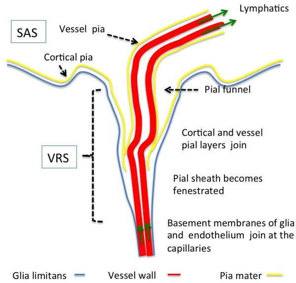
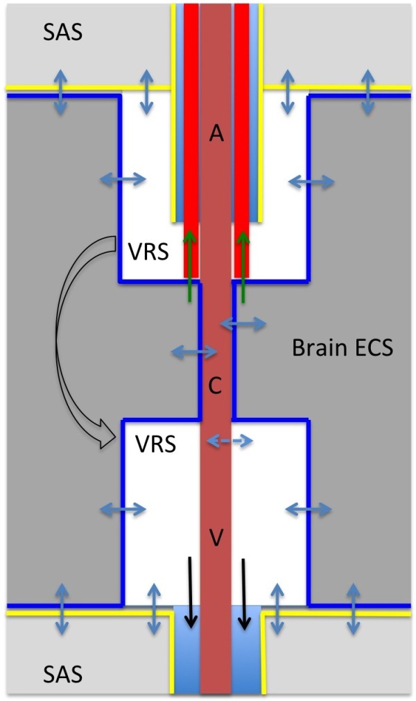
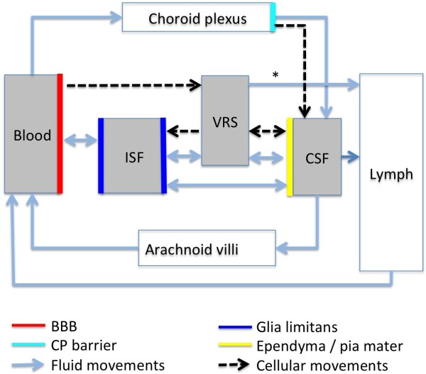
According to the traditional understanding of cerebrospinal fluid (CSF) physiology, the majority of CSF is produced by the choroid plexus, circulates through the ventricles, the cisterns, and the subarachnoid space to be absorbed into the blood by the arachnoid villi. This review surveys key developments leading to the traditional concept. Challenging this concept are novel insights utilizing molecular and cellular biology as well as neuroimaging, which indicate that CSF physiology may be much more complex than previously believed. The CSF circulation comprises not only a directed flow of CSF, but in addition a pulsatile to and fro movement throughout the entire brain with local fluid exchange between blood, interstitial fluid, and CSF. Astrocytes, aquaporins, and other membrane transporters are key elements in brain water and CSF homeostasis. A continuous bidirectional fluid exchange at the blood brain barrier produces flow rates, which exceed the choroidal CSF production rate by far. The CSF circulation around blood vessels penetrating from the subarachnoid space into the Virchow Robin spaces provides both a drainage pathway for the clearance of waste molecules from the brain and a site for the interaction of the systemic immune system with that of the brain. Important physiological functions, for example the regeneration of the brain during sleep, may depend on CSF circulation.
Overview
The claims and statistics of this paper haven’t been checked yet.
Run the check — free, no account needed. It reads the open-access text and checks the sources it cites.
Integrity signals
Registry
Cites
Statistics
Figures
Verification
Claims the paper makes
The claims and statistics of this paper haven’t been checked yet.
Run the check — free, no account needed. It reads the open-access text and checks the sources it cites.
Claims other papers make about this one
Each is checked against this paper’s own text: does it say what the citing paper says it does?
The cited source says what the claim attributes to it.
In the citing paper
“Uptake into arachnoid villi, as proposed more than one hundred years ago, seems to be of minor importance in humans, as reviewed by Brinker et al. (50) and as indicated in our recent study (13).”
From Brain-wide glymphatic enhancement and clearance in humans assessed with MRI. (2018) · cites this paper as [50]
Self-check
Percentages, sums and reported differences re-computed from the paper’s own numbers.
The self-check needs the paper’s full text.
Citations
Cites
Iliff, J. J., Wang, M., Liao, Y., Plogg, B. A., Peng, W., Gundersen, G. A., Benveniste, H., Vates, G. E., Deane, R., Goldman, S. A., et al. (2012) · Science Translational Medicine · doi:10.1126/scitranslmed.3003748 · Cited by 5,824
Sleep Drives Metabolite Clearance from the Adult Brain
Xie, L., Kang, H., Xu, Q., Chen, M. J., Liao, Y., Thiyagarajan, M., O’Donnell, J., Christensen, D. J., Nicholson, C., Iliff, J. J., et al. (2013) · Science · doi:10.1126/science.1241224 · Cited by 5,411
Structure and function of the blood–brain barrier
Abbott, N. J., Patabendige, A. A. K., Dolman, D. E. M., Yusof, S. R., & Begley, D. J. (2010) · Neurobiology of Disease · doi:10.1016/j.nbd.2009.07.030 · Cited by 5,185
Daneman, R., & Prat, A. (2015) · Cold Spring Harbor Perspectives in Biology · doi:10.1101/cshperspect.a020412 · Cited by 3,437
Aquaporin-4 deletion in mice reduces brain edema after acute water intoxication and ischemic stroke
Manley, G. T., Fujimura, M., Ma, T., Noshita, N., Filiz, F., Bollen, A. W., Chan, P. H., & Verkman, A. S. (2000) · Nature Medicine · doi:10.1038/72256 · Cited by 1,501
Diffusion in Brain Extracellular Space
Syková, E., & Nicholson, C. (2008) · Physiological Reviews · doi:10.1152/physrev.00027.2007 · Cited by 1,481
Nielsen, S. M., Nagelhus, E. A., Amiry-Moghaddam, M. R., Bourque, C. W., Agre, P. C., & Ottersen, O. P. (1997) · Journal of Neuroscience · doi:10.1523/jneurosci.17-01-00171.1997 · Cited by 1,440
Brain-wide pathway for waste clearance captured by contrast-enhanced MRI
Iliff, J. J., Lee, H., Yu, M., Feng, T., Logan, J., Nedergaard, M., & Benveniste, H. (2013) · Journal of Clinical Investigation · doi:10.1172/jci67677 · Cited by 1,148
Blood–brain barrier structure and function and the challenges for CNS drug delivery
Abbott, N. J. (2013) · Journal of Inherited Metabolic Disease · doi:10.1007/s10545-013-9608-0 · Cited by 951
Mathiisen, T. M., Lehre, K. P., Danbolt, N. C., & Ottersen, O. (2010) · Glia · doi:10.1002/glia.20990 · Cited by 893
Research works printed but not linked: 1
- 10.1186/1743-8454-5-10 · DOI · Paper page
Cited by
Sorted by citations
Items 801–821 of 821
Rasschaert, M. (2019) · theses.fr (ABES) · doi:10.70675/24be134az3f46z4a30za655z8252290d88a8
Rodrigues, K. S., Yamashita, R., & Katada, S. (2026) · Frontiers in Neural Circuits · doi:10.3389/fncir.2026.1818927
Intrathecal drug delivery—minimally invasive spine techniques: modern approaches
Sahni, S., Oehlermarx, W., Podgorski, E. M., & Kalia, H. (2026) · Elsevier eBooks · doi:10.1016/b978-0-443-27568-5.00014-2
Namaghi, S. S., Gajendran, M. K., Sokol, J. A., Krachmalnick, S., & Mehdizadeh, A. (2026) · Annals of Biomedical Engineering · doi:10.1007/s10439-026-04126-5
The glymphatic system and hydrocephalus: emerging insights into pathophysiological mechanisms
Geng, Z., Yang, Z., Li, P., Mao, Y., & Wei, J. (2026) · Reviews in the Neurosciences · doi:10.1515/revneuro-2026-0033 · Cited by 1
Chavan, S. S., Karande, G. S., & Patil, S. R. (2026) · Cureus · doi:10.7759/cureus.108726
Generative AI for CSF multi-omics: Learning strategies for small-cohort classification
Wuyart, A., & Millot, J. (2026) · Neuroscience Informatics · doi:10.1016/j.neuri.2026.100278
APOE and Proteomics in Chronic Inflammation
Shvetcov, A., & Finney, C. A. (2026) · Springer eBooks · doi:10.1007/978-3-031-96089-5_82
Ventricular System and CSF Flow
Palmiero, H. D. O. M. (2026) · Elsevier eBooks · doi:10.1016/b978-0-443-49243-3.00026-4
Olkhova, E. B., & Shandra, A. G. (2026) · Radiology - Practice · doi:10.52560/2713-0118-2026-3-73-87
Hyperandrogenism and polycystic ovary syndrome phenotypes in idiopathic intracranial hypertension
Savaşcı, D., Toydemir, H. E., Ekin, M., & Yayla, V. (2026) · Irish Journal of Medical Science (1971 -) · doi:10.1007/s11845-026-04552-3
Sun, L., He, L., Jian, Z., Lu, T., Miao, S., Zhou, R., Li, T., Yan, M., Zhang, Y., Yin, Y. J., et al. (2026) · medRxiv · doi:10.64898/2026.07.29.26359135
Cerebrospinal Fluid as a Window into Neuroimmune Responses During Brain Infection
Singh, G., & Rohlwink, U. K. (2026) · Cells · doi:10.3390/cells15181675
Akiyama, O., Shimizu, Y., Miyahara, R., Sakamoto, K., Ogino, I., Kodama, T., & Kondo, A. (2026) · World Neurosurgery · doi:10.1016/j.wneu.2026.125339
Wu, C., Yu, K., Shui, X., Lin, X., Fan, Y., & Chen, Y. (2026) · Frontiers in Neurology · doi:10.3389/fneur.2026.1861928
Lalou, A. D., Qvarlander, S., Behndig, S., Žižka, J., Malm, J., & Eklund, A. (2026) · Research Square · doi:10.21203/rs.3.rs-11018297/v1
Hyde, M., Cunningham, M. L., Chai, X., Peng, M., Hu, H. A., Lu, C., & Vasanthakumar, A. (2026) · BMC Biotechnology · doi:10.1186/s12896-026-01144-y
Medulloblastoma on the Move: How Tumor Cells Colonize the Leptomeninges – and How to Stop Them
Koubourli, D. V., Thompson, E. A., Gallitto, M., & Wechsler‐Reya, R. J. (2026) · Cellular and Molecular Bioengineering · doi:10.1007/s12195-026-00958-2
Bates, D., Wang, Q., Lang, J., Nathan, R., & Wu, Q. (2026) · Physics of Fluids · doi:10.1063/5.0334208
Metadata
- Authors
- Brinker, T., Stopa, E., Morrison, J., & Klinge, P.
- Affiliations
- Brown University (US)
- Corresponding author
- Brinker, Thomas
- ORCID
- Klinge, Petra
- Venue
- Fluids and Barriers of the CNS
- Published
- 2014
- Volume
- 11
- Issue
- 1
- Pages
- 10
- ISSN
- 2045-8118 (ISSN-L)
- PMC
- PMC4016637
- OpenAlex
- W2157256907
- Cited by
- 819
- References
- 167
- Citations by year
- 2026: 32 · 2025: 63 · 2024: 66 · 2023: 72 · 2022: 105 · 2021: 72 · 2020: 70 · 2019: 91 · 2018: 74 · 2017: 68 · 2016: 48 · 2015: 42 · 2014: 13
- FWCI
- 6.51
- Type
- journal article
- Language
- en
- Open access
- gold
- doi.org
- Licence
- cc-by
- Locations
- Fluids and Barriers of the CNS (publishedVersion, cc-by)
- Europe PMC (PubMed Central) (publishedVersion, cc-by)
- PubMed Central (publishedVersion, cc-by)
- PubMed (publishedVersion)
- Topics
- Health Sciences › Medicine › Clinical Neurology › Cerebrospinal fluid and hydrocephalus
- Life Sciences › Biochemistry, Genetics and Molecular Biology › Molecular Biology › Ion Transport and Channel Regulation
- Life Sciences › Neuroscience › Neurology › Barrier Structure and Function Studies
- Keywords
- cerebrospinal fluid flow, choroid plexus, arachnoid villi, CSF absorption, interstitial fluid, blood-CSF barrier, astrocytes, aquaporins, brain water homeostasis, perivascular spaces, cerebrospinal fluid drainage
About these checks
Registry, retraction, Cites and statistics results are computed, not judged, with one exception: a printed reference that no index lists is read once by a model before it counts as a citation error. Claims are extracted by a model and kept only when their quote appears verbatim in the text; each cited source is then checked, with a model’s help, for whether it says what the claim relies on. Those verdicts can be wrong — read the claim’s quote and the cited source before relying on one.
Statistics and arithmetic are recomputed, not judged.
Confidence bands are shown only for fields calibrated on our annotated test set. None is calibrated yet, so no band is shown.
What counts as a claim ›How we measure confidence ›How we check this ›
Are you an author, or do you see a problem with this page? Request a review
