

The DOI matches its Crossref record; 167 of the 167 references it cites were found in a registry or an index.
A new look at cerebrospinal fluid circulation
Open accessAbstract
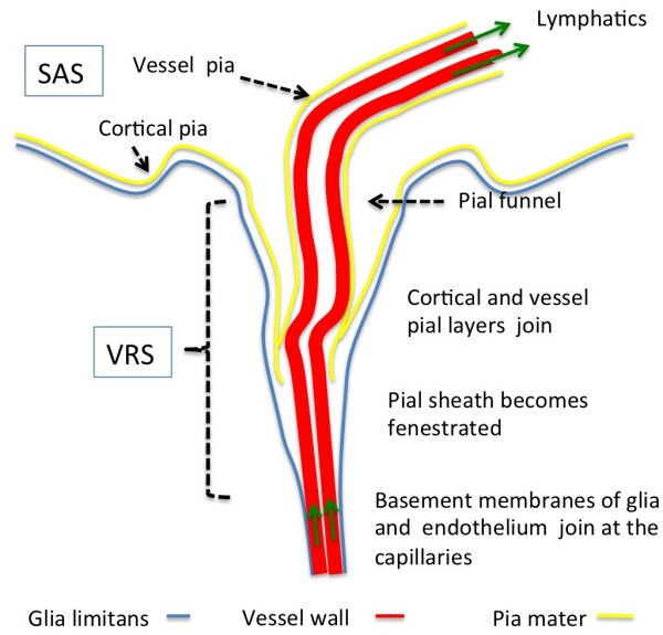
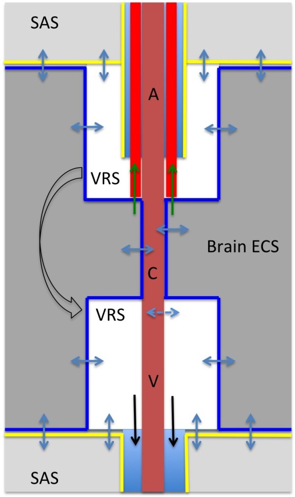
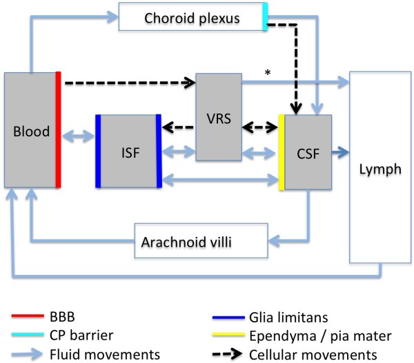
According to the traditional understanding of cerebrospinal fluid (CSF) physiology, the majority of CSF is produced by the choroid plexus, circulates through the ventricles, the cisterns, and the subarachnoid space to be absorbed into the blood by the arachnoid villi. This review surveys key developments leading to the traditional concept. Challenging this concept are novel insights utilizing molecular and cellular biology as well as neuroimaging, which indicate that CSF physiology may be much more complex than previously believed. The CSF circulation comprises not only a directed flow of CSF, but in addition a pulsatile to and fro movement throughout the entire brain with local fluid exchange between blood, interstitial fluid, and CSF. Astrocytes, aquaporins, and other membrane transporters are key elements in brain water and CSF homeostasis. A continuous bidirectional fluid exchange at the blood brain barrier produces flow rates, which exceed the choroidal CSF production rate by far. The CSF circulation around blood vessels penetrating from the subarachnoid space into the Virchow Robin spaces provides both a drainage pathway for the clearance of waste molecules from the brain and a site for the interaction of the systemic immune system with that of the brain. Important physiological functions, for example the regeneration of the brain during sleep, may depend on CSF circulation.
Overview
The claims and statistics of this paper haven’t been checked yet.
Run the check — free, no account needed. It reads the open-access text and checks the sources it cites.
Integrity signals
Registry
Cites
Statistics
Figures
Verification
Claims the paper makes
The claims and statistics of this paper haven’t been checked yet.
Run the check — free, no account needed. It reads the open-access text and checks the sources it cites.
Claims other papers make about this one
Each is checked against this paper’s own text: does it say what the citing paper says it does?
The cited source says what the claim attributes to it.
In the citing paper
“Uptake into arachnoid villi, as proposed more than one hundred years ago, seems to be of minor importance in humans, as reviewed by Brinker et al. (50) and as indicated in our recent study (13).”
From Brain-wide glymphatic enhancement and clearance in humans assessed with MRI. (2018) · cites this paper as [50]
Self-check
Percentages, sums and reported differences re-computed from the paper’s own numbers.
The self-check needs the paper’s full text.
Citations
Cites
Iliff, J. J., Wang, M., Liao, Y., Plogg, B. A., Peng, W., Gundersen, G. A., Benveniste, H., Vates, G. E., Deane, R., Goldman, S. A., et al. (2012) · Science Translational Medicine · doi:10.1126/scitranslmed.3003748 · Cited by 5,824
Sleep Drives Metabolite Clearance from the Adult Brain
Xie, L., Kang, H., Xu, Q., Chen, M. J., Liao, Y., Thiyagarajan, M., O’Donnell, J., Christensen, D. J., Nicholson, C., Iliff, J. J., et al. (2013) · Science · doi:10.1126/science.1241224 · Cited by 5,411
Structure and function of the blood–brain barrier
Abbott, N. J., Patabendige, A. A. K., Dolman, D. E. M., Yusof, S. R., & Begley, D. J. (2010) · Neurobiology of Disease · doi:10.1016/j.nbd.2009.07.030 · Cited by 5,185
Daneman, R., & Prat, A. (2015) · Cold Spring Harbor Perspectives in Biology · doi:10.1101/cshperspect.a020412 · Cited by 3,437
Aquaporin-4 deletion in mice reduces brain edema after acute water intoxication and ischemic stroke
Manley, G. T., Fujimura, M., Ma, T., Noshita, N., Filiz, F., Bollen, A. W., Chan, P. H., & Verkman, A. S. (2000) · Nature Medicine · doi:10.1038/72256 · Cited by 1,501
Diffusion in Brain Extracellular Space
Syková, E., & Nicholson, C. (2008) · Physiological Reviews · doi:10.1152/physrev.00027.2007 · Cited by 1,481
Nielsen, S. M., Nagelhus, E. A., Amiry-Moghaddam, M. R., Bourque, C. W., Agre, P. C., & Ottersen, O. P. (1997) · Journal of Neuroscience · doi:10.1523/jneurosci.17-01-00171.1997 · Cited by 1,440
Brain-wide pathway for waste clearance captured by contrast-enhanced MRI
Iliff, J. J., Lee, H., Yu, M., Feng, T., Logan, J., Nedergaard, M., & Benveniste, H. (2013) · Journal of Clinical Investigation · doi:10.1172/jci67677 · Cited by 1,148
Blood–brain barrier structure and function and the challenges for CNS drug delivery
Abbott, N. J. (2013) · Journal of Inherited Metabolic Disease · doi:10.1007/s10545-013-9608-0 · Cited by 951
Mathiisen, T. M., Lehre, K. P., Danbolt, N. C., & Ottersen, O. (2010) · Glia · doi:10.1002/glia.20990 · Cited by 893
Research works printed but not linked: 1
- 10.1186/1743-8454-5-10 · DOI · Paper page
Cited by
Sorted by citations
Items 201–300 of 821
Modulation of lymphatic transport in the central nervous system
Li, W., Chen, D., Liu, N., Luan, Y., Zhu, S., & Wang, H. (2022) · Theranostics · doi:10.7150/thno.66026 · Cited by 42
Horie, T., Kajihara, N., Matsumae, M., Obara, M., Hayashi, N., Hirayama, A., Takizawa, K., Takahara, T., Yatsushiro, S., & Kuroda, K. (2016) · World Neurosurgery · doi:10.1016/j.wneu.2016.07.062 · Cited by 41
Schön, M., Kovaničová, Z., Košutzká, Z., Nemec, M., Tomková, M., Jacková, L., Máderová, D., Slobodová, L., Valkovič, P., Ukropec, J., et al. (2019) · Scientific Reports · doi:10.1038/s41598-018-38201-2 · Cited by 40
Regulation of choroid plexus development and its functions
Kompaníková, P., & Bryja, V. (2022) · Cellular and Molecular Life Sciences · doi:10.1007/s00018-022-04314-1 · Cited by 42
Chen, J. C., Luu, A. R., Wise, N., Angelis, R. D., Agrawal, V., Mangini, L., Vincelette, J., Handyside, B., Sterling, H. J., Lo, M. J., et al. (2019) · Journal of Biological Chemistry · doi:10.1074/jbc.ra119.009811 · Cited by 41
Interactions of brain, blood, and CSF: a novel mathematical model of cerebral edema
Doron, O., Zadka, Y., Barnea, O., & Rosenthal, G. (2021) · Fluids and Barriers of the CNS · doi:10.1186/s12987-021-00274-z · Cited by 40
SCO-spondin, a giant matricellular protein that regulates cerebrospinal fluid activity
Sepúlveda, V., Maurelia, F., González, M., Aguayo, J., & Caprile, T. (2021) · Fluids and Barriers of the CNS · doi:10.1186/s12987-021-00277-w · Cited by 45
Targeting cerebrospinal fluid for discovery of brain cancer biomarkers
Shalaby, T., Achini, F., & Grotzer, M. A. (2016) · Journal of Cancer Metastasis and Treatment · doi:10.20517/2394-4722.2016.12 · Cited by 40
The use of cerebrospinal fluid in biomarker studies
Teunissen, C. E., Verheul, C., & Willemse, E. A. J. (2017) · Handbook of clinical neurology · doi:10.1016/b978-0-12-804279-3.00001-0 · Cited by 40
Multi-omic analysis elucidates the genetic basis of hydrocephalus
Hale, A. T., Bastarache, L. A., Morales, D. M., Wellons, J. C., Limbrick, D. D., & Gamazon, E. R. (2021) · Cell Reports · doi:10.1016/j.celrep.2021.109085 · Cited by 39
Lumbar Puncture Complications: A Review of Current Literature
Reis, A. E., Spano, M., Davis-Hayes, C., & Salama, G. R. (2024) · Current Pain and Headache Reports · doi:10.1007/s11916-024-01262-2 · Cited by 38
Gholampour, S., Bahmani, M., & Shariati, A. (2018) · Basic and Clinical Neuroscience Journal · doi:10.32598/bcn.9.10.285 · Cited by 38
Enhanced in vitro model of the CSF dynamics
Benninghaus, A., Balédent, O., Lokossou, A., Wembers, C. C., Leonhardt, S. K., & Radermacher, K. (2019) · Fluids and Barriers of the CNS · doi:10.1186/s12987-019-0131-z · Cited by 38
Navigating the ventricles: Novel insights into the pathogenesis of hydrocephalus
Bramall, A. N., Anton, E. S., Kahle, K. T., & Fecci, P. E. (2022) · EBioMedicine · doi:10.1016/j.ebiom.2022.103931 · Cited by 38
Fibrinogen, Fibrinogen-like 1 and Fibrinogen-like 2 Proteins, and Their Effects
Sulimai, N., Brown, J., & Lominadze, D. G. (2022) · Biomedicines · doi:10.3390/biomedicines10071712 · Cited by 38
Demeestere, D., Libert, C., & Vandenbroucke, R. E. (2015) · Brain Behavior and Immunity · doi:10.1016/j.bbi.2015.06.010 · Cited by 43
Nakatsuka, Y., Kawakita, F., Yasuda, R., Umeda, Y., Toma, N., Sakaida, H., & Suzuki, H. (2016) · Journal of neurosurgery · doi:10.3171/2016.5.jns152907 · Cited by 38
Choi, S., Jang, D. C., Chung, G., & Kim, S. K. (2022) · Cells · doi:10.3390/cells11193019 · Cited by 39
Infantile and Childhood Hydrocephalus
Whitehead, W. E., & Weiner, H. L. (2022) · New England Journal of Medicine · doi:10.1056/nejmra2116504 · Cited by 37
Mechanisms of cerebrospinal fluid and brain interstitial fluid production
Xiang, J., Hua, Y., Xi, G., & Keep, R. F. (2023) · Neurobiology of Disease · doi:10.1016/j.nbd.2023.106159 · Cited by 37
Lathe, R., Schultek, N. M., Balin, B. J., Ehrlich, G. D., Alberi, L. A., Perry, G. L., Breitschwerdt, E. B., Corry, D. B., Doty, R. L., Rissman, R. A., et al. (2023) · Alzheimer s & Dementia · doi:10.1002/alz.13076 · Cited by 39
Clinical magnetic resonance imaging evaluation of glymphatic function
Boyd, E., Kaur, J., Ding, G., Chopp, M., & Jiang, Q. (2024) · NMR in Biomedicine · doi:10.1002/nbm.5132 · Cited by 39
Ma, S., Björninen, T., Sydanheimo, L. T., Voutilainen, M. H., & Ukkonen, L. (2020) · IEEE Transactions on Antennas and Propagation · doi:10.1109/tap.2020.3016459 · Cited by 37
Tertiary Lymphoid Structures in the Central Nervous System: Implications for Glioblastoma
van de Walle, T., Vaccaro, A., Ramachandran, M., Pietilä, I., Essand, M., & Dimberg, A. (2021) · Frontiers in Immunology · doi:10.3389/fimmu.2021.724739 · Cited by 38
Flotillin is a Novel Diagnostic Blood Marker of Alzheimer’s Disease
Abdullah, M., Kimura, N., Akatsu, H., Hashizume, Y., Taslima, F., Tachita, T., Iida, S., Zou, K., Matsubara, E., & Michikawa, M. (2019) · Journal of Alzheimer s Disease · doi:10.3233/jad-190908 · Cited by 36
Hypertension, blood–brain barrier disruption and changes in intracranial pressure
Colombari, E., Biancardi, V. C., Colombari, D. S. A., Katayama, P. L., Medeiros, F., Aitken, A. V., Xavier, C. H., Pedrino, G. R., & Bragin, D. E. (2025) · The Journal of Physiology · doi:10.1113/jp285058 · Cited by 37
Cerebrospinal fluid proteomics in multiple sclerosis
Kroksveen, A. C., Opsahl, J. A., Guldbrandsen, A., Myhr, K., Oveland, E., Torkildsen, Ø., & Berven, F. S. (2014) · Biochimica et Biophysica Acta (BBA) - Proteins and Proteomics · doi:10.1016/j.bbapap.2014.12.013 · Cited by 35
Wilson, R., Osborne, C., & Halsey, C. (2018) · Pediatric Drugs · doi:10.1007/s40272-018-0298-9 · Cited by 35
Sepp, A., Meno‐Tetang, G. M. L., Weber, A. D., Sanderson, A., Schön, O., & Berges, A. (2019) · Journal of Pharmacokinetics and Pharmacodynamics · doi:10.1007/s10928-019-09640-9 · Cited by 35
Rasmussen, J. C., Kwon, S., Pinal, A., Bareis, A., Velasquez, F. C., Janssen, C. F., Morrow, J. R., Fife, C. E., Karni, R. J., & Sevick-Muraca, E. M. (2020) · Physiological Reports · doi:10.14814/phy2.14375 · Cited by 36
Rethinking the Conditions and Mechanism for Glymphatic Clearance
Ferris, C. F. (2021) · Frontiers in Neuroscience · doi:10.3389/fnins.2021.624690 · Cited by 36
Trillo-Contreras, J. L., Ramírez‐Lorca, R., Villadiego, J., & Echevarrı́a, M. (2022) · Biomolecules · doi:10.3390/biom12040530 · Cited by 35
Barami, K., & Sood, S. (2016) · Child s Nervous System · doi:10.1007/s00381-015-3010-1 · Cited by 35
Knirsch, W., Heye, K., Scheer, I., Tuura, R., Schranz, D., Hahn, A., Wetterling, K., Beck, I., Latal, B., & Reich, B. (2016) · European Journal of Cardio-Thoracic Surgery · doi:10.1093/ejcts/ezw399 · Cited by 35
Agarwal, N., Contarino, C., & Toro, E. F. (2019) · Veins and Lymphatics · doi:10.4081/vl.2019.8470 · Cited by 36
GALECTIN-8 Is a Neuroprotective Factor in the Brain that Can Be Neutralized by Human Autoantibodies
Pardo, E., Barake, F., Godoy, J. A., Oyanadel, C., Espinoza, S., Metz, C., Retamal, C., Massardo, L., Tapia‐Rojas, C., Inestrosa, N. C., et al. (2019) · Molecular Neurobiology · doi:10.1007/s12035-019-1621-3 · Cited by 34
Bandara, E. M. S., Asih, P. R., Pedrini, S., Hone, E., Fernando, W. M. A. D. B., & Martins, R. N. (2025) · Molecular Neurobiology · doi:10.1007/s12035-025-05219-3 · Cited by 37
Physiological Considerations for Modeling in vivo Antibody-Target Interactions
Dunlap, T. C., & Cao, Y. (2022) · Frontiers in Pharmacology · doi:10.3389/fphar.2022.856961 · Cited by 36
Intrathecal delivery and its applications in leptomeningeal disease
Khang, M., Bindra, R. S., & Saltzman, W. M. (2022) · Advanced Drug Delivery Reviews · doi:10.1016/j.addr.2022.114338 · Cited by 33
Ocular Lymphatic and Glymphatic Systems: Implications for Retinal Health and Disease
Uddin, M. N., & Rutar, M. (2022) · International Journal of Molecular Sciences · doi:10.3390/ijms231710139 · Cited by 35
Méndez-García, L. A., Escobedo, G., Minguer-Uribe, A. G., Viurcos-Sanabria, R., Aguayo-Guerrero, J. A., Carrillo-Ruiz, J. D., & Solleiro-Villavicencio, H. (2022) · Frontiers in Cellular Neuroscience · doi:10.3389/fncel.2022.977039 · Cited by 35
Islam, A., Sakamoto, T., Zhai, Q., Rahman, M. M., Mamun, M. A., Takahashi, Y., Kahyo, T., & Setou, M. (2022) · Pharmaceuticals · doi:10.3390/ph15111314 · Cited by 34
Amyotrophic lateral sclerosis: Is the spinal fluid pathway involved in seeding and spread?
Smith, R. A., Myers, K. J., Ravits, J., & Bowser, R. P. (2015) · Medical Hypotheses · doi:10.1016/j.mehy.2015.07.014 · Cited by 34
MRI investigation of glymphatic responses to Gd‐DTPA infusion rates
Ding, G., Chopp, M., Li, L., Zhang, L., Davoodi‐Bojd, E., Li, Q., Zhang, Z., & Jiang, Q. (2018) · Journal of Neuroscience Research · doi:10.1002/jnr.24325 · Cited by 33
The escape of retrobulbar cerebrospinal fluid in the astronaut’s eye: mission impossible?
Wostyn, P., Mader, T. H., Gibson, C. R., & Killer, H. E. (2019) · Eye · doi:10.1038/s41433-019-0453-8 · Cited by 32
Review of Cerebrospinal Fluid Physiology and Dynamics: A Call for Medical Education Reform
Atchley, T. J., Vukić, B., Vukić, M., & Walters, B. C. (2022) · Neurosurgery · doi:10.1227/neu.0000000000002000 · Cited by 32
Cerebrospinal Fluid Biomarkers of Neurodegeneration Are Decreased or Normal in Narcolepsy
Jennum, P., Pedersen, L. Ø., Bahl, J. M. C., Modvig, S., Fog, K., Holm, A., Kornum, B. R., & Gammeltoft, S. (2016) · SLEEP · doi:10.1093/sleep/zsw006 · Cited by 33
Continuous intrathecal orexin delivery inhibits cataplexy in a murine model of narcolepsy
Kaushik, M. K., Aritake, K., Imanishi, A., Kanbayashi, T., Ichikawa, T., Shimizu, T., Urade, Y., & Yanagisawa, M. (2018) · Proceedings of the National Academy of Sciences · doi:10.1073/pnas.1722686115 · Cited by 34
Central nervous system lymphatic unit, immunity, and epilepsy: Is there a link?
Noè, F. M., & Marchi, N. (2019) · Epilepsia Open · doi:10.1002/epi4.12302 · Cited by 33
Intrathecal delivery of Macromolecules: Clinical status and emerging technologies
Ajeeb, R., & Clegg, J. R. (2023) · Advanced Drug Delivery Reviews · doi:10.1016/j.addr.2023.114949 · Cited by 31
The Cryptococcus neoformans Titan Cell: From In Vivo Phenomenon to In Vitro Model
Zhou, X., & Ballou, E. R. (2018) · Current Clinical Microbiology Reports · doi:10.1007/s40588-018-0107-9 · Cited by 31
Sindeeva, O. A., Verkhovskii, R. A., Sarimollaoglu, M., Афанасьева, Г. А., Fedonnikov, A., Osintsev, E. Y., Kurochkina, E. N., Gorin, D. A., Deyev, S. M., Zharov, V. P., et al. (2019) · Cells · doi:10.3390/cells8101195 · Cited by 30
Characterization of the circadian oscillator in the choroid plexus of rats
Yamaguchi, T., Hamada, T., Matsuzaki, T., & Iijima, N. (2020) · Biochemical and Biophysical Research Communications · doi:10.1016/j.bbrc.2020.01.125 · Cited by 30
Taoka, T., Ito, R., Nakamichi, R., Nakane, T., Kawai, H., & Naganawa, S. (2022) · Magnetic Resonance in Medical Sciences · doi:10.2463/mrms.rev.2022-0012 · Cited by 30
Levels of amyloid-beta-42 and CSF pressure are directly related in patients with Alzheimer’s disease
Schirinzi, T., Lazzaro, G. D., Sancesario, G. M., Colona, V. L., Scaricamazza, E., Mercuri, N. B., Martorana, A., & Sancesario, G. (2017) · Journal of Neural Transmission · doi:10.1007/s00702-017-1786-8 · Cited by 31
Tuță, S. (2022) · Life · doi:10.3390/life12060854 · Cited by 29
Hebisch, M., Klostermeier, S., Wolf, K., Boccaccini, A. R., Wolf, S. E., Tanzi, R. E., & Kim, D. Y. (2023) · Advanced Science · doi:10.1002/advs.202205037 · Cited by 31
Engineering choroid plexus-on-a-chip with oscillatory flow for modeling brain metastasis
Lim, J., Rhee, S., Choi, H., Lee, J., Kuttappan, S., Nguyen, T. T., Choi, S., Kim, Y., & Jeon, N. L. (2023) · Materials Today Bio · doi:10.1016/j.mtbio.2023.100773 · Cited by 29
Flanagan, M. F. (2015) · Neurology Research International · doi:10.1155/2015/794829 · Cited by 29
Impaired cerebrospinal fluid dynamics along the entire optic nerve in normal‐tension glaucoma
Pircher, A., Montali, M., Wostyn, P., Pircher, J., Berberat, J., Remonda, L., & Killer, H. E. (2018) · Acta Ophthalmologica · doi:10.1111/aos.13647 · Cited by 29
Barrier dysfunction or drainage reduction: differentiating causes of CSF protein increase
Asgari, M., de Zélicourt, D., & Kurtcuoglu, V. (2017) · Fluids and Barriers of the CNS · doi:10.1186/s12987-017-0063-4 · Cited by 28
Systematic analysis of the cerebrospinal fluid proteome of fibromyalgia patients
Khoonsari, P. E., Musunri, S., Herman, S., Svensson, C. I., Tanum, L., Gordh, T., & Kultima, K. (2018) · Journal of Proteomics · doi:10.1016/j.jprot.2018.04.014 · Cited by 28
How does the brain remove its waste metabolites from within?
Cheng, Y., & Haorah, J. (2019) · PubMed Central · Cited by 28
Özöner, B., Kilic, M., Aydın, L., Aydın, S., Arslan, Y. K., Müslüman, A. M., & Yılmaz, A. (2020) · European Journal of Trauma and Emergency Surgery · doi:10.1007/s00068-020-01409-x · Cited by 28
Cerebrospinal fluid turnover as a driver of brain clearance
Smets, N. G., Strijkers, G. J., Vinje, V., & Bakker, E. N. T. P. (2023) · NMR in Biomedicine · doi:10.1002/nbm.5029 · Cited by 28
The Glymphatic-Lymphatic Continuum: Opportunities for Osteopathic Manipulative Medicine
Hitscherich, K. J., Smith, K., Cuoco, J. A., Ruvolo, K. E., Mancini, J., Leheste, J. R., & Torres, G. (2016) · Journal of Osteopathic Medicine · doi:10.7556/jaoa.2016.033 · Cited by 30
Shear-Induced Amyloid Formation in the Brain: I. Potential Vascular and Parenchymal Processes
Trumbore, C. N. (2016) · Journal of Alzheimer's Disease · doi:10.3233/jad-160027 · Cited by 27
Li, Q., Aalling, N., Förstera, B., Ertürk, A., Nedergaard, M., Møllgård, K., & Xavier, A. L. R. (2020) · Fluids and Barriers of the CNS · doi:10.1186/s12987-020-0176-z · Cited by 27
Stadio, A. D., Ralli, M., Roccamatisi, D., Scarpa, A., Volpe, A. D., Cassandro, C., Ricci, G., Greco, A., & Bernitsas, E. (2021) · Neurological Sciences · doi:10.1007/s10072-020-04948-8 · Cited by 27
Choi, H., Mun, S., Joo, E.-J., Lee, K. Y., Kang, H., & Lee, J. (2021) · International Journal of Biological Macromolecules · doi:10.1016/j.ijbiomac.2021.05.172 · Cited by 27
Yang, Y., & Zhan, W. (2022) · Pharmaceutical Research · doi:10.1007/s11095-022-03261-7 · Cited by 27
Human parasagittal dura is a potential neuroimmune interface
Melin, E., Ringstad, G. A., Valnes, L. M., & Eide, P. K. (2023) · Communications Biology · doi:10.1038/s42003-023-04634-3 · Cited by 27
Sennfält, S., Thrippleton, M. J., Stringer, M. S., Arteaga, C., Chappell, F. M., Doubal, F., García, D. J., Zhang, J., Cheng, Y., & Wardlaw, J. M. (2023) · Journal of Cerebral Blood Flow & Metabolism · doi:10.1177/0271678x231179555 · Cited by 27
Agnati, L. F., Marcoli, M., Leo, G., Maura, G., & Guidolin, D. (2017) · International Journal of Molecular Medicine · doi:10.3892/ijmm.2017.2874 · Cited by 27
Rodgers, J., Bradley, B., & Kennedy, P. G. E. (2017) · Methods · doi:10.1016/j.ymeth.2017.06.015 · Cited by 27
Emerging Insights and New Perspectives on the Nature of Hydrocephalus
Tomycz, L. D., Hale, A. T., & George, T. M. (2017) · Pediatric Neurosurgery · doi:10.1159/000484173 · Cited by 27
Targeting the Choroid Plexuses for Protein Drug Delivery
Bryniarski, M. A., Ren, T., Rizvi, A. R., Snyder, A. M., & Morris, M. E. (2020) · Pharmaceutics · doi:10.3390/pharmaceutics12100963 · Cited by 28
Endothelin-1 mediated vasoconstriction leads to memory impairment and synaptic dysfunction
Diwakar, L., Gowaikar, R., Chithanathan, K., Gnanabharathi, B., Tomar, D. S., & Ravindranath, V. (2021) · Scientific Reports · doi:10.1038/s41598-021-84258-x · Cited by 28
Recent Update on Pharmacokinetics and Drug Metabolism in CNS-based Drug Discovery
Sanap, S. N., Bisen, A. C., Kedar, A. S., Agrawal, S., & Bhatta, R. S. (2023) · Current Pharmaceutical Design · doi:10.2174/1381612829666230707121415 · Cited by 26
Retrolaminar Migration of Intraocular Silicone Oil
Boren, R. A., Cloy, C. D., Gupta, A. S., Dewan, V. N., & Hogan, R. N. (2016) · Journal of Neuro-Ophthalmology · doi:10.1097/wno.0000000000000440 · Cited by 26
Djukic, M., Lange, P., Erbguth, F., & Nau, R. (2022) · Journal of Neuroinflammation · doi:10.1186/s12974-022-02538-3 · Cited by 25
A hitchhiker’s guide to cerebrospinal fluid biomarkers for neuro-oncology
Riviere-Cazaux, C., Keough, M. B., Zuccato, J., Kumar, R. R., Schulz, S. C., Warrington, A. E., Ruff, M. W., Ellingson, B. M., Sanai, N., Campian, J. L., et al. (2024) · Neuro-Oncology · doi:10.1093/neuonc/noae276 · Cited by 25
Schmidt, M. J., Rummel, C. D., Hauer, J., Kolecka, M., Ondreka, N., McClure, V., & Roth, J. (2016) · Fluids and Barriers of the CNS · doi:10.1186/s12987-016-0034-1 · Cited by 25
Targeting choroid plexus epithelium as a novel therapeutic strategy for hydrocephalus
Yang, Y., He, J. W., Wang, Y.-C., Wang, C., Tan, C., Liao, J., Tong, L., & Xiao, G. (2022) · Journal of Neuroinflammation · doi:10.1186/s12974-022-02500-3 · Cited by 26
Vandenbulcke, S., Pauw, T. D., Dewaele, F., Degroote, J., & Segers, P. (2022) · Frontiers in Bioengineering and Biotechnology · doi:10.3389/fbioe.2022.1040517 · Cited by 25
Electrokinetic Convection-Enhanced Delivery of Solutes to the Brain
Faraji, A. H., Jaquins‐Gerstl, A., Valenta, A. C., Ou, Y., & Weber, S. G. (2020) · ACS Chemical Neuroscience · doi:10.1021/acschemneuro.0c00037 · Cited by 25
Hydrocephalus: An update on latest progress in pathophysiological and therapeutic research
Anwar, F., Zhang, K., Sun, C., Pang, M., Zhou, W., Li, H., He, R., Liu, X., & Ming, D. (2024) · Biomedicine & Pharmacotherapy · doi:10.1016/j.biopha.2024.117702 · Cited by 24
Simats, A., García‐Berrocoso, T., Ramiro, L., Giralt, D., Gill, N., Peñalba, A., Bustamante, A., Rosell, A., & Montaner, J. (2018) · Scientific Reports · doi:10.1038/s41598-018-26237-3 · Cited by 24
Cao, D., Kang, N., Pillai, J. J., Miao, X., Paez, A. G., Xu, X., Xu, J., Xu, L., Qin, Q., van Zijl, P. C. M., et al. (2020) · Magnetic Resonance in Medicine · doi:10.1002/mrm.28389 · Cited by 25
[Retracted] Glymphatic System: Emerging Therapeutic Target for Neurological Diseases
Xuan, X., Zhou, G., Chen, C., Shao, A., Zhou, Y., Li, X., & Zhou, J. (2022) · Oxidative Medicine and Cellular Longevity · doi:10.1155/2022/6189170 · Cited by 25
RetractedAβ Clearance, “hub” of Multiple Deficiencies Leading to Alzheimer Disease
Gallina, P., Scollato, A., Conti, R., Lorenzo, N. D., & Porfirio, B. (2015) · Frontiers in Aging Neuroscience · doi:10.3389/fnagi.2015.00200 · Cited by 25
Zhang, C., Lin, J., Wei, F., Song, J. H., Chen, W., Shan, L., Xue, R., Wang, G., Tao, J., Zhang, G., et al. (2018) · Life Sciences · doi:10.1016/j.lfs.2018.03.057 · Cited by 24
Joseph, C. R., Benhatzel, C. M., Stern, L. J., Hopper, O. M., & Lockwood, M. D. (2020) · Magnetic Resonance Materials in Physics Biology and Medicine · doi:10.1007/s10334-019-00818-3 · Cited by 24
Robert, S. M., Reeves, B. C., Marlier, A., Duy, P. Q., DeSpenza, T., Kundishora, A. J., Kiziltug, E., Singh, A. K., Allington, G., Alper, S. L., et al. (2021) · Child s Nervous System · doi:10.1007/s00381-021-05255-z · Cited by 24
Basic principles of neuroimmunology
Yoshida, T. M., Wang, A., & Hafler, D. A. (2022) · Seminars in Immunopathology · doi:10.1007/s00281-022-00951-7 · Cited by 27
Ellenbogen, Y., Brar, K., Yang, K., Lee, Y., & Ajani, O. (2020) · Journal of Neurosurgery Pediatrics · doi:10.3171/2020.4.peds19720 · Cited by 23
Decoupling Between Brain Activity and Cerebrospinal Fluid Movement in Neurological Disorders
Liu, X. (2023) · Journal of Magnetic Resonance Imaging · doi:10.1002/jmri.29148 · Cited by 23
The role of astrocytes in the glymphatic network: a narrative review
Das, N., Dhamija, R., & Sarkar, S. (2023) · Metabolic Brain Disease · doi:10.1007/s11011-023-01327-y · Cited by 23
Immunological Barriers to Stem Cell Therapy in the Central Nervous System
Tullis, G. E., Spears, K., & Kirk, M. D. (2014) · Stem Cells International · doi:10.1155/2014/507905 · Cited by 23
Szpecht, D., Gadzinowski, J., Seremak‐Mrozikiewicz, A., Kurzawińska, G., & Szymankiewicz, M. (2017) · Scientific Reports · doi:10.1038/srep42541 · Cited by 23
Metadata
- Authors
- Brinker, T., Stopa, E., Morrison, J., & Klinge, P.
- Affiliations
- Brown University (US)
- Corresponding author
- Brinker, Thomas
- ORCID
- Klinge, Petra
- Venue
- Fluids and Barriers of the CNS
- Published
- 2014
- Volume
- 11
- Issue
- 1
- Pages
- 10
- ISSN
- 2045-8118 (ISSN-L)
- PMC
- PMC4016637
- OpenAlex
- W2157256907
- Cited by
- 819
- References
- 167
- Citations by year
- 2026: 32 · 2025: 63 · 2024: 66 · 2023: 72 · 2022: 105 · 2021: 72 · 2020: 70 · 2019: 91 · 2018: 74 · 2017: 68 · 2016: 48 · 2015: 42 · 2014: 13
- FWCI
- 6.51
- Type
- journal article
- Language
- en
- Open access
- gold
- doi.org
- Licence
- cc-by
- Locations
- Fluids and Barriers of the CNS (publishedVersion, cc-by)
- Europe PMC (PubMed Central) (publishedVersion, cc-by)
- PubMed Central (publishedVersion, cc-by)
- PubMed (publishedVersion)
- Topics
- Health Sciences › Medicine › Clinical Neurology › Cerebrospinal fluid and hydrocephalus
- Life Sciences › Biochemistry, Genetics and Molecular Biology › Molecular Biology › Ion Transport and Channel Regulation
- Life Sciences › Neuroscience › Neurology › Barrier Structure and Function Studies
- Keywords
- cerebrospinal fluid flow, choroid plexus, arachnoid villi, CSF absorption, interstitial fluid, blood-CSF barrier, astrocytes, aquaporins, brain water homeostasis, perivascular spaces, cerebrospinal fluid drainage
About these checks
Registry, retraction, Cites and statistics results are computed, not judged, with one exception: a printed reference that no index lists is read once by a model before it counts as a citation error. Claims are extracted by a model and kept only when their quote appears verbatim in the text; each cited source is then checked, with a model’s help, for whether it says what the claim relies on. Those verdicts can be wrong — read the claim’s quote and the cited source before relying on one.
Statistics and arithmetic are recomputed, not judged.
Confidence bands are shown only for fields calibrated on our annotated test set. None is calibrated yet, so no band is shown.
What counts as a claim ›How we measure confidence ›How we check this ›
Are you an author, or do you see a problem with this page? Request a review
