

The DOI matches its Crossref record; 167 of the 167 references it cites were found in a registry or an index.
A new look at cerebrospinal fluid circulation
Open accessAbstract
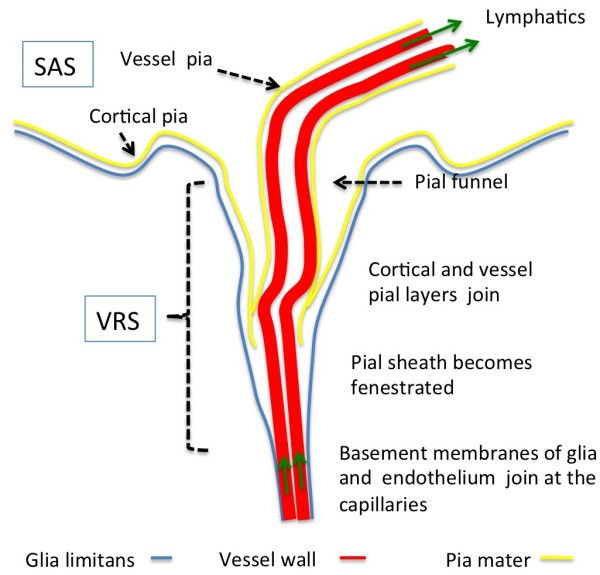
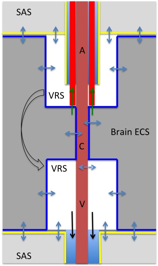
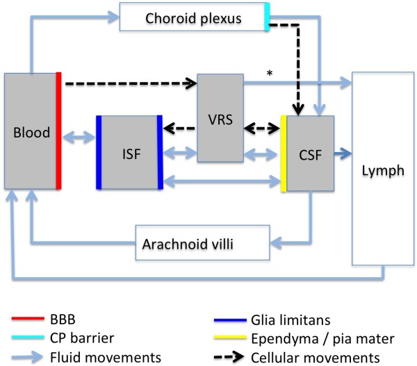
According to the traditional understanding of cerebrospinal fluid (CSF) physiology, the majority of CSF is produced by the choroid plexus, circulates through the ventricles, the cisterns, and the subarachnoid space to be absorbed into the blood by the arachnoid villi. This review surveys key developments leading to the traditional concept. Challenging this concept are novel insights utilizing molecular and cellular biology as well as neuroimaging, which indicate that CSF physiology may be much more complex than previously believed. The CSF circulation comprises not only a directed flow of CSF, but in addition a pulsatile to and fro movement throughout the entire brain with local fluid exchange between blood, interstitial fluid, and CSF. Astrocytes, aquaporins, and other membrane transporters are key elements in brain water and CSF homeostasis. A continuous bidirectional fluid exchange at the blood brain barrier produces flow rates, which exceed the choroidal CSF production rate by far. The CSF circulation around blood vessels penetrating from the subarachnoid space into the Virchow Robin spaces provides both a drainage pathway for the clearance of waste molecules from the brain and a site for the interaction of the systemic immune system with that of the brain. Important physiological functions, for example the regeneration of the brain during sleep, may depend on CSF circulation.
Overview
The claims and statistics of this paper haven’t been checked yet.
Run the check — free, no account needed. It reads the open-access text and checks the sources it cites.
Integrity signals
Registry
Cites
Statistics
Figures
Verification
Claims the paper makes
The claims and statistics of this paper haven’t been checked yet.
Run the check — free, no account needed. It reads the open-access text and checks the sources it cites.
Claims other papers make about this one
Each is checked against this paper’s own text: does it say what the citing paper says it does?
The cited source says what the claim attributes to it.
In the citing paper
“Uptake into arachnoid villi, as proposed more than one hundred years ago, seems to be of minor importance in humans, as reviewed by Brinker et al. (50) and as indicated in our recent study (13).”
From Brain-wide glymphatic enhancement and clearance in humans assessed with MRI. (2018) · cites this paper as [50]
Self-check
Percentages, sums and reported differences re-computed from the paper’s own numbers.
The self-check needs the paper’s full text.
Citations
Cites
Iliff, J. J., Wang, M., Liao, Y., Plogg, B. A., Peng, W., Gundersen, G. A., Benveniste, H., Vates, G. E., Deane, R., Goldman, S. A., et al. (2012) · Science Translational Medicine · doi:10.1126/scitranslmed.3003748 · Cited by 5,824
Sleep Drives Metabolite Clearance from the Adult Brain
Xie, L., Kang, H., Xu, Q., Chen, M. J., Liao, Y., Thiyagarajan, M., O’Donnell, J., Christensen, D. J., Nicholson, C., Iliff, J. J., et al. (2013) · Science · doi:10.1126/science.1241224 · Cited by 5,411
Structure and function of the blood–brain barrier
Abbott, N. J., Patabendige, A. A. K., Dolman, D. E. M., Yusof, S. R., & Begley, D. J. (2010) · Neurobiology of Disease · doi:10.1016/j.nbd.2009.07.030 · Cited by 5,185
Daneman, R., & Prat, A. (2015) · Cold Spring Harbor Perspectives in Biology · doi:10.1101/cshperspect.a020412 · Cited by 3,437
Aquaporin-4 deletion in mice reduces brain edema after acute water intoxication and ischemic stroke
Manley, G. T., Fujimura, M., Ma, T., Noshita, N., Filiz, F., Bollen, A. W., Chan, P. H., & Verkman, A. S. (2000) · Nature Medicine · doi:10.1038/72256 · Cited by 1,501
Diffusion in Brain Extracellular Space
Syková, E., & Nicholson, C. (2008) · Physiological Reviews · doi:10.1152/physrev.00027.2007 · Cited by 1,481
Nielsen, S. M., Nagelhus, E. A., Amiry-Moghaddam, M. R., Bourque, C. W., Agre, P. C., & Ottersen, O. P. (1997) · Journal of Neuroscience · doi:10.1523/jneurosci.17-01-00171.1997 · Cited by 1,440
Brain-wide pathway for waste clearance captured by contrast-enhanced MRI
Iliff, J. J., Lee, H., Yu, M., Feng, T., Logan, J., Nedergaard, M., & Benveniste, H. (2013) · Journal of Clinical Investigation · doi:10.1172/jci67677 · Cited by 1,148
Blood–brain barrier structure and function and the challenges for CNS drug delivery
Abbott, N. J. (2013) · Journal of Inherited Metabolic Disease · doi:10.1007/s10545-013-9608-0 · Cited by 951
Mathiisen, T. M., Lehre, K. P., Danbolt, N. C., & Ottersen, O. (2010) · Glia · doi:10.1002/glia.20990 · Cited by 893
Research works printed but not linked: 1
- 10.1186/1743-8454-5-10 · DOI · Paper page
Cited by
Sorted by citations
Flow of cerebrospinal fluid is driven by arterial pulsations and is reduced in hypertension.
Mestre, H., Tithof, J., Du, T., Song, W., Peng, W., Sweeney, A. M., Olveda, G., Thomas, J. H., Nedergaard, M., & Kelley, D. H. (2018) · Nature Communications · doi:10.1038/s41467-018-07318-3 · Cited by 1,110
CNS lymphatic drainage and neuroinflammation are regulated by meningeal lymphatic vasculature.
Louveau, A., Herz, J., Alme, M. N., Salvador, A. F., Dong, M. Q., Viar, K. E., Herod, S. G., Knopp, J., Setliff, J. C., Lupi, A. L., et al. (2018) · Nature Neuroscience · doi:10.1038/s41593-018-0227-9 · Cited by 956
Proal, A. D., & VanElzakker, M. B. (2021) · Frontiers in Microbiology · doi:10.3389/fmicb.2021.698169 · Cited by 921
Blood GFAP as an emerging biomarker in brain and spinal cord disorders
Abdelhak, A., Foschi, M., Abu-Rumeileh, S., Yue, J. K., D’Anna, L., Huss, A., Oeckl, P., Ludolph, A. C., Kuhle, J., Petzold, A., et al. (2022) · Nature Reviews Neurology · doi:10.1038/s41582-021-00616-3 · Cited by 807
Mechanisms of fluid movement into, through and out of the brain: evaluation of the evidence
Hladky, S. B., & Barrand, M. A. (2014) · Fluids and Barriers of the CNS · doi:10.1186/2045-8118-11-26 · Cited by 633
Rasmussen, M. K., Mestre, H., & Nedergaard, M. (2021) · Physiological Reviews · doi:10.1152/physrev.00031.2020 · Cited by 642
Intranasal Oxytocin: Myths and Delusions
Leng, G., & Ludwig, M. (2016) · Biological Psychiatry · doi:10.1016/j.biopsych.2015.05.003 · Cited by 616
Molecular pathophysiology of cerebral edema
Stokum, J. A., Gerzanich, V., & Simard, J. M. (2015) · Journal of Cerebral Blood Flow & Metabolism · doi:10.1177/0271678x15617172 · Cited by 613
The role of brain barriers in fluid movement in the CNS: is there a ‘glymphatic’ system?
Abbott, N. J., Pizzo, M. E., Preston, J. E., Janigro, D., & Thorne, R. G. (2018) · Acta Neuropathologica · doi:10.1007/s00401-018-1812-4 · Cited by 593
Brain-wide glymphatic enhancement and clearance in humans assessed with MRI.
Ringstad, G., Valnes, L. M., Dale, A. M., Pripp, A. H., Vatnehol, S.-A. S., Emblem, K. E., Mardal, K.-A., & Eide, P. K. (2018) · JCI Insight · doi:10.1172/jci.insight.121537 · Cited by 533
“005 Basal ganglia [11, 12, 13, 50, 51, 52] –3 ± 8 –6 ± 14 1 ± 25 –4 ± 14 4 ± 18 32 –8 ± 18 0.”
The Glymphatic System and Waste Clearance with Brain Aging: A Review
Benveniste, H., Liu, X., Koundal, S., Sanggaard, S., Lee, H., & Wardlaw, J. (2019) · Gerontology · doi:10.1159/000490349 · Cited by 517
Gänger, S., & Schindowski, K. (2018) · Pharmaceutics · doi:10.3390/pharmaceutics10030116 · Cited by 447
A balanced view of the cerebrospinal fluid composition and functions: Focus on adult humans
Spector, R., Snodgrass, S. R., & Johanson, C. E. (2015) · Experimental Neurology · doi:10.1016/j.expneurol.2015.07.027 · Cited by 467
Proulx, S. T. (2021) · Cellular and Molecular Life Sciences · doi:10.1007/s00018-020-03706-5 · Cited by 412
Cho, A.-N., Jin, Y., An, Y., Kim, J., Choi, Y. S., Lee, J. S., Kim, J., Choi, W.-Y., Koo, D.-J., Yu, W., et al. (2021) · Nature Communications · doi:10.1038/s41467-021-24775-5 · Cited by 397
The development and functions of multiciliated epithelia
Spassky, N., & Meunier, A. (2017) · Nature Reviews Molecular Cell Biology · doi:10.1038/nrm.2017.21 · Cited by 390
Deer, T. R., Pope, J. E., Hayek, S. M., Bux, A., Buchser, E., Eldabe, S. S., Andrés, J. A. D., Erdek, M. A., Patin, D. J., Grider, J. S., et al. (2017) · Neuromodulation Technology at the Neural Interface · doi:10.1111/ner.12538 · Cited by 366
Bakker, E. N. T. P., Bacskai, B. J., Arbel-Ornath, M., Aldea, R., Bedussi, B., Morris, A. W. J., Weller, R. O., & Carare, R. O. (2016) · Cellular and Molecular Neurobiology · doi:10.1007/s10571-015-0273-8 · Cited by 353
Faraco, G., Sugiyama, Y., Lane, D., Garcia-Bonilla, L., Chang, H., Santisteban, M. M., Racchumi, G., Murphy, M., Van Rooijen, N., Anrather, J., et al. (2016) · Journal of Clinical Investigation · doi:10.1172/jci86950 · Cited by 350
Simon, M. J., & Iliff, J. J. (2015) · Biochimica et Biophysica Acta (BBA) - Molecular Basis of Disease · doi:10.1016/j.bbadis.2015.10.014 · Cited by 349
Cerebrospinal fluid dynamics and intracranial pressure elevation in neurological diseases
Bothwell, S. W., Janigro, D., & Patabendige, A. (2019) · Fluids and Barriers of the CNS · doi:10.1186/s12987-019-0129-6 · Cited by 348
The blood–brain barrier in psychosis
Pollak, T. A., Drndarski, S., Stone, J. M., David, A. S., McGuire, P. K., & Abbott, N. J. (2017) · The Lancet Psychiatry · doi:10.1016/s2215-0366(17)30293-6 · Cited by 311
A journey into the brain: insight into how bacterial pathogens cross blood–brain barriers
Coureuil, M., Lécuyer, H., Bourdoulous, S., & Nassif, X. (2017) · Nature Reviews Microbiology · doi:10.1038/nrmicro.2016.178 · Cited by 305
Hladky, S. B., & Barrand, M. A. (2016) · Fluids and Barriers of the CNS · doi:10.1186/s12987-016-0040-3 · Cited by 304
Vascular basement membranes as pathways for the passage of fluid into and out of the brain
Morris, A. W. J., Sharp, M. M., Albargothy, N. J., Fernandes, R., Hawkes, C. A., Verma, A., Weller, R. O., & Carare, R. O. (2016) · Acta Neuropathologica · doi:10.1007/s00401-016-1555-z · Cited by 289
Brain aging mechanisms with mechanical manifestations
Blinkouskaya, Y., Caçoilo, A., Gollamudi, T., Jalalian, S., & Weickenmeier, J. (2021) · Mechanisms of Ageing and Development · doi:10.1016/j.mad.2021.111575 · Cited by 306
Lymphatic drainage system of the brain: A novel target for intervention of neurological diseases
Sun, B., Wang, L.-H., Yang, T., Sun, J., Mao, L., Yang, M., Yuan, H., Colvin, R. A., & Yang, X. (2017) · Progress in Neurobiology · doi:10.1016/j.pneurobio.2017.08.007 · Cited by 264
Achariyar, T. M., Li, B., Peng, W., Verghese, P. B., Shi, Y., McConnell, E., Benraiss, A., Kasper, T., Song, W., Takano, T., et al. (2016) · Molecular Neurodegeneration · doi:10.1186/s13024-016-0138-8 · Cited by 256
Non-invasive MR imaging of human brain lymphatic networks with connections to cervical lymph nodes
Albayram, S., Smith, G., Tufan, F., Tuna, İ. S., Bostancıklıoğlu, M., Zile, M. R., & Albayram, Ö. (2022) · Nature Communications · doi:10.1038/s41467-021-27887-0 · Cited by 246
RETINAL VASCULAR CYSTOID MACULAR EDEMA
Spaide, R. F. (2016) · Retina · doi:10.1097/iae.0000000000001158 · Cited by 236
Inflammation in acquired hydrocephalus: pathogenic mechanisms and therapeutic targets
Karimy, J. K., Reeves, B. C., Damisah, E. C., Duy, P. Q., Antwi, P., David, W. B., Wang, K. X.-L., Schiff, S. J., Limbrick, D. D., Alper, S. L., et al. (2020) · Nature Reviews Neurology · doi:10.1038/s41582-020-0321-y · Cited by 237
Zhang, L., & Hargens, A. R. (2017) · Physiological Reviews · doi:10.1152/physrev.00017.2016 · Cited by 234
A Historical Review of Brain Drug Delivery
Pardridge, W. M. (2022) · Pharmaceutics · doi:10.3390/pharmaceutics14061283 · Cited by 219
A balanced view of choroid plexus structure and function: Focus on adult humans
Spector, R., Keep, R. F., Snodgrass, S. R., Smith, Q. R., & Johanson, C. E. (2015) · Experimental Neurology · doi:10.1016/j.expneurol.2015.02.032 · Cited by 232
The brain interstitial system: Anatomy, modeling, in vivo measurement, and applications
Lei, Y., Han, H., Yuan, F., Javeed, A., & Zhao, Y. L. (2016) · Progress in Neurobiology · doi:10.1016/j.pneurobio.2015.12.007 · Cited by 228
The Glymphatic System (En)during Inflammation
Mogensen, F. L., Delle, C., & Nedergaard, M. (2021) · International Journal of Molecular Sciences · doi:10.3390/ijms22147491 · Cited by 222
Obermeier, B., Verma, A., & Ransohoff, R. M. (2016) · Handbook of clinical neurology · doi:10.1016/b978-0-444-63432-0.00003-7 · Cited by 216
The cerebrospinal fluid and barriers – anatomic and physiologic considerations
Tumani, H., Huss, A. M., & Bachhuber, F. (2017) · Handbook of clinical neurology · doi:10.1016/b978-0-12-804279-3.00002-2 · Cited by 217
Hladky, S. B., & Barrand, M. A. (2018) · Fluids and Barriers of the CNS · doi:10.1186/s12987-018-0113-6 · Cited by 206
Olstad, E. W., Ringers, C., Hansen, J. N., Wens, A., Brandt, C., Wachten, D., Yaksi, E., & Jurisch‐Yaksi, N. (2018) · Current Biology · doi:10.1016/j.cub.2018.11.059 · Cited by 214
The Glymphatic Pathway: Waste Removal from the CNS via Cerebrospinal Fluid Transport
Benveniste, H., Lee, H., & Volkow, N. D. (2017) · The Neuroscientist · doi:10.1177/1073858417691030 · Cited by 205
Hydrocephalus: historical analysis and considerations for treatment
Hochstetler, A., Raskin, J. S., & Blazer‐Yost, B. L. (2022) · European journal of medical research · doi:10.1186/s40001-022-00798-6 · Cited by 190
General Anesthesia Inhibits the Activity of the “Glymphatic System”
Gakuba, C., Gaberel, T., Goursaud, S., Bourges, J., Di Palma, C., Quenault, A., de Lizarrondo, S. M., Vivien, D., & Gauberti, M. (2018) · Theranostics · doi:10.7150/thno.19154 · Cited by 190
The choroid plexus is an important circadian clock component
Myung, J., Schmal, C., Hong, S., Tsukizawa, Y., Rose, P., Zhang, Y., Holtzman, M. J., Schutter, E. D., Herzel, H., Bordyugov, G., et al. (2018) · Nature Communications · doi:10.1038/s41467-018-03507-2 · Cited by 193
Intrathecal drug delivery in the era of nanomedicine
Fowler, M. J., Cotter, J. D., Knight, B. E., Sevick-Muraca, E. M., Sandberg, D. I., & Sirianni, R. W. (2020) · Advanced Drug Delivery Reviews · doi:10.1016/j.addr.2020.02.006 · Cited by 187
Paravascular spaces at the brain surface: Low resistance pathways for cerebrospinal fluid flow
Bedussi, B., Almasian, M., de Vos, J., VanBavel, E., & Bakker, E. N. (2018) · Journal of Cerebral Blood Flow & Metabolism · doi:10.1177/0271678x17737984 · Cited by 181
How Do Meningeal Lymphatic Vessels Drain the CNS?
Raper, D. M. S., Louveau, A., & Kipnis, J. (2016) · Trends in Neurosciences · doi:10.1016/j.tins.2016.07.001 · Cited by 184
Lun, M. P., Johnson, M. B., Broadbelt, K. G., Watanabe, M., Kang, Y., Chau, K. F., Springel, M. W., Malesz, A., Sousa, A. M. M., Pletikos, M., et al. (2015) · Journal of Neuroscience · doi:10.1523/jneurosci.3081-14.2015 · Cited by 187
Akiguchi, I., Pallàs, M., Budka, H., Akiyama, H., Ueno, M., Han, J., Yagi, H., Tomohumi, N., Chiba, Y., Sugiyama, H., et al. (2017) · Neuropathology · doi:10.1111/neup.12373 · Cited by 175
Evidence for Cerebrospinal Fluid Entry Into the Optic Nerve via a Glymphatic Pathway
Mathieu, E., Gupta, N., Ahari, A., Zhou, X., Hanna, J., & Yücel, Y. H. (2017) · Investigative Ophthalmology & Visual Science · doi:10.1167/iovs.17-22290 · Cited by 176
Harrison, I. F., Siow, B., Akilo, A. B., Evans, P. G., Ismail, O., Ohene, Y., Nahavandi, P., Thomas, D. L., Lythgoe, M. F., & Wells, J. A. (2018) · eLife · doi:10.7554/elife.34028 · Cited by 170
Bacyinski, A., Xu, M., Wang, W., & Hu, J. (2017) · Frontiers in Neuroanatomy · doi:10.3389/fnana.2017.00101 · Cited by 170
Lizano, P. L., Lutz, O., Ling, G., Lee, A. M., Eum, S., Bishop, J. R., Kelly, S., Pasternak, O., Clementz, B. A., Pearlson, G. D., et al. (2019) · American Journal of Psychiatry · doi:10.1176/appi.ajp.2019.18070825 · Cited by 164
Torralva, R., & Janowsky, A. J. (2019) · Journal of Pharmacology and Experimental Therapeutics · doi:10.1124/jpet.119.258566 · Cited by 161
Cerebrospinal fluid circulation: What do we know and how do we know it?
Harris, C., Khasawneh, A., & Garling, R. J. (2018) · Brain Circulation · doi:10.4103/bc.bc_3_18 · Cited by 160
Jošt, G., Lenhard, D. C., Sieber, M. A., Lohrke, J., Frenzel, T., & Pietsch, H. (2015) · Investigative Radiology · doi:10.1097/rli.0000000000000242 · Cited by 159
Fluid dynamics of cerebrospinal fluid flow in perivascular spaces
Thomas, J. H. (2019) · Journal of The Royal Society Interface · doi:10.1098/rsif.2019.0572 · Cited by 155
Current Evidence for a Role of the Kynurenine Pathway of Tryptophan Metabolism in Multiple Sclerosis
Lovelace, M. D., Varney, B., Sundaram, G., Franco, N. F., Ng, M. L., Pai, S., Lim, C. K., Guillemin, G. J., & Brew, B. J. (2016) · Frontiers in Immunology · doi:10.3389/fimmu.2016.00246 · Cited by 161
Mechanisms of Astrocyte-Mediated Cerebral Edema
Stokum, J. A., Kurland, D. B., Gerzanich, V., & Simard, J. M. (2014) · Neurochemical Research · doi:10.1007/s11064-014-1374-3 · Cited by 158
Sundaram, S., Hughes, R. L., Peterson, E., Müller-Oehring, E. M., Brontë-Stewart, H. M., Poston, K. L., Faerman, A., Bhowmick, C., & Schulte, T. (2019) · Neuroscience & Biobehavioral Reviews · doi:10.1016/j.neubiorev.2019.05.016 · Cited by 152
Chen, S., Shao, L., & Ma, L. (2021) · Frontiers in Cellular Neuroscience · doi:10.3389/fncel.2021.716825 · Cited by 154
Klotho controls the brain-immune system interface in the choroid plexus
Zhu, L., Stein, L. R., Kim, D., Ho, K., Yu, G.-Q., Zhan, L., Larsson, T. E., & Mucke, L. (2018) · Proceedings of the National Academy of Sciences · doi:10.1073/pnas.1808609115 · Cited by 148
Nonsurgical therapy for hydrocephalus: a comprehensive and critical review
Bigio, M. R. D., & Curzio, D. L. D. (2015) · Fluids and Barriers of the CNS · doi:10.1186/s12987-016-0025-2 · Cited by 144
The action radius of oxytocin release in the mammalian CNS: from single vesicles to behavior
Chini, B., Verhage, M., & Grinevich, V. (2017) · Trends in Pharmacological Sciences · doi:10.1016/j.tips.2017.08.005 · Cited by 141
The choroid plexus: a missing link in our understanding of brain development and function
Saunders, N. R., Dzięgielewska, K. M., Fame, R. M., Lehtinen, M. K., & Liddelow, S. A. (2022) · Physiological Reviews · doi:10.1152/physrev.00060.2021 · Cited by 145
Normal-pressure hydrocephalus: A critical review
Oliveira, L. M., Nitríni, R., & Román, G. C. (2019) · Dementia & Neuropsychologia · doi:10.1590/1980-57642018dn13-020001 · Cited by 136
Mechanisms of Pathogen Invasion into the Central Nervous System
Cain, M. D., Salimi, H., Diamond, M., & Klein, R. S. (2019) · Neuron · doi:10.1016/j.neuron.2019.07.015 · Cited by 137
A new glaucoma hypothesis: a role of glymphatic system dysfunction
Wostyn, P., Dam, D. V., Audenaert, K., Killer, H. E., Deyn, P. P. D., & Groot, V. D. (2015) · Fluids and Barriers of the CNS · doi:10.1186/s12987-015-0012-z · Cited by 137
Role of choroid plexus in cerebrospinal fluid hydrodynamics
Orešković, D., Radoš, M., & Klarica, M. (2017) · Neuroscience · doi:10.1016/j.neuroscience.2017.04.025 · Cited by 133
Direct Measurement of Cerebrospinal Fluid Production in Mice
Liu, G., Mestre, H., Sweeney, A. M., Sun, Q., Weikop, P., Du, T., & Nedergaard, M. (2020) · Cell Reports · doi:10.1016/j.celrep.2020.108524 · Cited by 131
Brain-to-cervical lymph node signaling after stroke.
Esposito, E., Ahn, B. J., Shi, J., Nakamura, Y., Park, J. H., Mandeville, E. T., Yu, Z., Chan, S. J., Desai, R., Hayakawa, A., et al. (2019) · Nature Communications · doi:10.1038/s41467-019-13324-w · Cited by 123
Coles, J. A., Myburgh, E., Brewer, J. M., & McMenamin, P. G. (2017) · Progress in Neurobiology · doi:10.1016/j.pneurobio.2017.05.002 · Cited by 123
Non-Invasive MRI of Blood–Cerebrospinal Fluid Barrier Function
Evans, P., Sokolska, M., Alves, A., Harrison, I. F., Ohene, Y., Nahavandi, P., Ismail, O., Miranda, E., Lythgoe, M. F., Thomas, D. L., et al. (2020) · Nature Communications · doi:10.1038/s41467-020-16002-4 · Cited by 122
Headache arising from idiopathic changes in CSF pressure
Ducros, A., & Biousse, V. (2015) · The Lancet Neurology · doi:10.1016/s1474-4422(15)00015-0 · Cited by 120
The Glymphatic System in Humans: Investigations With Magnetic Resonance Imaging
Naganawa, S., Taoka, T., Ito, R., & Kawamura, M. (2023) · Investigative Radiology · doi:10.1097/rli.0000000000000969 · Cited by 120
The Interstitial System of the Brain in Health and Disease
Shetty, A. K., & Zanirati, G. (2020) · Aging and Disease · doi:10.14336/ad.2020.0103 · Cited by 121
Lindstrøm, E. K., Ringstad, G. A., Mardal, K., & Eide, P. K. (2018) · NeuroImage Clinical · doi:10.1016/j.nicl.2018.09.006 · Cited by 113
Kan, S., Aoyagi-Scharber, M., Le, S. Q., Vincelette, J., Ohmi, K., Bullens, S., Wendt, D. J., Christianson, T., Tiger, P. M. N., Brown, J. R.-M., et al. (2014) · Proceedings of the National Academy of Sciences · doi:10.1073/pnas.1416660111 · Cited by 111
The choroid plexus and its role in the pathogenesis of neurological infections
Thompson, D., Brissette, C. A., & Watt, J. A. (2022) · Fluids and Barriers of the CNS · doi:10.1186/s12987-022-00372-6 · Cited by 116
Antisense drugs make sense for neurological diseases
Bennett, C. F., Kordasiewicz, H. B., & Cleveland, D. W. (2021) · Annual Review of Pharmacology and Toxicology · doi:10.1146/annurev-pharmtox-010919-023738 · Cited by 108
Meningeal Lymphatics: From Anatomy to Central Nervous System Immune Surveillance
Papadopoulos, Z., Herz, J., & Kipnis, J. (2020) · The Journal of Immunology · doi:10.4049/jimmunol.1900838 · Cited by 114
Convection-Enhanced Delivery: Connection to and Impact of Interstitial Fluid Flow
Stine, C. A., & Munson, J. M. (2019) · Frontiers in Oncology · doi:10.3389/fonc.2019.00966 · Cited by 110
Kyrtsos, C. R., & Baras, J. S. (2015) · PLoS ONE · doi:10.1371/journal.pone.0139574 · Cited by 108
Decreased CSF clearance and increased brain amyloid in Alzheimer’s disease
Li, Y., Rusinek, H., Butler, T., Glodzik, L., Pirraglia, E., Babich, J. W., Mozley, P. D., Nehmeh, S. A., Pahlajani, S. S., Wang, X. H., et al. (2022) · Fluids and Barriers of the CNS · doi:10.1186/s12987-022-00318-y · Cited by 110
Glymphatic Cerebrospinal Fluid and Solute Transport Quantified by MRI and PET Imaging
Benveniste, H., Lee, H., Ozturk, B., Chen, X., Koundal, S., Vaska, P., Tannenbaum, A., & Volkow, N. D. (2020) · Neuroscience · doi:10.1016/j.neuroscience.2020.11.014 · Cited by 106
Cerebral Vessels: An Overview of Anatomy, Physiology, and Role in the Drainage of Fluids and Solutes
Agarwal, N., & Carare, R. O. (2021) · Frontiers in Neurology · doi:10.3389/fneur.2020.611485 · Cited by 119
Kaur, J., Fahmy, L. M., Davoodi‐Bojd, E., Zhang, L., Ding, G., Hu, J., Zhang, Z., Chopp, M., & Jiang, Q. (2021) · Frontiers in Neuroanatomy · doi:10.3389/fnana.2021.665803 · Cited by 108
Outer brain barriers in rat and human development
Brøchner, C. B., Holst, C. B., & Møllgård, K. (2015) · Frontiers in Neuroscience · doi:10.3389/fnins.2015.00075 · Cited by 110
Clearance from the mouse brain by convection of interstitial fluid towards the ventricular system
Bedussi, B., van Lier, M. G. J. T. B., Bartstra, J. W., de Vos, J., Siebes, M., VanBavel, E., & Bakker, E. N. T. P. (2015) · Fluids and Barriers of the CNS · doi:10.1186/s12987-015-0019-5 · Cited by 102
A new look at cerebrospinal fluid movement
Orešković, D., & Klarica, M. (2014) · Fluids and Barriers of the CNS · doi:10.1186/2045-8118-11-16 · Cited by 104
Fülöp, G., Tarantini, S., Yabluchanskiy, A., Molnár, A., Prodan, C. I., Kiss, T., Csípő, T., Lipécz, Á., Balasubramanian, P., Farkas, E., et al. (2019) · American Journal of Physiology-Heart and Circulatory Physiology · doi:10.1152/ajpheart.00776.2018 · Cited by 101
Solar, P., Zamani, A., Lakatosová, K., & Joukal, M. (2022) · Fluids and Barriers of the CNS · doi:10.1186/s12987-022-00312-4 · Cited by 103
Cerebrospinal Fluid–Basic Concepts Review
Czarniak, N., Kamińska, J., Matowicka-Karna, J., & Koper‐Lenkiewicz, O. M. (2023) · Biomedicines · doi:10.3390/biomedicines11051461 · Cited by 110
Meningeal lymphatic drainage: novel insights into central nervous system disease
Zhang, Q., Niu, Y., Li, Y., Xia, C., Chen, Z., Chen, Y., & Feng, H. (2025) · Signal Transduction and Targeted Therapy · doi:10.1038/s41392-025-02177-z · Cited by 100
Emami, A., Tepper, J., Short, B. G., Yaksh, T. L., Bendele, A. M., Ramani, T., Cisternas, A. F., Chang, J., & Mellon, R. D. (2017) · International Journal of Toxicology · doi:10.1177/1091581817741840 · Cited by 97
Wagner, K., Unger, L., Salman, M. M., Kitchen, P., Bill, R. M., & Yool, A. J. (2022) · International Journal of Molecular Sciences · doi:10.3390/ijms23031388 · Cited by 97
Harnessing cerebrospinal fluid circulation for drug delivery to brain tissues
Naseri Kouzehgarani, G., Feldsien, T., Engelhard, H. H., Mirakhur, K. K., Phipps, C., Nimmrich, V., Clausznitzer, D., & Lefebvre, D. R. (2021) · Advanced Drug Delivery Reviews · doi:10.1016/j.addr.2021.03.002 · Cited by 95
Mestre, H., Verma, N., Greene, T. D., Lin, L. A., Ladrón-de-Guevara, A., Sweeney, A. M., Liu, G., Thomas, V. K., Galloway, C. A., Bentley, K. L. D. M., et al. (2022) · Nature Communications · doi:10.1038/s41467-022-31257-9 · Cited by 97
Liquid biopsies in patients with diffuse glioma
Best, M. G., Sol, N., Zijl, S., Reijneveld, J. C., Wesseling, P., & Würdinger, T. (2015) · Acta Neuropathologica · doi:10.1007/s00401-015-1399-y · Cited by 94
Korbecki, A., Zimny, A., Podgórski, P., Sąsiadek, M. J., & Bladowska, J. (2019) · Polish Journal of Radiology · doi:10.5114/pjr.2019.86881 · Cited by 92
Metadata
- Authors
- Brinker, T., Stopa, E., Morrison, J., & Klinge, P.
- Affiliations
- Brown University (US)
- Corresponding author
- Brinker, Thomas
- ORCID
- Klinge, Petra
- Venue
- Fluids and Barriers of the CNS
- Published
- 2014
- Volume
- 11
- Issue
- 1
- Pages
- 10
- ISSN
- 2045-8118 (ISSN-L)
- PMC
- PMC4016637
- OpenAlex
- W2157256907
- Cited by
- 819
- References
- 167
- Citations by year
- 2026: 32 · 2025: 63 · 2024: 66 · 2023: 72 · 2022: 105 · 2021: 72 · 2020: 70 · 2019: 91 · 2018: 74 · 2017: 68 · 2016: 48 · 2015: 42 · 2014: 13
- FWCI
- 6.51
- Type
- journal article
- Language
- en
- Open access
- gold
- doi.org
- Licence
- cc-by
- Locations
- Fluids and Barriers of the CNS (publishedVersion, cc-by)
- Europe PMC (PubMed Central) (publishedVersion, cc-by)
- PubMed Central (publishedVersion, cc-by)
- PubMed (publishedVersion)
- Topics
- Health Sciences › Medicine › Clinical Neurology › Cerebrospinal fluid and hydrocephalus
- Life Sciences › Biochemistry, Genetics and Molecular Biology › Molecular Biology › Ion Transport and Channel Regulation
- Life Sciences › Neuroscience › Neurology › Barrier Structure and Function Studies
- Keywords
- cerebrospinal fluid flow, choroid plexus, arachnoid villi, CSF absorption, interstitial fluid, blood-CSF barrier, astrocytes, aquaporins, brain water homeostasis, perivascular spaces, cerebrospinal fluid drainage
About these checks
Registry, retraction, Cites and statistics results are computed, not judged, with one exception: a printed reference that no index lists is read once by a model before it counts as a citation error. Claims are extracted by a model and kept only when their quote appears verbatim in the text; each cited source is then checked, with a model’s help, for whether it says what the claim relies on. Those verdicts can be wrong — read the claim’s quote and the cited source before relying on one.
Statistics and arithmetic are recomputed, not judged.
Confidence bands are shown only for fields calibrated on our annotated test set. None is calibrated yet, so no band is shown.
What counts as a claim ›How we measure confidence ›How we check this ›
Are you an author, or do you see a problem with this page? Request a review
