

The DOI matches its Crossref record; 167 of the 167 references it cites were found in a registry or an index.
A new look at cerebrospinal fluid circulation
Open accessAbstract
According to the traditional understanding of cerebrospinal fluid (CSF) physiology, the majority of CSF is produced by the choroid plexus, circulates through the ventricles, the cisterns, and the subarachnoid space to be absorbed into the blood by the arachnoid villi. This review surveys key developments leading to the traditional concept. Challenging this concept are novel insights utilizing molecular and cellular biology as well as neuroimaging, which indicate that CSF physiology may be much more complex than previously believed. The CSF circulation comprises not only a directed flow of CSF, but in addition a pulsatile to and fro movement throughout the entire brain with local fluid exchange between blood, interstitial fluid, and CSF. Astrocytes, aquaporins, and other membrane transporters are key elements in brain water and CSF homeostasis. A continuous bidirectional fluid exchange at the blood brain barrier produces flow rates, which exceed the choroidal CSF production rate by far. The CSF circulation around blood vessels penetrating from the subarachnoid space into the Virchow Robin spaces provides both a drainage pathway for the clearance of waste molecules from the brain and a site for the interaction of the systemic immune system with that of the brain. Important physiological functions, for example the regeneration of the brain during sleep, may depend on CSF circulation.
Overview
The claims and statistics of this paper haven’t been checked yet.
Run the check — free, no account needed. It reads the open-access text and checks the sources it cites.
Integrity signals
Registry
Cites
Statistics
Figures
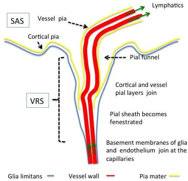
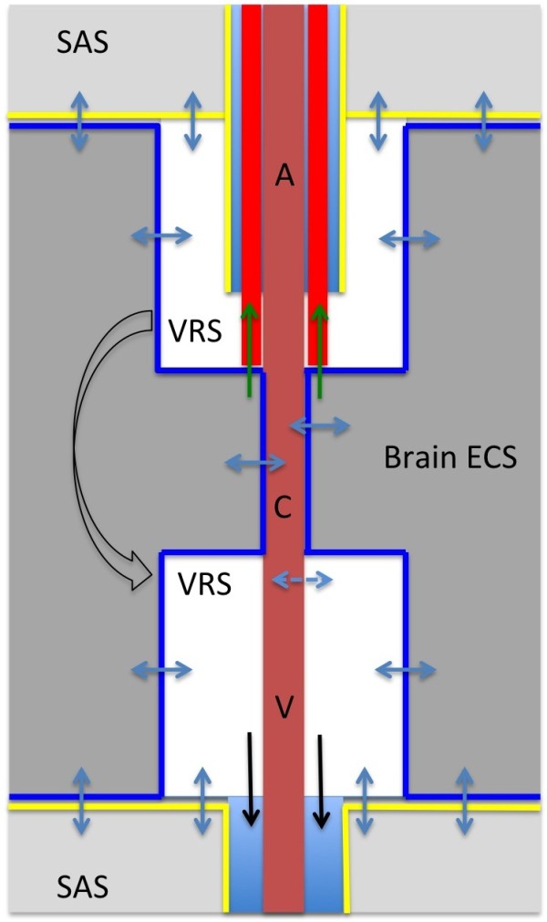
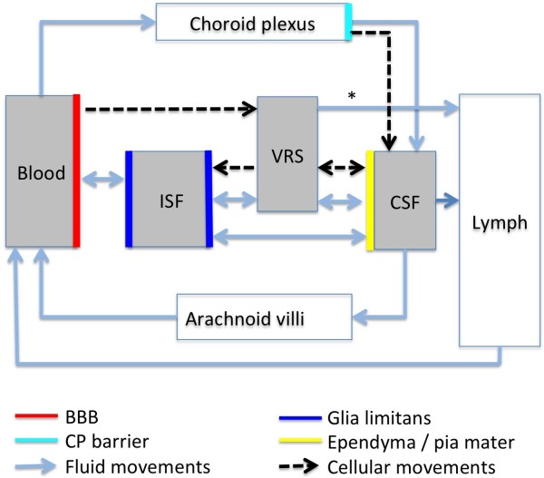
Verification
Claims the paper makes
The claims and statistics of this paper haven’t been checked yet.
Run the check — free, no account needed. It reads the open-access text and checks the sources it cites.
Claims other papers make about this one
Each is checked against this paper’s own text: does it say what the citing paper says it does?
The cited source says what the claim attributes to it.
In the citing paper
“Uptake into arachnoid villi, as proposed more than one hundred years ago, seems to be of minor importance in humans, as reviewed by Brinker et al. (50) and as indicated in our recent study (13).”
From Brain-wide glymphatic enhancement and clearance in humans assessed with MRI. (2018) · cites this paper as [50]
Self-check
Percentages, sums and reported differences re-computed from the paper’s own numbers.
The self-check needs the paper’s full text.
Citations
Cites
Iliff, J. J., Wang, M., Liao, Y., Plogg, B. A., Peng, W., Gundersen, G. A., Benveniste, H., Vates, G. E., Deane, R., Goldman, S. A., et al. (2012) · Science Translational Medicine · doi:10.1126/scitranslmed.3003748 · Cited by 5,824
Sleep Drives Metabolite Clearance from the Adult Brain
Xie, L., Kang, H., Xu, Q., Chen, M. J., Liao, Y., Thiyagarajan, M., O’Donnell, J., Christensen, D. J., Nicholson, C., Iliff, J. J., et al. (2013) · Science · doi:10.1126/science.1241224 · Cited by 5,411
Structure and function of the blood–brain barrier
Abbott, N. J., Patabendige, A. A. K., Dolman, D. E. M., Yusof, S. R., & Begley, D. J. (2010) · Neurobiology of Disease · doi:10.1016/j.nbd.2009.07.030 · Cited by 5,185
Daneman, R., & Prat, A. (2015) · Cold Spring Harbor Perspectives in Biology · doi:10.1101/cshperspect.a020412 · Cited by 3,437
Aquaporin-4 deletion in mice reduces brain edema after acute water intoxication and ischemic stroke
Manley, G. T., Fujimura, M., Ma, T., Noshita, N., Filiz, F., Bollen, A. W., Chan, P. H., & Verkman, A. S. (2000) · Nature Medicine · doi:10.1038/72256 · Cited by 1,501
Diffusion in Brain Extracellular Space
Syková, E., & Nicholson, C. (2008) · Physiological Reviews · doi:10.1152/physrev.00027.2007 · Cited by 1,481
Nielsen, S. M., Nagelhus, E. A., Amiry-Moghaddam, M. R., Bourque, C. W., Agre, P. C., & Ottersen, O. P. (1997) · Journal of Neuroscience · doi:10.1523/jneurosci.17-01-00171.1997 · Cited by 1,440
Brain-wide pathway for waste clearance captured by contrast-enhanced MRI
Iliff, J. J., Lee, H., Yu, M., Feng, T., Logan, J., Nedergaard, M., & Benveniste, H. (2013) · Journal of Clinical Investigation · doi:10.1172/jci67677 · Cited by 1,148
Blood–brain barrier structure and function and the challenges for CNS drug delivery
Abbott, N. J. (2013) · Journal of Inherited Metabolic Disease · doi:10.1007/s10545-013-9608-0 · Cited by 951
Mathiisen, T. M., Lehre, K. P., Danbolt, N. C., & Ottersen, O. (2010) · Glia · doi:10.1002/glia.20990 · Cited by 893
Research works printed but not linked: 1
- 10.1186/1743-8454-5-10 · DOI · Paper page
Cited by
Sorted by citations
Items 101–200 of 821
Taoka, T., & Naganawa, S. (2020) · Japanese Journal of Radiology · doi:10.1007/s11604-020-01017-0 · Cited by 88
(2020) · International Review of Neurobiology · doi:10.1016/bs.irn.2020.02.006 · Cited by 87
Cerebrospinal fluid hypersecretion in pediatric hydrocephalus
Karimy, J. K., Durán, D., Hu, J. K., Furey, C. G., Gaillard, J. R., Bayri, Y., Rice, H., DiLuna, M. L., Gerzanich, V., Simard, J. M., et al. (2016) · Neurosurgical FOCUS · doi:10.3171/2016.8.focus16278 · Cited by 90
Spontaneous intracranial hypotension: review and expert opinion
Ferrante, E., Trimboli, M., & Rubino, F. (2019) · Acta Neurologica Belgica · doi:10.1007/s13760-019-01166-8 · Cited by 87
A translational platform PBPK model for antibody disposition in the brain
Chang, H.-Y., Wu, S., Meno‐Tetang, G. M. L., & Shah, D. K. (2019) · Journal of Pharmacokinetics and Pharmacodynamics · doi:10.1007/s10928-019-09641-8 · Cited by 87
Therapeutic Options for Mucopolysaccharidoses: Current and Emerging Treatments
Sawamoto, K., Stapleton, M., Alméciga-Díaz, C. J., Espejo-Mojica, A. J., Losada, J. C., Súarez, D. A., & Tomatsu, S. (2019) · Drugs · doi:10.1007/s40265-019-01147-4 · Cited by 86
Antibody pharmacokinetics in rat brain determined using microdialysis
Chang, H.-Y., Morrow, K., Bonacquisti, E. E., Zhang, W., & Shah, D. K. (2018) · mAbs · doi:10.1080/19420862.2018.1473910 · Cited by 87
The need for mathematical modelling of spatial drug distribution within the brain
Vendel, E., Rottschäfer, V., & de Lange, E. C. M. (2019) · Fluids and Barriers of the CNS · doi:10.1186/s12987-019-0133-x · Cited by 87
Matsumae, M., Sato, O., Hirayama, A., Hayashi, N., TAKIZAWA, K., Atsumi, H., & Sorimachi, T. (2016) · Neurologia medico-chirurgica · doi:10.2176/nmc.ra.2016-0020 · Cited by 84
Neuroinflammatory signals drive spinal curve formation in zebrafish models of idiopathic scoliosis
Gennip, J. L. M. V., Boswell, C. W., & Ciruna, B. G. (2018) · Science Advances · doi:10.1126/sciadv.aav1781 · Cited by 84
Eide, P. K., Valnes, L. M., Pripp, A. H., Mardal, K., & Ringstad, G. A. (2019) · Journal of Cerebral Blood Flow & Metabolism · doi:10.1177/0271678x19874790 · Cited by 83
Fate of nanoparticles in the central nervous system after intrathecal injection in healthy mice
Householder, K. T., Dharmaraj, S., Sandberg, D. I., Wechsler-Reya, R. J., & Sirianni, R. W. (2019) · Scientific Reports · doi:10.1038/s41598-019-49028-w · Cited by 83
Gregoriades, J. M. C., Madaris, A. T., Álvarez, F. J., & Alvarez-Leefmans, F. J. (2018) · American Journal of Physiology-Cell Physiology · doi:10.1152/ajpcell.00026.2018 · Cited by 81
In Vivo Stability of Therapeutic Proteins
Schuster, J., Koulov, A. V., Mahler, H., Detampel, P., Huwyler, J., Singh, S. K., & Mathaes, R. (2020) · Pharmaceutical Research · doi:10.1007/s11095-019-2689-1 · Cited by 81
Tokuda, E., Takei, Y.-I., Ohara, S., Fujiwara, N., Hozumi, I., & Furukawa, Y. (2019) · Molecular Neurodegeneration · doi:10.1186/s13024-019-0341-5 · Cited by 81
Decreased Cerebrospinal Fluid Flow Is Associated With Cognitive Deficit in Elderly Patients
Attier‐Zmudka, J., Sérot, J., Valluy, J., Saffarini, M., Macaret, A.-S., Diouf, M., Dao, S., Douadi, Y., Malinowski, K. P., & Balédent, O. (2019) · Frontiers in Aging Neuroscience · doi:10.3389/fnagi.2019.00087 · Cited by 79
Liu, G., Ladrón-de-Guevara, A., Izhiman, Y., Nedergaard, M., & Du, T. (2022) · Fluids and Barriers of the CNS · doi:10.1186/s12987-022-00382-4 · Cited by 80
The role of T cells in the pathogenesis of Parkinson’s disease
Chen, Z., Chen, S., & Liu, J. (2018) · Progress in Neurobiology · doi:10.1016/j.pneurobio.2018.08.002 · Cited by 81
Idiopathic Normal Pressure Hydrocephalus
Nassar, B. R., & Lippa, C. F. (2016) · Gerontology and Geriatric Medicine · doi:10.1177/2333721416643702 · Cited by 77
Transport Across the Blood-Brain Barrier
Fu, B. M., & Akli, L. F. A. (2018) · Advances in experimental medicine and biology · doi:10.1007/978-3-032-22285-5_15 · Cited by 76
Col1a1+ perivascular cells in the brain are a source of retinoic acid following stroke.
Kelly, K. K., MacPherson, A. M., Grewal, H., Strnad, F., Jones, J. W., Yu, J., Pierzchalski, K., Kane, M. A., Herson, P. S., & Siegenthaler, J. A. (2016) · BMC Neuroscience · doi:10.1186/s12868-016-0284-5 · Cited by 75
Insulin action in the brain: Roles in energy and glucose homeostasis
Dodd, G. T., & Tiganis, T. (2017) · Journal of Neuroendocrinology · doi:10.1111/jne.12513 · Cited by 77
Cerebrospinal fluid circulation and hydrocephalus
Leinonen, V., Vanninen, R. L., & Rauramaa, T. H. (2017) · Handbook of clinical neurology · doi:10.1016/b978-0-12-802395-2.00005-5 · Cited by 78
The Lateral Ventricles: A Detailed Review of Anatomy, Development, and Anatomic Variations
Scelsi, C. L., Rahim, T., Morris, J. A., Kramer, G. J., Gilbert, B. C., & Forseen, S. E. (2020) · American Journal of Neuroradiology · doi:10.3174/ajnr.a6456 · Cited by 73
Idiopathic intracranial hypertension: a step change in understanding the disease mechanisms
Yiangou, A., Mollan, S. P., & Sinclair, A. J. (2023) · Nature Reviews Neurology · doi:10.1038/s41582-023-00893-0 · Cited by 72
Wang, Q., Delva, L., Weinreb, P. H., Pepinsky, R. B., Graham, D. L., Veizaj, E., Cheung, A. E., Chen, W., Nestorov, I. A., Rohde, E., et al. (2018) · Fluids and Barriers of the CNS · doi:10.1186/s12987-018-0093-6 · Cited by 71
Chuntova, P., Downey, K., Hegde, B., Almeida, N. D., & Okada, H. (2019) · Frontiers in Immunology · doi:10.3389/fimmu.2018.03062 · Cited by 71
Wostyn, P., Groot, V. D., Dam, D. V., Audenaert, K., Killer, H. E., & Deyn, P. P. D. (2017) · BioMed Research International · doi:10.1155/2017/5123148 · Cited by 70
Elucidating the Neuropathologic Mechanisms of SARS-CoV-2 Infection
Pacheco‐Herrero, M., Soto-Rojas, L. O., Harrington, C. R., Flores-Martinez, Y. M., Villegas-Rojas, M. M., León-Aguilar, A. M., Martínez-Gómez, P. A., Campa-Córdoba, B. B., Apátiga-Pérez, R., Corniel-Taveras, C. N., et al. (2021) · Frontiers in Neurology · doi:10.3389/fneur.2021.660087 · Cited by 71
Eide, P. K., Valnes, L. M., Lindstrøm, E. K., Mardal, K., & Ringstad, G. A. (2021) · Fluids and Barriers of the CNS · doi:10.1186/s12987-021-00251-6 · Cited by 68
Chen, X., Lim, D. A., Lawlor, M. W., Dimmock, D., Vite, C. H., Lester, T. L., Tavakkoli, F., Sadhu, C., Prasad, S., & Gray, S. J. (2023) · Human Gene Therapy · doi:10.1089/hum.2022.163 · Cited by 70
A novel method to study cerebrospinal fluid dynamics in rats
Karimy, J. K., Kahle, K. T., Kurland, D. B., Yu, E., Gerzanich, V., & Simard, J. M. (2014) · Journal of Neuroscience Methods · doi:10.1016/j.jneumeth.2014.12.015 · Cited by 69
Kalinina, J., Ahn, J. Y., Devi, N. S., Wang, L., Li, Y., Olson, J. J., Glantz, M., Smith, T. W., Kim, E. L., Giese, A., et al. (2016) · Clinical Cancer Research · doi:10.1158/1078-0432.ccr-15-2965 · Cited by 72
Diagnosis and Differential Diagnosis of Hydrocephalus in Adults
Langner, S., Fleck, S. K., Baldauf, J., Mensel, B., Kühn, J., & Kirsch, M. (2017) · RöFo - Fortschritte auf dem Gebiet der Röntgenstrahlen und der bildgebenden Verfahren · doi:10.1055/s-0043-108550 · Cited by 68
The molecular anatomy and functions of the choroid plexus in healthy and diseased brain
Kratzer, I., Ek, C. J., & Stolp, H. B. (2020) · Biochimica et Biophysica Acta (BBA) - Biomembranes · doi:10.1016/j.bbamem.2020.183430 · Cited by 74
Wen, Q., Tong, Y., Zhou, X., Džemidžić, M., Ho, C. Y., & Wu, Y. (2022) · NeuroImage · doi:10.1016/j.neuroimage.2022.119464 · Cited by 67
The choroid plexus as a sex hormone target: Functional implications
Santos, C. R. A., Duarte, A. C., Quintela, T., Tomás, J., Albuquerque, T., Marques, F., Palha, J. A., & Gonçalves, I. (2016) · Frontiers in Neuroendocrinology · doi:10.1016/j.yfrne.2016.12.002 · Cited by 71
Guerra, M. M., González, C., Caprile, T., Jara, M., Vío, K., Muñoz, R. I., Rodríguez, S., & Rodríguez, E. M. (2015) · Frontiers in Cellular Neuroscience · doi:10.3389/fncel.2015.00480 · Cited by 65
Eide, P. K., & Pripp, A. H. (2014) · Fluids and Barriers of the CNS · doi:10.1186/2045-8118-11-19 · Cited by 65
Eide, P. K., Mariussen, E., Uggerud, H. T., Pripp, A. H., Lashkarivand, A., Hassel, B., Christensen, H., Hovd, M. H., & Ringstad, G. A. (2021) · JCI Insight · doi:10.1172/jci.insight.147063 · Cited by 65
Klarica, M., Radoš, M., & Orešković, D. (2019) · Neuroscience · doi:10.1016/j.neuroscience.2019.06.032 · Cited by 66
Choroid plexus perfusion and bulk cerebrospinal fluid flow across the adult lifespan
Eisma, J. J., McKnight, C. D., Hett, K., Elenberger, J., Song, A. K., Stark, A. J., Claassen, D. O., & Donahue, M. J. (2022) · Journal of Cerebral Blood Flow & Metabolism · doi:10.1177/0271678x221129101 · Cited by 63
Ringstad, G. A., Emblem, K. E., & Eide, P. K. (2015) · Journal of neurosurgery · doi:10.3171/2015.6.jns15496 · Cited by 63
Orchestration of dynamic copper navigation – new and missing pieces
Öhrvik, H., Aaseth, J., & Horn, N. (2017) · Metallomics · doi:10.1039/c7mt00010c · Cited by 62
Román, G. C., Jackson, R. E., Fung, S. H., Zhang, Y. J., & Verma, A. K. (2019) · Current Neurology and Neuroscience Reports · doi:10.1007/s11910-019-0952-9 · Cited by 62
Spiegelberg, A., Preuß, M., & Kurtcuoglu, V. (2016) · Interdisciplinary Neurosurgery · doi:10.1016/j.inat.2016.03.004 · Cited by 60
Konjikusic, M. J., Yeetong, P., Boswell, C. W., Lee, C., Roberson, E. C., Ittiwut, R., Suphapeetiporn, K., Ciruna, B. G., Gurnett, C. A., Wallingford, J. B., et al. (2018) · PLoS Genetics · doi:10.1371/journal.pgen.1007817 · Cited by 62
Fame, R. M., Cortés‐Campos, C., & Sive, H. L. (2020) · BioEssays · doi:10.1002/bies.201900186 · Cited by 64
Yamada, S., Otani, T., Ii, S., Kawano, H., Nozaki, K., Wada, S., Oshima, M., & Watanabe, Y. (2023) · European Radiology · doi:10.1007/s00330-023-09632-x · Cited by 60
Recommendations for reproducibility of cerebrospinal fluid extracellular vesicle studies
Sandau, U. S., Magaña, S. M., Costa, J., Nolan, J. P., Ikezu, T., Vella, L. J., Jackson, H. K., Moreira, L. R., Palacio, P. L., Hill, A. F., et al. (2023) · Journal of Extracellular Vesicles · doi:10.1002/jev2.12397 · Cited by 60
Luu, K. T., Norris, D. A., Gunawan, R., Henry, S. P., Geary, R. S., & Wang, Y. (2017) · The Journal of Clinical Pharmacology · doi:10.1002/jcph.884 · Cited by 59
The Brain-Nose Interface: A Potential Cerebrospinal Fluid Clearance Site in Humans
Mehta, N. H., Sherbansky, J., Kamer, A. R., Carare, R. O., Butler, T., Rusinek, H., Chiang, G. C.-Y., Li, Y., Strauß, S., Saint-Louis, L. A., et al. (2022) · Frontiers in Physiology · doi:10.3389/fphys.2021.769948 · Cited by 58
Braun, C., Sakamoto, A., Fuchs, H., Ishiguro, N., Suzuki, S., Cui, Y., Klinder, K., Watanabe, M., Terasaki, T., & Sauer, A. (2017) · Molecular Pharmaceutics · doi:10.1021/acs.molpharmaceut.7b00449 · Cited by 58
Presentation and Progression of Papilledema in Cerebral Venous Sinus Thrombosis
Liu, K. C., Bhatti, M. T., Chen, J. J.-W., Fairbanks, A. M., Foroozan, R., McClelland, C. M., Lee, M. S., Satija, C. E., Francis, C. E., Wildes, M. T., et al. (2020) · American Journal of Ophthalmology · doi:10.1016/j.ajo.2019.12.022 · Cited by 58
Wang, M. X., Ray, L., Tanaka, K. F., Iliff, J. J., & Heys, J. J. (2020) · Glia · doi:10.1002/glia.23923 · Cited by 62
A computational model of cerebrospinal fluid production and reabsorption driven by Starling forces
Buishas, J., Gould, I. G., & Linninger, A. A. (2014) · Croatian Medical Journal · doi:10.3325/cmj.2014.55.481 · Cited by 59
The effect of aging on brain barriers and the consequences for Alzheimer’s disease development
Gorlé, N., Cauwenberghe, C. V., Libert, C., & Vandenbroucke, R. E. (2016) · Mammalian Genome · doi:10.1007/s00335-016-9637-8 · Cited by 57
Blood-Based Biomarkers of Neuroinflammation in Alzheimer’s Disease: A Central Role for Periphery?
Angiulli, F., Conti, E., Zoia, C., Re, F. D., Appollonio, I. M., Ferrarese, C., & Tremolizzo, L. (2021) · Diagnostics · doi:10.3390/diagnostics11091525 · Cited by 55
Disruption of the Blood-Brain Barrier During Neuroinflammatory and Neuroinfectious Diseases
Salimi, H., & Klein, R. S. (2019) · Contemporary clinical neuroscience · doi:10.1007/978-3-030-19515-1_7 · Cited by 56
Looking for novel, brain-derived, peripheral biomarkers of neurological disorders
Chmielewska, N., Szyndler, J., Makowska, K., Wojtyna, D., Maciejak, P., & Płaźnik, A. (2018) · Neurologia i Neurochirurgia Polska · doi:10.1016/j.pjnns.2018.02.002 · Cited by 55
Top-down proteomic profiling of human saliva in multiple sclerosis patients
Manconi, B., Liori, B., Cabras, T., Vincenzoni, F., Iavarone, F., Lorefice, L., Cocco, E. E., Castagnola, M., Messana, I., & Olianas, A. (2018) · Journal of Proteomics · doi:10.1016/j.jprot.2018.07.019 · Cited by 54
Fang, Y., Huang, L., Wang, X., Si, X., Lenahan, C., Shi, H., Shao, A., Tang, J., Chen, S., Zhang, J., et al. (2021) · Journal of Cerebral Blood Flow & Metabolism · doi:10.1177/0271678x211045748 · Cited by 52
Drug Transporters in the Central Nervous System
Stieger, B., & Gao, B. (2015) · Clinical Pharmacokinetics · doi:10.1007/s40262-015-0241-y · Cited by 51
New means to assess neonatal inflammatory brain injury
Jin, C., Londoño, I., Mallard, C. E., & Lodygensky, G. A. (2015) · Journal of Neuroinflammation · doi:10.1186/s12974-015-0397-2 · Cited by 54
The Glymphatic System in Diabetes-Induced Dementia
Kim, Y., Nam, K. I., & Song, J. (2018) · Frontiers in Neurology · doi:10.3389/fneur.2018.00867 · Cited by 54
Eide, P. K., Lashkarivand, A., Hagen-Kersten, Å. A., Gjertsen, Ø., Nedregaard, B., Sletteberg, R., Løvland, G., Vatnehol, S. A. S., Pripp, A. H., Valnes, L. M., et al. (2022) · Frontiers in Neurology · doi:10.3389/fneur.2022.857328 · Cited by 53
Mehta, N. H., Suss, R. A., Dyke, J. P., Theise, N. D., Chiang, G. C.-Y., Strauß, S., Saint-Louis, L. A., Li, Y., Pahlajani, S. S., Babaria, V., et al. (2022) · Neurobiology of Disease · doi:10.1016/j.nbd.2022.105776 · Cited by 51
Coutu, J., (ADNI), T. A. D. N. I., Goldblatt, A. E. A., Rosas, H. D., & Salat, D. H. (2015) · Journal of Alzheimer s Disease · doi:10.3233/jad-150306 · Cited by 51
Libien, J. M., Kupersmith, M. J., Blaner, W. S., McDermott, M., Gao, S., Liu, Y., Corbett, J. J., & Wall, M. (2016) · Journal of the Neurological Sciences · doi:10.1016/j.jns.2016.11.014 · Cited by 51
Hovd, M. H., Mariussen, E., Uggerud, H. T., Lashkarivand, A., Christensen, H., Ringstad, G. A., & Eide, P. K. (2022) · Fluids and Barriers of the CNS · doi:10.1186/s12987-022-00352-w · Cited by 52
Fluid outflow in the rat spinal cord: the role of perivascular and paravascular pathways
Liu, S., Lam, M. A., Sial, A. W., Hemley, S. J., Bilston, L. E., & Stoodley, M. A. (2018) · Fluids and Barriers of the CNS · doi:10.1186/s12987-018-0098-1 · Cited by 52
Choroid plexus volume after stroke
Egorova, N., Gottlieb, E., Khlif, M. S., Spratt, N. J., & Brodtmann, A. G. (2019) · International Journal of Stroke · doi:10.1177/1747493019851277 · Cited by 50
Intrathecal Inflammation in Progressive Multiple Sclerosis
Monaco, S., Nicholas, R. S., Reynolds, R., & Magliozzi, R. (2020) · International Journal of Molecular Sciences · doi:10.3390/ijms21218217 · Cited by 51
Cranioplasty Following Severe Traumatic Brain Injury: Role in Neurorecovery
Özöner, B. (2021) · Current Neurology and Neuroscience Reports · doi:10.1007/s11910-021-01147-6 · Cited by 49
Sleep reverts changes in human gray and white matter caused by wake-dependent training
Bernardi, G., Cecchetti, L., Siclari, F., Buchmann, A., Yu, X., Handjaras, G., Bellesi, M., Ricciardi, E., Kecskemeti, S., Riedner, B. A., et al. (2016) · NeuroImage · doi:10.1016/j.neuroimage.2016.01.020 · Cited by 50
Directional cerebrospinal fluid movement between brain ventricles in larval zebrafish
Fame, R. M., Chang, J. T., Hong, A., Aponte-Santiago, N. A., & Sive, H. L. (2016) · Fluids and Barriers of the CNS · doi:10.1186/s12987-016-0036-z · Cited by 49
Zhang, Q., Chen, Y., Li, Y., Feng, Z., Liang, L., Hao, X., Kang, W., Zhang, Z., Zhang, X., Hu, R., et al. (2024) · Theranostics · doi:10.7150/thno.91653 · Cited by 49
Computational modelling of fluid and solute transport in the brain
Martinac, A. D., & Bilston, L. E. (2019) · Biomechanics and Modeling in Mechanobiology · doi:10.1007/s10237-019-01253-y · Cited by 49
Non-invasive measurement of choroid plexus apparent blood flow with arterial spin labeling
Zhao, L., Taso, M., Dai, W., Press, D. Z., & Alsop, D. C. (2020) · Fluids and Barriers of the CNS · doi:10.1186/s12987-020-00218-z · Cited by 48
Liu, K. C., Fleischman, D., Lee, A. G., Killer, H. E., Chen, J. J.-W., & Bhatti, M. T. (2019) · Survey of Ophthalmology · doi:10.1016/j.survophthal.2019.08.005 · Cited by 48
Sullivan, J. M., Mazur, C., Wolf, D. A., Horky, L. L., Currier, N. V., Fitzsimmons, B., Hesterman, J. Y., Pauplis, R., Haller, S., Powers, B. E., et al. (2020) · Journal of Translational Medicine · doi:10.1186/s12967-020-02461-2 · Cited by 47
Matsumoto, K., Chiba, Y., Fujihara, R., Kubo, H., Sakamoto, H., & Ueno, M. (2015) · Histochemistry and Cell Biology · doi:10.1007/s00418-015-1366-7 · Cited by 46
Intracranial Hypotension and Cerebrospinal Fluid Leak
Chan, S. M., Chodakiewitz, Y. G., Maya, M. M., Schievink, W. I., & Moser, F. G. (2019) · Neuroimaging Clinics of North America · doi:10.1016/j.nic.2019.01.002 · Cited by 46
Tominaga, M., Michiue, T., Ishikawa, T., Inamori-Kawamoto, O., Oritani, S., & Maeda, H. (2015) · Forensic Science International · doi:10.1016/j.forsciint.2015.07.005 · Cited by 45
Non-Neuronal Cells in the Hypothalamic Adaptation to Metabolic Signals
Freire‐Regatillo, A., Argente-Arizón, P., Argente, J., García‐Segura, L. M., & Chowen, J. A. (2017) · Frontiers in Endocrinology · doi:10.3389/fendo.2017.00051 · Cited by 48
Neuroimaging in Pediatric Hydrocephalus
Krishnan, P., Raybaud, C., Palasamudram, S., & Shroff, M. M. (2019) · The Indian Journal of Pediatrics · doi:10.1007/s12098-019-02962-z · Cited by 47
Skripka, A., Méndez-González, D., Marin, R., Ximendes, E. C., del Rosal, B., Jaque, D., & Rodríguez‐Sevilla, P. (2021) · Nanoscale Advances · doi:10.1039/d1na00502b · Cited by 45
Low b‐value diffusion tensor imaging for measuring pseudorandom flow of cerebrospinal fluid
Bito, Y., Harada, K., Ochi, H., & Kudo, K. (2021) · Magnetic Resonance in Medicine · doi:10.1002/mrm.28806 · Cited by 44
Rahman, M. M., Lee, J., Kim, Y. H., & Park, C. (2023) · Biomedicines · doi:10.3390/biomedicines11051413 · Cited by 43
Yamada, S., Ishikawa, M., Iwamuro, Y., & Yamamoto, K. (2016) · Scientific Reports · doi:10.1038/srep39070 · Cited by 43
Characterization of Cerebrospinal Fluid via Data-Independent Acquisition Mass Spectrometry
Barkovits, K., Linden, A., Galozzi, S., Schilde, L. M., Pacharra, S., Mollenhauer, B., Stoepel, N., Steinbach, S., May, C., Uszkoreit, J., et al. (2018) · Journal of Proteome Research · doi:10.1021/acs.jproteome.8b00308 · Cited by 43
Translational Approaches for Brain Delivery of Biologics via Cerebrospinal Fluid
Sadekar, S., Bowen, M., Cai, H., Jamalian, S., Rafidi, H., Shatz-Binder, W., Lafrance‐Vanasse, J., Chan, P., Meilandt, W. J., Oldendorp, A., et al. (2022) · Clinical Pharmacology & Therapeutics · doi:10.1002/cpt.2531 · Cited by 44
Wan, J., Zhou, S., Mea, H. J., Guo, Y., Ku, H., & Urbina, B. (2022) · Chemical Reviews · doi:10.1021/acs.chemrev.1c00480 · Cited by 45
Tincu, C.-E., Andrițoiu, C. V., Popa, M., & Ochiuz, L. (2023) · Polymers · doi:10.3390/polym15193969 · Cited by 43
MR assessment of pediatric hydrocephalus: a road map
Raybaud, C. (2015) · Child s Nervous System · doi:10.1007/s00381-015-2888-y · Cited by 42
Lower body negative pressure reduces optic nerve sheath diameter during head-down tilt
Marshall-Goebel, K. J., Terlević, R., Gerlach, D. A., Kühn, S., Mulder, E. R., & Rittweger, J. (2017) · Journal of Applied Physiology · doi:10.1152/japplphysiol.00256.2017 · Cited by 42
Preventing Post-Lumbar Puncture Headache
Cognat, E., Koehl, B., Lilamand, M., Goutagny, S., Belbachir, A., de Charentenay, L., Guiddir, T., Zetlaoui, P. J., Roos, C., & Paquet, C. (2021) · Annals of Emergency Medicine · doi:10.1016/j.annemergmed.2021.02.019 · Cited by 42
Wolf, D. A., Hesterman, J. Y., Sullivan, J. M., Orcutt, K. D., Silva, M. D., Lobo, M. R., Wellman, T. J., Hoppin, J., & Verma, A. (2016) · JCI Insight · doi:10.1172/jci.insight.85311 · Cited by 42
Cerebrospinal fluid dynamics along the optic nerve
Sheng, J., Li, Q., Liu, T., & Wang, X. (2022) · Frontiers in Neurology · doi:10.3389/fneur.2022.931523 · Cited by 41
Bridging the Gap Between Fluid Biomarkers for Alzheimer’s Disease, Model Systems, and Patients
Bjorkli, C., Sandvig, A., & Sandvig, I. (2020) · Frontiers in Aging Neuroscience · doi:10.3389/fnagi.2020.00272 · Cited by 41
Metadata
- Authors
- Brinker, T., Stopa, E., Morrison, J., & Klinge, P.
- Affiliations
- Brown University (US)
- Corresponding author
- Brinker, Thomas
- ORCID
- Klinge, Petra
- Venue
- Fluids and Barriers of the CNS
- Published
- 2014
- Volume
- 11
- Issue
- 1
- Pages
- 10
- ISSN
- 2045-8118 (ISSN-L)
- PMC
- PMC4016637
- OpenAlex
- W2157256907
- Cited by
- 819
- References
- 167
- Citations by year
- 2026: 32 · 2025: 63 · 2024: 66 · 2023: 72 · 2022: 105 · 2021: 72 · 2020: 70 · 2019: 91 · 2018: 74 · 2017: 68 · 2016: 48 · 2015: 42 · 2014: 13
- FWCI
- 6.51
- Type
- journal article
- Language
- en
- Open access
- gold
- doi.org
- Licence
- cc-by
- Locations
- Fluids and Barriers of the CNS (publishedVersion, cc-by)
- Europe PMC (PubMed Central) (publishedVersion, cc-by)
- PubMed Central (publishedVersion, cc-by)
- PubMed (publishedVersion)
- Topics
- Health Sciences › Medicine › Clinical Neurology › Cerebrospinal fluid and hydrocephalus
- Life Sciences › Biochemistry, Genetics and Molecular Biology › Molecular Biology › Ion Transport and Channel Regulation
- Life Sciences › Neuroscience › Neurology › Barrier Structure and Function Studies
- Keywords
- cerebrospinal fluid flow, choroid plexus, arachnoid villi, CSF absorption, interstitial fluid, blood-CSF barrier, astrocytes, aquaporins, brain water homeostasis, perivascular spaces, cerebrospinal fluid drainage
About these checks
Registry, retraction, Cites and statistics results are computed, not judged, with one exception: a printed reference that no index lists is read once by a model before it counts as a citation error. Claims are extracted by a model and kept only when their quote appears verbatim in the text; each cited source is then checked, with a model’s help, for whether it says what the claim relies on. Those verdicts can be wrong — read the claim’s quote and the cited source before relying on one.
Statistics and arithmetic are recomputed, not judged.
Confidence bands are shown only for fields calibrated on our annotated test set. None is calibrated yet, so no band is shown.
What counts as a claim ›How we measure confidence ›How we check this ›
Are you an author, or do you see a problem with this page? Request a review
