

The DOI matches its Crossref record; 8 of the 8 references it cites were found in a registry or an index.
Keratinocytes drive the epithelial hyperplasia key to sea lice resistance in coho salmon
Open accessPublished version of a preprint
Abstract
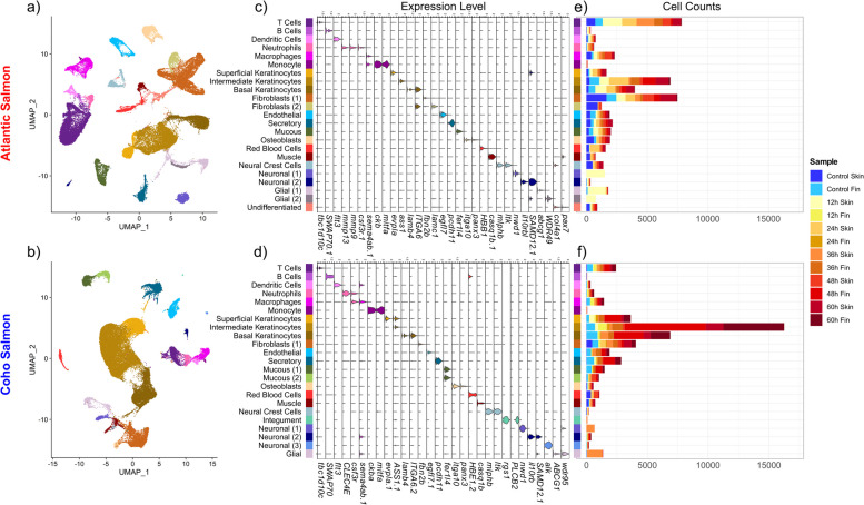
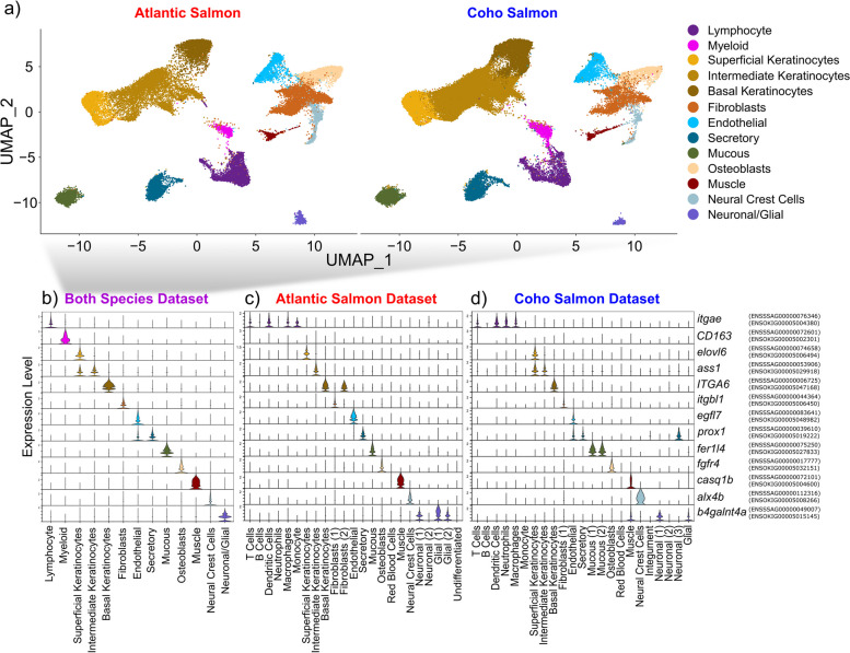
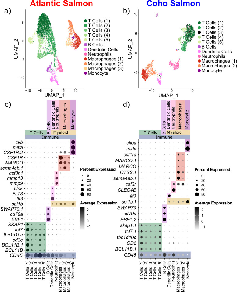
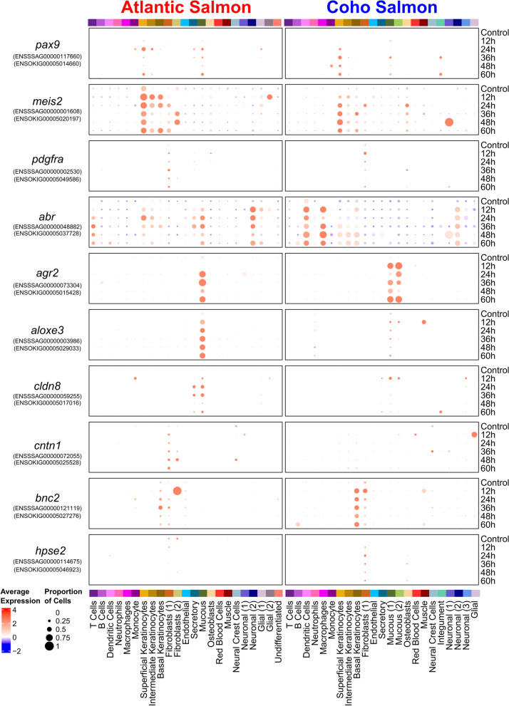
Background: Salmonid species have followed markedly divergent evolutionary trajectories in their interactions with sea lice. While sea lice parasitism poses significant economic, environmental, and animal welfare challenges for Atlantic salmon ( Salmo salar ) aquaculture, coho salmon ( Oncorhynchus kisutch ) exhibit near-complete resistance to sea lice, achieved through a potent epithelial hyperplasia response leading to rapid louse detachment. The molecular mechanisms underlying these divergent responses to sea lice are unknown. Results: We characterized the cellular and molecular responses of Atlantic salmon and coho salmon to sea lice using single-nuclei RNA sequencing. Juvenile fish were exposed to copepodid sea lice ( Lepeophtheirus salmonis ), and lice-attached pelvic fin and skin samples were collected 12 h, 24 h, 36 h, 48 h, and 60 h after exposure, along with control samples. Comparative analysis of control and treatment samples revealed an immune and wound-healing response that was common to both species, but attenuated in Atlantic salmon, potentially reflecting greater sea louse immunomodulation. Our results revealed unique but complementary roles of three layers of keratinocytes in the epithelial hyperplasia response leading to rapid sea lice rejection in coho salmon. Our results suggest that basal keratinocytes direct the expansion and mobility of intermediate and, especially, superficial keratinocytes, which eventually encapsulate the parasite. Conclusions: Our results highlight the key role of keratinocytes in coho salmon’s sea lice resistance and the diverged biological response of the two salmonid host species when interacting with this parasite. This study has identified key pathways and candidate genes that could be manipulated using various biotechnological solutions to improve Atlantic salmon sea lice resistance.
Overview
The claims and statistics of this paper haven’t been checked yet.
Run the check — free, no account needed. It reads the open-access text and checks the sources it cites.
Integrity signals
Registry
Cites
Statistics
Figures
Verification
Claims the paper makes
The claims and statistics of this paper haven’t been checked yet.
Run the check — free, no account needed. It reads the open-access text and checks the sources it cites.
Self-check
Percentages, sums and reported differences re-computed from the paper’s own numbers.
The self-check needs the paper’s full text.
Citations
Cites
OrthoFinder: phylogenetic orthology inference for comparative genomics
Emms, D. M., & Kelly, S. (2019) · Genome Biology · doi:10.1186/s13059-019-1832-y · Cited by 7,676
Epidermal Growth Factor Receptor Cell Proliferation Signaling Pathways
Wee, P., & Wang, Z. (2017) · Cancers · doi:10.3390/cancers9050052 · Cited by 2,027
Iron homeostasis in host defence and inflammation
Ganz, T., & Nemeth, E. (2015) · Nature Reviews Immunology · doi:10.1038/nri3863 · Cited by 843
Nutritional immunity: the battle for nutrient metals at the host–pathogen interface
Murdoch, C. C., & Skaar, E. P. (2022) · Nature Reviews Microbiology · doi:10.1038/s41579-022-00745-6 · Cited by 701 · cited 2×
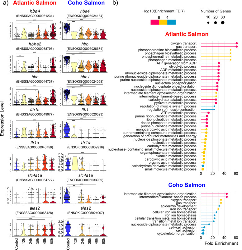
“Nutritional immunity, where hosts reduce the availability of iron in their tissues, is commonly employed to dissuade iron-seeking pathogens [27].”
BackgroundSOCS3, a Major Regulator of Infection and Inflammation
Carow, B., & Rottenberg, M. E. (2014) · Frontiers in Immunology · doi:10.3389/fimmu.2014.00058 · Cited by 555 · cited 3×
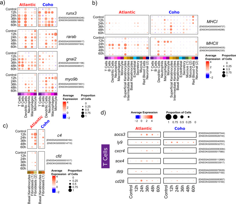
“For example, we speculate that the upregulation of the inflammation-dampening socs3 [119] in Atlantic salmon T cells may be a result of sea louse immunomodulation.”
BackgroundViral infection and iron metabolism
Drakesmith, H., & Prentice, A. (2008) · Nature Reviews Microbiology · doi:10.1038/nrmicro1930 · Cited by 469
Lipid abnormalities in atopic skin are driven by type 2 cytokines
Berdyshev, E., Goleva, E., Bronova, I., Dyjack, N., Rios, C., Jung, J., Taylor, P., Jeong, M., Hall, C. F., Richers, B. N., et al. (2018) · JCI Insight · doi:10.1172/jci.insight.98006 · Cited by 283
Nechanitzky, R., Akbas, D., Scherer, S., Györy, I., Hoyler, T., Ramamoorthy, S., Diefenbach, A., & Grosschedl, R. (2013) · Nature Immunology · doi:10.1038/ni.2641 · Cited by 218
Research works printed but not linked: 178
- Brooker AJ, Skern-Mauritzen R, Bron JE. Production, mortality, and infectivity of planktonic larval sea lice, Lepeophtheirus salmonis (Krøyer, 1837): current knowledge and implications for epidemiological modelling. ICES J Mar Sci. 2018;75(4):1214–34. · DOI
- Boxshall GA, Bravo S. On the identity of the common Caligus (Copepoda: Siphonostomatoida: Caligidae) from salmonid netpen systems in southern Chile. Contrib Zool. 2000;69(1–2):137–46. · DOI
- Carvalho LA, Whyte SK, Braden LM, Purcell SL, Manning AJ, Muckle A, Fast MD. Impact of co-infection with Lepeophtheirus salmonis and Moritella viscosa on inflammatory and immune responses of Atlantic salmon (Salmo salar). J Fish Dis. 2020;43(4):459–73. · DOI
- Abolofia J, Asche F, Wilen JE. The cost of lice: quantifying the impacts of parasitic sea lice on farmed salmon. Mar Res Econ. 2017;32(3):329–49. · DOI
- Torrissen O, Jones S, Asche F, Guttormsen A, Skilbrei OT, Nilsen F, et al. Salmon lice–impact on wild salmonids and salmon aquaculture. J Fish Dis. 2013;36(3):171–94. · DOI
- Barrett LT, Oppedal F, Robinson N, Dempster T. Prevention not cure: a review of methods to avoid sea lice infestations in salmon aquaculture. Rev Aquac. 2020;12(4):2527–43. · DOI
- Burridge L, Weis JS, Cabello F, Pizarro J, Bostick K. Chemical use in salmon aquaculture: a review of current practices and possible environmental effects. Aquaculture. 2010;306(1–4):7–23. · DOI
- Glover KA, Aasmundstad T, Nilsen F, Storset A, Skaala Ø. Variation of Atlantic salmon families (Salmo salar L.) in susceptibility to the sea lice Lepeophtheirus salmonis and Caligus elongatus. Aquaculture. 2005;245(1–4):19–30. · DOI
- Gjerde B, Ødegård J, Thorland I. Estimates of genetic variation in the susceptibility of Atlantic salmon (Salmo salar) to the salmon louse Lepeophtheirus salmonis. Aquaculture. 2011;314(1–4):66–72. · DOI
- Holborn MK, Rochus CM, Ang KP, Elliott JA, Leadbeater S, Powell F, Boulding EG. Family-based genome wide association analysis for salmon lice (Lepeophtheirus salmonis) resistance in North American Atlantic salmon using a 50 K SNP array. Aquaculture. 2019;511:734215. · DOI
- Tsai HY, Hamilton A, Tinch AE, Guy DR, Bron JE, Taggart JB, et al. Genomic prediction of host resistance to sea lice in farmed Atlantic salmon populations. Genet Sel Evol. 2016;48(1):1–11. · DOI
- Correa K, Bangera R, Figueroa R, Lhorente JP, Yáñez JM. The use of genomic information increases the accuracy of breeding value predictions for sea louse (Caligus rogercresseyi) resistance in Atlantic salmon (Salmo salar). Genet Sel Evol. 2017;49(1):1–5. · DOI
- Ødegård J, Medina M, Torgersen JS, Korsvoll SA, Deerenberg R, Yáñez JM, et al. Genetic selection for reduced parasite load in Atlantic salmon: zero-sum game or a tool for group-level protection against sea lice? Aquaculture. 2024;581:740438. · DOI
- Correa K, Lhorente JP, Bassini L, López ME, Di Genova A, Maass A, et al. Genome wide association study for resistance to Caligus rogercresseyi in Atlantic salmon (Salmo salar L.) using a 50K SNP genotyping array. Aquaculture. 2017;472:61–5. · DOI
- Rochus CM, Holborn MK, Ang KP, Elliott JA, Glebe BD, Leadbeater S, et al. Genome-wide association analysis of salmon lice (Lepeophtheirus salmonis) resistance in a North American Atlantic salmon population. Aquac Res. 2018;49(3):1329–38. · DOI
- Robledo D, Gutiérrez AP, Barría A, Lhorente JP, Houston RD, Yáñez JM. Discovery and functional annotation of quantitative trait loci affecting resistance to sea lice in Atlantic salmon. Front Genet. 2019;10:56. · DOI
- Robinson NA, Robledo D, Sveen L, Ruiz Daniels R, Krasnov A, Coates A, et al. Applying genetic technologies to combat infectious diseases in aquaculture. Rev Aquac. 2023;15(2):491–535. · DOI
- Johnson SC, Albright LJ. Comparative susceptibility and histopathology of the response of naive Atlantic, chinook and coho salmon to experimental infection with Lepeophtheirus salmonis (Copepoda: Caligidae). Dis Aquat Organ. 1992;14(3):179–93. · DOI
- Fast MD. Fish immune responses to parasitic copepod (namely sea lice) infection. Dev Comp Immunol. 2014;43(2):300–12. · DOI
- Braden LM, Michaud D, Groman D, Byrne P, Hori TS, Fast MD. Rejection of Lepeophtheirus salmonis driven in part by chitin sensing is not impacted by seawater acclimitization in Coho salmon (Oncorhynchus kisutch). Sci Rep. 2023;13(1):1–19. · DOI
- Fast MD, Sims DE, Burka JF, Mustafa A, Ross NW. Skin morphology and humoral non-specific defence parameters of mucus and plasma in rainbow trout, coho and Atlantic salmon. Comp Biochem Physiol A Mol Integr Physiol. 2002;132(3):645–57. · DOI
- Johnson SC, Fast MD. Interactions between sea lice and their hosts. In: Wiegertjes GF, Flik G, editors. Host-parasite interactions. Abingdon, UK: Taylor & Francis; 2004. p. 131–59. · DOI
- Valenzuela-Muñoz V, Boltaña S, Gallardo-Escárate C. Comparative immunity of Salmo salar and Oncorhynchus kisutch during infestation with the sea louse Caligus rogercresseyi: an enrichment transcriptome analysis. Fish Shellfish Immunol. 2016;59:276–87. · DOI
- Fast MD, Braden LM. Salmon-louse interaction and immunological consequences for the host. In: Treasurer J, Bricknell IR, Bron JE, editors. Sea lice biology and control. Essex UK: 5M Books Ltd; 2022. p. 272–93.
- Braden LM, Koop BF, Jones SR. Signatures of resistance to Lepeophtheirus salmonis include a TH2-type response at the louse-salmon interface. Dev Comp Immunol. 2015;48(1):178–91. · DOI
- Valenzuela-Muñoz V, Boltaña S, Gallardo-Escárate C. Uncovering iron regulation with species-specific transcriptome patterns in Atlantic salmon and coho salmon during a Caligus rogercresseyi infestation. J Fish Dis. 2017;40(9):1169–84. · DOI
- Sveen LR, Robinson N, Krasnov A, Ruiz Daniels R, Vaadal M, Karlsen C, et al. Transcriptomic landscape of Atlantic salmon (Salmo salar L.) skin. G3. 2023;13(11):jkad215. · DOI
- Hawkes JW. The structure of fish skin: I. General organization Cell Tissue Res. 1974;149(2):147–58. · DOI
- Quilhac A, Sire JY. Spreading, proliferation, and differentiation of the epidermis after wounding a cichlid fish, Hemichromis bimaculatus. Anat Rec. 1999;254(3):435–51. · DOI
- Sveen L, Karlsen C, Ytteborg E. Mechanical induced wounds in fish–a review on models and healing mechanisms. Rev Aquac. 2020;12(4):2446–65. · DOI
- Elliott DG. THE SKIN| Functional morphology of the integumentary system in fishes. In: Farrell AP. Encyclopedia of fish physiology. Academic Press. 2011. p. 476–88.
- Elliott DG. Integumentary system. In: The laboratory fish. Academic Press; 2000. p. 271–306. · DOI
- Sehring IM, Weidinger G. Recent advancements in understanding fin regeneration in zebrafish. Wiley Interdiscip Rev Dev Biol. 2020;9(1):e367. · DOI
- Rasmussen JP, Vo NT, Sagasti A. Fish scales dictate the pattern of adult skin innervation and vascularization. Dev Cell. 2018;46(3):344–59. · DOI
- Esteban MÁ, Cerezuela R. Fish mucosal immunity: skin. In: Beck BH, Peatman E. Mucosal health in aquaculture. Academic Press. 2015. p. 67–92.
- Chen G, Ning B, Shi T. Single-cell RNA-seq technologies and related computational data analysis. Front Genet. 2019;10:317. · DOI
- Kolodziejczyk AA, Kim JK, Svensson V, Marioni JC, Teichmann SA. The technology and biology of single-cell RNA sequencing. Mol Cell. 2015;58(4):610–20. · DOI · Paper page
- Sun S, Liu Z, Jiang Q, Zou Y. Embryonic expression patterns of TBC1D10 subfamily genes in zebrafish. Gene Expr Patterns. 2022;43:119226. · DOI
- Carmona SJ, Teichmann SA, Ferreira L, Macaulay IC, Stubbington MJ, Cvejic A, et al. Single-cell transcriptome analysis of fish immune cells provides insight into the evolution of vertebrate immune cell types. Genome Res. 2017;27(3):451–61. · DOI
- Raab M, Wang H, Lu Y, Smith X, Wu Z, Strebhardt K, et al. T cell receptor “inside-out” pathway via signaling module SKAP1-RapL regulates T cell motility and interactions in lymph nodes. Immunity. 2010;32(4):541–56. · DOI
- Mason DY, Cordell J, Brown M, Pallesen G, Ralfkiaer E, Rothbard J, Crumpton M, Gatter KC. Detection of T cells in paraffin wax embedded tissue using antibodies against a peptide sequence from the CD3 antigen. J Clin Pathol. 1989;42(11):1194–200. · DOI
- Lang G, Wotton D, Owen MJ, Sewell WA, Brown MH, Mason DY, et al. The structure of the human CD2 gene and its expression in transgenic mice. EMBO J. 1988;7(6):1675–82. · DOI
- Borggrefe T, Keshavarzi S, Gross B, Wabl M, Jessberger R. Impaired IgE response in SWAP-70-deficient mice. Eur J Immunol. 2001;31(8):2467–75. · DOI
- Liu X, Li YS, Shinton SA, Rhodes J, Tang L, Feng H, et al. Zebrafish B cell development without a pre–B cell stage, revealed by CD79 fluorescence reporter transgenes. J Immunol. 2017;199(5):1706–15. · DOI
- Karsunky H, Merad M, Cozzio A, Weissman IL, Manz MG. Flt3 ligand regulates dendritic cell development from Flt3+ lymphoid and myeloid-committed progenitors to Flt3+ dendritic cells in vivo. J Exp Med. 2003;198(2):305–13. · DOI
- Marafioti T, Paterson JC, Ballabio E, Reichard KK, Tedoldi S, Hollowood K, et al. Novel markers of normal and neoplastic human plasmacytoid dendritic cells. Blood. 2008;111(7):3778–92. · DOI
- Liu F, Wu HY, Wesselschmidt R, Kornaga T, Link DC. Impaired production and increased apoptosis of neutrophils in granulocyte colony-stimulating factor receptor–deficient mice. Immunity. 1996;5(5):491–501. · DOI
- Taylor RS, Ruiz Daniels R, Dobie R, Naseer S, Clark TC, Henderson NC, et al. Single cell transcriptomics of Atlantic salmon (Salmo salar L.) liver reveals cellular heterogeneity and immunological responses to challenge by Aeromonas salmonicida. Front Immunol. 2022;13:984799. · DOI
- Sharma A, Rijavec M, Tomar S, Yamani A, Ganesan V, Krempski J, et al. Acute systemic myeloid inflammatory and stress response in severe food allergic reactions. Clin Exp Allergy. 2023;53(5):536–49. · DOI
- Ito D, Kumanogoh A. The role of Sema4A in angiogenesis, immune responses, carcinogenesis, and retinal systems. Cell Adh Migr. 2016;10(6):692–9. · DOI
- Sehgal A, Donaldson DS, Pridans C, Sauter KA, Hume DA, Mabbott NA. The role of CSF1R-dependent macrophages in control of the intestinal stem-cell niche. Nat Commun. 2018;9(1):1272. · DOI
- Farnsworth DR, Saunders LM, Miller AC. A single-cell transcriptome atlas for zebrafish development. Dev Biol. 2020;459(2):100–8. · DOI
- Liuzzo JP, Petanceska SS, Moscatelli D, Devi LA. Inflammatory mediators regulate cathepsin S in macrophages and microglia: a role in attenuating heparan sulfate interactions. Mol Med. 1999;5:320–33. · DOI
- Anbazhagan K, Duroux-Richard I, Jorgensen C, Apparailly F. Transcriptomic network support distinct roles of classical and non-classical monocytes in human. Int Rev Immunol. 2014;33(6):470–89. · DOI
- Subkhankulova T, Camargo Sosa K, Uroshlev LA, Nikaido M, Shriever N, Kasianov AS, et al. Zebrafish pigment cells develop directly from persistent highly multipotent progenitors. Nat Commun. 2023;14(1):1258. · DOI
- Ruhrberg C, Hajibagheri MN, Parry DA, Watt FM. Periplakin, a novel component of cornified envelopes and desmosomes that belongs to the plakin family and forms complexes with envoplakin. J Cell Biol. 1997;139(7):1835–49. · DOI
- Perez CJ, Jaubert J, Guénet JL, Barnhart KF, Ross-Inta CM, Quintanilla VC, Aubin I, et al. Two hypomorphic alleles of mouse Ass1 as a new animal model of citrullinemia type I and other hyperammonemic syndromes. Am J Pathol. 2010;177(4):1958–68. · DOI
- Rizzolio F, Bione S, Villa A, Berti E, Cassetti A, Bulfone A, et al. Spatial and temporal expression of POF1B, a gene expressed in epithelia. Gene Expr Patterns. 2007;7(4):529–34. · DOI
- Malaisse J, Hermant M, Hayez A, Poumay Y, Lambert de Rouvroit C. Meaning of relative gene expression in multilayered cultures of epidermal keratinocytes. Exp Dermatol. 2014;23(10):754–6. · DOI
- Brinckmann J, Hunzelmann N, Kahle B, Rohwedel J, Kramer J, Gibson MA, et al. Enhanced fibrillin-2 expression is a general feature of wound healing and sclerosis: potential alteration of cell attachment and storage of TGF-β. Lab Invest. 2010;90(5):739–52. · DOI
- Koch M, Bohrmann B, Matthison M, Hagios C, Trueb B, Chiquet M. Large and small splice variants of collagen XII: differential expression and ligand binding. J Cell Biol. 1995;130(4):1005–14. · DOI
- Usuba R, Pauty J, Soncin F, Matsunaga YT. EGFL7 regulates sprouting angiogenesis and endothelial integrity in a human blood vessel model. Biomaterials. 2019;197:305–16. · DOI
- Ruppert AL, Keshavarz M, Winterberg S, Oberwinkler J, Kummer W, Schütz B. Advillin is a tuft cell marker in the mouse alimentary tract. J Mol Histol. 2020;51:421–35. · DOI
- Willms RJ, Jones LO, Hocking JC, Foley E. A cell atlas of microbe-responsive processes in the zebrafish intestine. Cell Rep. 2022;38(5):110311. · DOI
- Gerbe F, Sidot E, Smyth DJ, Ohmoto M, Matsumoto I, Dardalhon V, et al. Intestinal epithelial tuft cells initiate type 2 mucosal immunity to helminth parasites. Nature. 2016;529(7585):226–30. · DOI
- Middelhoff M, Nienhüser H, Valenti G, Maurer HC, Hayakawa Y, Takahashi R, et al. Prox1-positive cells monitor and sustain the murine intestinal epithelial cholinergic niche. Nat Commun. 2020;11(1):111. · DOI
- Song Y, Meng Z, Zhang S, Li N, Hu W, Li H. miR-4739/ITGA10/PI3K signaling regulates differentiation and apoptosis of osteoblast. Regen Ther. 2022;21:342–50. · DOI
- Cool S, Jackson R, Pincus P, Dickinson I, Nurcombe V. Fibroblast growth factor receptor 4 (FGFR4) expression in newborn murine calvaria and primary osteoblast cultures. Int J Dev Biol. 2004;46(4):519–23.
- Furlan S, Mosole S, Murgia M, Nagaraj N, Argenton F, Volpe P, Nori A. Calsequestrins in skeletal and cardiac muscle from adult Danio rerio. J Muscle Res Cell Motil. 2016;37:27–39. · DOI
- Yoshimoto Y, Ikemoto-Uezumi M, Hitachi K, Fukada SI, Uezumi A. Methods for accurate assessment of myofiber maturity during skeletal muscle regeneration. Front Cell Dev Biol. 2020;8:267. · DOI
- Van der Loop FT, Van Eys GJ, Schaart G, Ramaekers FC. Titin expression as an early indication of heart and skeletal muscle differentiation in vitro. Developmental re-organisation in relation to cytoskeletal constituents. J Muscle Res Cell Motil. 1996;17:23–36. · DOI
- Kimura T, Takehana Y, Naruse K. pnp4a is the causal gene of the medaka iridophore mutant guanineless. G3: Genes. 2017;7(4):1357–63. · DOI · Paper page
- Santos ME, Braasch I, Boileau N, Meyer BS, Sauteur L, Böhne A, Belting HG, Affolter M, Salzburger W. The evolution of cichlid fish egg-spots is linked with a cis-regulatory change. Nat Commun. 2014;5(1):5149. · DOI
- Jang HS, Chen Y, Ge J, Wilkening AN, Hou Y, Lee HJ, Choi YR, Lowdon RF, Xing X, Li D, Kaufman CK. Epigenetic dynamics shaping melanophore and iridophore cell fate in zebrafish. Genome Biol. 2021;22:1–18. · DOI
- Yamada S, Sakakibara SI. Expression profile of the STAND protein Nwd1 in the developing and mature mouse central nervous system. J Comp Neurol. 2018;526(13):2099–114. · DOI
- Fernandez T, Morgan T, Davis N, Klin A, Morris A, Farhi A, Lifton RP. Disruption of contactin 4 (CNTN4) results in developmental delay and other features of 3p deletion syndrome. Am J Hum Genet. 2004;74(6):1286–93. · DOI
- Maroilley T, Tsai MH, Mascarenhas R, Diao C, Khanbabaei M, Kaya S, Depienne C, Tarailo-Graovac M, Klein KM. A novel FAME1 repeat configuration in a European family identified using a combined genomics approach. Epilepsia Open. 2023;8(2):659–65. · DOI
- Zhou Z, Peng X, Insolera R, Fink DJ, Mata M. Interleukin-10 provides direct trophic support to neurons. J Neurochem. 2009;110(5):1617–27. · DOI
- Joseph DB, Henry GH, Malewska A, Reese JC, Mauck RJ, Gahan JC, Hutchinson RC, Malladi VS, Roehrborn CG, Vezina CM, Strand DW. Single-cell analysis of mouse and human prostate reveals novel fibroblasts with specialized distribution and microenvironment interactions. J Pathol. 2021;255(2):141–54. · DOI
- He H, Suryawanshi H, Morozov P, Gay-Mimbrera J, Del Duca E, Kim HJ, et al. Single-cell transcriptome analysis of human skin identifies novel fibroblast subpopulation and enrichment of immune subsets in atopic dermatitis. J Allergy Clin Immunol. 2020;145(6):1615–28. · DOI
- Song H, Sun W, Ye G, Ding X, Liu Z, Zhang S, et al. Long non-coding RNA expression profile in human gastric cancer and its clinical significances. J Transl Med. 2013;11(1):1–10. · DOI
- Park KS, Korfhagen TR, Bruno MD, Kitzmiller JA, Wan H, Wert SE, et al. SPDEF regulates goblet cell hyperplasia in the airway epithelium. J Clin Invest. 2007;117(4):978–88. · DOI
- Ma J, Rubin BK, Voynow JA. Mucins, mucus, and goblet cells. Chest. 2018;154(1):169–76. · DOI
- Tarr PT, Edwards PA. ABCG1 and ABCG4 are coexpressed in neurons and astrocytes of the CNS and regulate cholesterol homeostasis through SREBP-2. J Lipid Res. 2008;49(1):169–82. · DOI
- Gerrits E, Giannini LA, Brouwer N, Melhem S, Seilhean D, Le Ber I, et al. Neurovascular dysfunction in GRN-associated frontotemporal dementia identified by single-nucleus RNA sequencing of human cerebral cortex. Nat Neurosci. 2022;25(8):1034–48. · DOI · Paper page
- Seale P, Sabourin LA, Girgis-Gabardo A, Mansouri A, Gruss P, Rudnicki MA. Pax7 is required for the specification of myogenic satellite cells. Cell. 2000;102(6):777–86. · DOI
- Kuo DS, Labelle-Dumais C, Gould DB. COL4A1 and COL4A2 mutations and disease: insights into pathogenic mechanisms and potential therapeutic targets. Hum Mol Genet. 2012;21(R1):R97–110. · DOI · Paper page
- Vernersson E, Khoo NK, Henriksson ML, Roos G, Palmer RH, Hallberg B. Characterization of the expression of the ALK receptor tyrosine kinase in mice. Gene Expr Patterns. 2006;6(5):448–61. · DOI
- Mo ES, Cheng Q, Reshetnyak AV, Schlessinger J, Nicoli S. Alk and Ltk ligands are essential for iridophore development in zebrafish mediated by the receptor tyrosine kinase Ltk. PNAS. 2017;114(45):12027–32. · DOI
- Sveen LR, Timmerhaus G, Krasnov A, Takle H, Handeland S, Ytteborg E. Wound healing in post-smolt Atlantic salmon (Salmo salar L.). Sci Rep. 2019;9(1):3565. · DOI
- Uss E, Rowshani AT, Hooibrink B, Lardy NM, van Lier RA, ten Berge IJ. CD103 is a marker for alloantigen-induced regulatory CD8+ T cells. J Immunol. 2006;177(5):2775–83. · DOI
- Lau SK, Chu PG, Weiss LM. CD163: a specific marker of macrophages in paraffin-embedded tissue samples. Am J Clin Pathol. 2004;122(5):794–801. · DOI
- Gerbe F, Jay P. Intestinal tuft cells: epithelial sentinels linking luminal cues to the immune system. Mucosal Immunol. 2016;9(6):1353–9. · DOI
- Rheinländer A, Schraven B, Bommhardt U. CD45 in human physiology and clinical medicine. Immunol Lett. 2018;196:22–32. · DOI
- Zhu J, Yamane H, Cote-Sierra J, Guo L, Paul WE. GATA-3 promotes Th2 responses through three different mechanisms: induction of Th2 cytokine production, selective growth of Th2 cells and inhibition of Th1 cell-specific factors. Cell Res. 2006;16(1):3–10. · DOI
- Mise T, Iijima M, Inohaya K, Kudo A, Wada H. Function of Pax1 and Pax9 in the sclerotome of medaka fish. Genesis. 2008;46(4):185–92. · DOI
- Douglas G, Cho MT, Telegrafi A, Winter S, Carmichael J, Zackai EH, et al. De novo missense variants in MEIS2 recapitulate the microdeletion phenotype of cardiac and palate abnormalities, developmental delay, intellectual disability and dysmorphic features. Am J Med Genet A. 2018;176(9):1845–51. · DOI
- Ushakumary MG, Riccetti M, Perl AK. Resident interstitial lung fibroblasts and their role in alveolar stem cell niche development, homeostasis, injury, and regeneration. Stem Cells Transl Med. 2021;10(7):1021–32. · DOI · Paper page
- Chou MY, Li HC. Genomic organization and characterization of the human type XXI collagen (COL21A1) gene. Genomics. 2002;79(3):395–401. · DOI
- Vaughan EM, Miller AL, Hoi-Ying EY, Bement WM. Control of local Rho GTPase crosstalk by Abr. Curr Biol. 2011;21(4):270–7. · DOI
- Bergström JH, Berg KA, Rodríguez-Piñeiro AM, Stecher B, Johansson ME, Hansson GC. AGR2, an endoplasmic reticulum protein, is secreted into the gastrointestinal mucus. PLoS ONE. 2014;9(8):e104186. · DOI
- Umasuthan N, Xue X, Caballero-Solares A, Kumar S, Westcott JD, Chen Z, et al. Transcriptomic profiling in fins of Atlantic salmon parasitized with sea lice: evidence for an early imbalance between chalimus-induced immunomodulation and the host’s defense response. Int J Mol Sci. 2020;21(7):2417. · DOI
- Ullah R, Ansar M, Durrani ZU, Lee K, Santos-Cortez RL, Muhammad D, et al. Novel mutations in the genes TGM 1 and ALOXE 3 underlying autosomal recessive congenital ichthyosis. Int J Dermatol. 2016;55(5):524–30. · DOI
- Zeissig S, Bürgel N, Günzel D, Richter J, Mankertz J, Wahnschaffe U, et al. Changes in expression and distribution of claudin 2, 5 and 8 lead to discontinuous tight junctions and barrier dysfunction in active Crohn’s disease. Gut. 2007;56(1):61–72. · DOI
- Houreld NN, Ayuk SM, Abrahamse H. Cell adhesion molecules are mediated by photobiomodulation at 660 nm in diabetic wounded fibroblast cells. Cells. 2018;7(4):30. · DOI
- Bobowski-Gerard M, Boulet C, Zummo FP, Dubois-Chevalier J, Gheeraert C, Bou Saleh M, et al. Functional genomics uncovers the transcription factor BNC2 as required for myofibroblastic activation in fibrosis. Nat Commun. 2022;13(1):5324. · DOI
- Patterson LB, Parichy DM. Interactions with iridophores and the tissue environment required for patterning melanophores and xanthophores during zebrafish adult pigment stripe formation. PLoS Genet. 2013;9(5):e1003561. · DOI · Paper page
- Peretti T, Waisberg J, Mader AM, de Matos LL, da Costa RB, Conceição GM, et al. Heparanase-2, syndecan-1, and extracellular matrix remodeling in colorectal carcinoma. Eur J Gastroenterol Hepatol. 2008;20(8):756–65. · DOI
- Puig-Kröger A, Corbí A. RUNX3: a new player in myeloid gene expression and immune response. J Cell Biochem. 2006;98(4):744–56. · DOI
- Raverdeau M, Mills KH. Modulation of T cell and innate immune responses by retinoic acid. J Immunol. 2014;192(7):2953–8. · DOI
- Han SB, Moratz C, Huang NN, Kelsall B, Cho H, Shi CS, et al. Rgs1 and Gnai2 regulate the entrance of B lymphocytes into lymph nodes and B cell motility within lymph node follicles. Immunity. 2005;22(3):343–54. · DOI
- Maravillas-Montero JL, Santos-Argumedo L. The myosin family: unconventional roles of actin-dependent molecular motors in immune cells. J Leukoc Biol. 2012;91(1):35–46. · DOI
- Kuwahara M, Yamashita M, Shinoda K, Tofukuji S, Onodera A, Shinnakasu R, et al. The transcription factor Sox4 is a downstream target of signaling by the cytokine TGF-β and suppresses TH2 differentiation. Nat Immunol. 2012;13(8):778–86. · DOI
- Kumar A, Humphreys TD, Kremer KN, Bramati PS, Bradfield L, Edgar CE, Hedin KE. CXCR4 physically associates with the T cell receptor to signal in T cells. Immunity. 2006;25(2):213–24. · DOI
- Mathieson BJ, Sharrow SO, Bottomly K, Fowlkes BJ. Ly 9, an alloantigenic marker of lymphocyte differentiation. J Immunol. 1980;125(5):2127–36. · DOI
- Martínez D, Oyarzún-Salazar R, Quilapi AM, Coronado J, Enriquez R, Vargas-Lagos C, et al. Live and inactivated Piscirickettsia salmonis activated nutritional immunity in Atlantic salmon (Salmo salar). Front Immunol. 2023;14:1187209. · DOI
- Jennings ML. Cell physiology and molecular mechanism of anion transport by erythrocyte band 3/AE1. Am J Physiol Cell Physiol. 2021;321(6):C1028–59. · DOI
- Huang SG, Zhang LL, Niu Q, Xiang GM, Liu LL, Jiang DN, et al. Hypoxia promotes epithelial-mesenchymal transition of hepatocellular carcinoma cells via inducing GLIPR-2 expression. PLoS ONE. 2013;8(10):e77497. · DOI
- Mou Y, Zhang L, Liu Z, Song X. Abundant expression of ferroptosis-related SAT1 is related to unfavorable outcome and immune cell infiltration in low-grade glioma. BMC Cancer. 2022;22(1):1–15. · DOI
- Chen J, Ingham N, Kelly J, Jadeja S, Goulding D, Pass J, et al. Spinster homolog 2 (spns2) deficiency causes early onset progressive hearing loss. PLoS Genet. 2014;10(10): e1004688. · DOI
- Caldwell JM, Collins MH, Kemme KA, Sherrill JD, Wen T, Rochman M, et al. Cadherin 26 is an alpha integrin-binding epithelial receptor regulated during allergic inflammation. Mucosal Immunol. 2017;10(5):1190–201. · DOI
- Wiche G. Commentary: role of plectin in cytoskeleton organization and dynamics. J Cell Sci. 1998;111(17):2477–86. · DOI
- Granadeiro L, Dirks RP, Ortiz-Delgado JB, Gavaia PJ, Sarasquete C, Laize V, et al. Warfarin-exposed zebrafish embryos resembles human warfarin embryopathy in a dose and developmental-time dependent manner–from molecular mechanisms to environmental concerns. Ecotoxicol Environ Saf. 2019;181:559–71. · DOI
- Qian M, Chen Z, Wang S, Guo X, Zhang Z, Qiu W, et al. PLEKHG5 is a novel prognostic biomarker in glioma patients. Int J Clin Oncol. 2019;24:1350–8. · DOI
- Daggett DF, Boyd CA, Gautier P, Bryson-Richardson RJ, Thisse C, Thisse B, et al. Developmentally restricted actin-regulatory molecules control morphogenetic cell movements in the zebrafish gastrula. Curr Biol. 2004;14(18):1632–8. · DOI
- Ma AC, Ward AC, Liang R, Leung AY. The role of jak2a in zebrafish hematopoiesis. Blood. 2007;110(6):1824–30. · DOI
- Wu H, Huang M, Cao P, Wang T, Shu Y, Liu P. MiR-135a targets JAK2 and inhibits gastric cancer cell proliferation. Cancer Biol Ther. 2012;13(5):281–8. · DOI
- Sasaki A, Yasukawa H, Shouda T, Kitamura T, Dikic I, Yoshimura A. CIS3/SOCS-3 suppresses erythropoietin (EPO) signaling by binding the EPO receptor and JAK2. J Biol Chem. 2000;275(38):29338–47. · DOI
- Podobnik M, Kisovec M, Anderluh G. Molecular mechanism of pore formation by aerolysin-like proteins. Philos Trans R Soc B: Biol Sci. 2017;372(1726):20160209. · DOI
- Chen LL, Xie J, Cao DD, Jia N, Li YJ, Sun H, et al. The pore-forming protein Aep1 is an innate immune molecule that prevents zebrafish from bacterial infection. Dev Comp Immunol. 2018;82:49–54. · DOI
- Xiong Y, Dan C, Ren F, Su Z, Zhang Y, Mei J. Proteomic profiling of yellow catfish (Pelteobagrus fulvidraco) skin mucus identifies differentially-expressed proteins in response to Edwardsiella ictaluri infection. Fish Shellfish Immunol. 2020;100:98–108. · DOI
- Lee RT, Asharani PV, Carney TJ. Basal keratinocytes contribute to all strata of the adult zebrafish epidermis. PLoS ONE. 2014;9(1):e84858. · DOI
- Leclerc-Mercier S, Lemoine R, Bigorgne AE, Sepulveda F, Leveau C, Fischer A, et al. Ichthyosis as the dermatological phenotype associated with TTC7A mutations. Br J Dermatol. 2016;175(5):1061–4. · DOI
- Subramanian K, Neill DR, Malak HA, Spelmink L, Khandaker S, Marchiori GDL, et al. Pneumolysin binds to the mannose receptor C type 1 (MRC-1) leading to anti-inflammatory responses and enhanced pneumococcal survival. Nat Microbiol. 2019;4(1):62–70. · DOI
- Robledo D, Gutiérrez AP, Barría A, Yáñez JM, Houston RD. Gene expression response to sea lice in Atlantic salmon skin: RNA sequencing comparison between resistant and susceptible animals. Front Genet. 2018;9:287. · DOI
- Palazón-Riquelme P, Worboys JD, Green J, Valera A, Martín-Sánchez F, Pellegrini C, Brough D, López-Castejón G. USP7 and USP47 deubiquitinases regulate NLRP3 inflammasome activation. EMBO Rep. 2018;19(10):e44766. · DOI
- Lecaudey LA, Schliewen UK, Osinov AG, Taylor EB, Bernatchez L, Weiss SJ. Inferring phylogenetic structure, hybridization and divergence times within Salmoninae (Teleostei: Salmonidae) using RAD-sequencing. Mol Phylogenet Evol. 2018;124:82–99. · DOI
- Pike AW. Sea lice—major pathogens of farmed Atlantic salmon. Parasitol Today. 1989;5(9):291–7. · DOI
- Fast MD, Burka JF, Johnson SC, Ross NW. Enzymes released from Lepeophtheirus salmonis in response to mucus from different salmonids. J Parasitol. 2003;89(1):7–13. · DOI
- Fast MD, Muise DM, Easy RE, Ross NW, Johnson SC. The effects of Lepeophtheirus salmonis infections on the stress response and immunological status of Atlantic salmon (Salmo salar). Fish Shellfish Immunol. 2006;21(3):228–41. · DOI
- Carr MM, McVittie E, Guy K, Gawkrodger DJ, Hunter JA. MHC class II antigen expression in normal human epidermis. Immunology. 1986;59(2):223.
- Tamoutounour S, Han SJ, Deckers J, Constantinides MG, Hurabielle C, Harrison OJ, et al. Keratinocyte-intrinsic MHCII expression controls microbiota-induced Th1 cell responses. PNAS. 2019;116(47):23643–52. · DOI
- Holm HJ, Skugor S, Bjelland AK, Radunovic S, Wadsworth S, Koppang EO, Evensen Ø. Contrasting expression of immune genes in scaled and scaleless skin of Atlantic salmon infected with young stages of Lepeophtheirus salmonis. Dev Comp Immunol. 2017;67:153–65. · DOI
- Barker JN, Griffiths CE, Nickoloff BJ, Mitra RS, Dixit VM. Keratinocytes as initiators of inflammation. Lancet. 1991;337(8735):211–4. · DOI
- Cai W, Kumar S, Navaneethaiyer U, Caballero-Solares A, Carvalho LA, Whyte SK, et al. Transcriptome analysis of Atlantic salmon (Salmo salar) skin in response to sea lice and infectious salmon anemia virus co-infection under different experimental functional diets. Front Immunol. 2022;12. https://doi.org/10.3389/fimmu.2021.787033. · DOI
- Robinson NA, Gjedrem T, Quillet E. Improvement of disease resistance by genetic methods. In: Jeney G. Fish diseases. Academic Press. 2017. p. 21–50.
- Skugor S, Glover KA, Nilsen F, Krasnov A. Local and systemic gene expression responses of Atlantic salmon (Salmo salar L.) to infection with the salmon louse (Lepeophtheirus salmonis). BMC Genomics. 2008;9:1–18. · DOI
- Swart M, Troeberg L. Effect of polarization and chronic inflammation on macrophage expression of heparan sulfate proteoglycans and biosynthesis enzymes. J Histochem Cytochem. 2019;67(1):9–27. · DOI
- McManus CM, Maizels RM. Regulatory T cells in parasite infections: susceptibility, specificity and specialisation. Trends Parasitol. 2023;39(7):547–62. · DOI
- Øvergård AC, Eichner C, Nuñez-Ortiz N, Kongshaug H, Borchel A, Dalvin S. Transcriptomic and targeted immune transcript analyses confirm localized skin immune responses in Atlantic salmon towards the salmon louse. Fish Shellfish Immunol. 2023;138:108835. · DOI
- Wang L, Liu Y, Dai Y, Tang X, Yin T, Wang C, et al. Single-cell RNA-seq analysis reveals BHLHE40-driven pro-tumour neutrophils with hyperactivated glycolysis in pancreatic tumour microenvironment. Gut. 2023;72(5):958–71. · DOI
- Fast MD, Johnson SC, Eddy TD, Pinto D, Ross NW. Lepeophtheirus salmonis secretory/excretory products and their effects on Atlantic salmon immune gene regulation. Parasite Immunol. 2007;29(4):179–89. · DOI
- Braden LM, Monaghan SJ, Fast MD. Salmon immunological defence and interplay with the modulatory capabilities of its ectoparasite Lepeophtheirus salmonis. Parasite Immunol. 2020;42(8):e12731. · DOI
- Núñez-Acuña G, Gonçalves AT, Valenzuela-Munoz V, Pino-Marambio J, Wadsworth S, Gallardo-Escárate C. Transcriptome immunomodulation of in-feed additives in Atlantic salmon Salmo salar infested with sea lice Caligus rogercresseyi. Fish Shellfish Immunol. 2015;47(1):450–60. · DOI
- Wang T, Gorgoglione B, Maehr T, Holland JW, Vecino JL, Wadsworth S, Secombes CJ. Fish suppressors of cytokine signaling (SOCS): gene discovery, modulation of expression and function. J Signal Transduct. 2011;905813. · DOI
- Zhang M, Xiao ZZ, Sun L. Suppressor of cytokine signaling 3 inhibits head kidney macrophage activation and cytokine expression in Scophthalmus maximus. Dev Comp Immunol. 2011;35(2):174–81. · DOI
- Kittilsen S, Johansen IB, Braastad BO, Øverli Ø. Pigments, parasites and personalitiy: towards a unifying role for steroid hormones? PLoS ONE. 2012;7(4):e34281. · DOI
- Weinberg ED. Iron availability and infection. Biochim Biophys Acta Gen Sub. 2009;1790(7):600–5. · DOI · Paper page
- Valenzuela-Muñoz V, Gallardo-Escárate C. Iron metabolism modulation in Atlantic salmon infested with the sea lice Lepeophtheirus salmonis and Caligus rogercresseyi: a matter of nutritional immunity? Fish Shellfish Immunol. 2017;60:97–102. · DOI
- Fast MD, Ross NW, Craft CA, Locke SJ, MacKinnon SL, Johnson SC. Lepeophtheirus salmonis: characterization of prostaglandin E2 in secretory products of the salmon louse by RP-HPLC and mass spectrometry. Exp Parasitol. 2004;107(1–2):5–13. · DOI
- Sutherland BJ, Koczka KW, Yasuike M, Jantzen SG, Yazawa R, Koop BF, et al. Comparative transcriptomics of Atlantic Salmo salar, chum Oncorhynchus keta and pink salmon O. gorbuscha during infections with salmon lice Lepeophtheirus salmonis. BMC Genomics. 2014;15:1–17. · DOI
- Burka JF, Fast MD, Revie CW. Lepeophtheirus salmonis and Caligus rogercresseyi. In: Woo PTK, Buchmann K, editors. Fish parasites: pathobiology and protection. Wallingford UK: Cabi; 2012. p. 350–70. · DOI
- Sheldon JR, Skaar EP. Metals as phagocyte antimicrobial effectors. Curr Opin Immunol. 2019;60:1–9. · DOI
- Caballero-Solares A, Umasuthan N, Xue X, Katan T, Kumar S, Westcott JD, et al. Interacting effects of sea louse (Lepeophtheirus salmonis) infection and formalin-killed Aeromonas salmonicida on Atlantic salmon skin transcriptome. Front Immunol. 2022;13. https://doi.org/10.3389/fimmu.2022.804987. · DOI
- Ruiz Daniels R, Taylor RS, Robledo D, Macqueen DJ. Single cell genomics as a transformative approach for aquaculture research and innovation. Rev Aquac. 2023;15(4):1618–37. · DOI
- Robinson NA, Østbye TKK, Kettunen AH, Coates A, Barrett LT, Robledo D, Dempster T. A guide to assess the use of gene editing in aquaculture. Rev Aquac. 2024;16(2):775–84. · DOI
- Ruiz Daniels R, Taylor RS, Dobie R, Salisbury S, Furniss JJ, Clark E, et al. A versatile nuclei extraction protocol for single nucleus sequencing in non-model species–optimization in various Atlantic salmon tissues. PLoS ONE. 2023;18(9):e0285020. · DOI
- Dobin A. STAR manual 2.7. 0a. Weill Cornell Medicine. 2019.
- Kaminow B, Yunusov D, Dobin A. STARsolo: accurate, fast and versatile mapping/quantification of single-cell and single-nucleus RNA-seq data. BioRxiv. 2021. https://doi.org/10.1101/2021.05.05.442755. · DOI
- Pertea G, Pertea M. GFF utilities: GffRead and GffCompare. F1000Research. 2020;9:ISCB Comm J-304. · DOI · Paper page
- Hao Y, Hao S, Andersen-Nissen E, Mauck WM, Zheng S, Butler A, et al. Integrated analysis of multimodal single-cell data. Cell. 2021;184(13):3573–87. · DOI · Paper page
- Ahlmann-Eltze C, Huber W. glmGamPoi: fitting Gamma-Poisson generalized linear models on single cell count data. Bioinformatics. 2020;36(24):5701–2. · DOI
- McGinnis CS, Murrow LM, Gartner ZJ. DoubletFinder: doublet detection in single-cell RNA sequencing data using artificial nearest neighbors. Cell Syst. 2019;8(4):329–37. · DOI · Paper page
- Maglott D, Ostell J, Pruitt KD, Tatusova T. Entrez Gene: gene-centered information at NCBI. Nucleic Acids Res. 2005;33(suppl_1):D54–8.
- UniProt Consortium. UniProt: a worldwide hub of protein knowledge. Nucleic Acids Res. 2019;47(D1):D506–15. · DOI · Paper page
- Smedley D, Haider S, Ballester B, Holland R, London D, Thorisson G, Kasprzyk A. BioMart–biological queries made easy. BMC Genomics. 2009;10(1):1–12. · DOI
- Ge SX, Jung D, Yao R. ShinyGO: a graphical gene-set enrichment tool for animals and plants. Bioinformatics. 2020;36(8):2628–9. · DOI · Paper page
- Salisbury SJ, Ruiz Daniels R, Monaghan SJ, Bron JE, Villamayor PR, Gervais O, et al. 2024. GEO https://www.ncbi.nlm.nih.gov/geo/query/acc.cgi?acc=GSE269132.
Metadata
- Authors
- Salisbury, S. J., Daniels, R. R., Monaghan, S. J., Bron, J. E., Villamayor, P. R., Gervais, O., Fast, M. D., Sveen, L., Houston, R. D., Robinson, N., & Robledo, D.
- Affiliations
- Roslin Institute (GB)
- University of Stirling (GB)
- University of Prince Edward Island (CA)
- Nofima (NO)
- Deakin University (AU)
- Corresponding author
- Salisbury, S. J.
- Robinson, N.
- Robledo, D.
- ORCID
- Venue
- BMC Biology
- Published
- 2024-07-29
- Volume
- 22
- Issue
- 1
- Pages
- 160-160
- ISSN
- 1741-7007 (ISSN-L)
- PMC
- PMC11287951
- OpenAlex
- W4401082749
- Cited by
- 12
- References
- 8
- Citations by year
- 2026: 7 · 2025: 5
- FWCI
- 1.99
- Type
- journal article
- Language
- en
- Open access
- gold
- doi.org
- Locations
- BMC Biology (publishedVersion, cc-by)
- PubMed Central (publishedVersion, cc-by)
- PubMed (publishedVersion)
- DOAJ (DOAJ: Directory of Open Access Journals) (submittedVersion, cc-by-sa)
- Stirling Online Research Repository (University of Stirling) (submittedVersion, cc-by)
- IslandScholar (University of Prince Edward Island) (submittedVersion, cc-by)
- Edinburgh Research Explorer (University of Edinburgh) (submittedVersion, cc-by)
- Funders
- Fiskeri - og havbruksnæringens forskningsfond (901631)
- Biotechnology and Biological Sciences Research Council (BB/V009818/1, BBS/E/D/20002172, BBS/E/D/30002275, BBS/E/D/10002070, BBS/E/RL/230002A, BB/V009990/1)
- Natural Sciences and Engineering Research Council of Canada
- Topics
- Life Sciences › Immunology and Microbiology › Immunology › Aquaculture disease management and microbiota
- Life Sciences › Immunology and Microbiology › Parasitology › Parasite Biology and Host Interactions
- Life Sciences › Immunology and Microbiology › Immunology › Invertebrate Immune Response Mechanisms
- Keywords
- coho salmon, Atlantic salmon, Lepeophtheirus salmonis, epithelial hyperplasia, keratinocytes, host-parasite interactions, wound healing, immune response, single-nucleus RNA sequencing, salmonid aquaculture
- MeSH terms
- Animals, Fish Diseases, Host-Parasite Interactions, Hyperplasia, Keratinocytes, Oncorhynchus kisutch, Salmo salar, Copepoda, Disease Resistance
About these checks
Registry, retraction, Cites and statistics results are computed, not judged, with one exception: a printed reference that no index lists is read once by a model before it counts as a citation error. Claims are extracted by a model and kept only when their quote appears verbatim in the text; each cited source is then checked, with a model’s help, for whether it says what the claim relies on. Those verdicts can be wrong — read the claim’s quote and the cited source before relying on one.
Statistics and arithmetic are recomputed, not judged.
Confidence bands are shown only for fields calibrated on our annotated test set. None is calibrated yet, so no band is shown.
What counts as a claim ›How we measure confidence ›How we check this ›
Are you an author, or do you see a problem with this page? Request a review
