

The DOI matches its Crossref record; 95 of the 95 references it cites were found in a registry or an index.
Ultrastructural evidence of microglial heterogeneity in Alzheimer’s disease amyloid pathology
Open accessAbstract
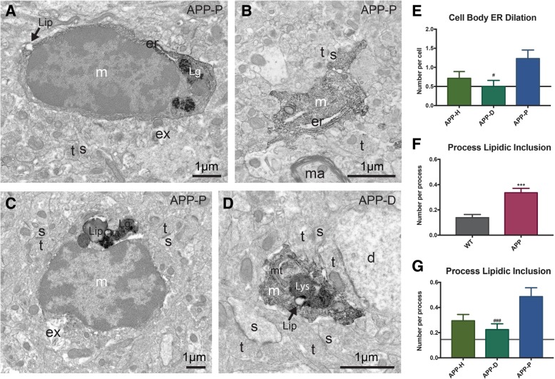
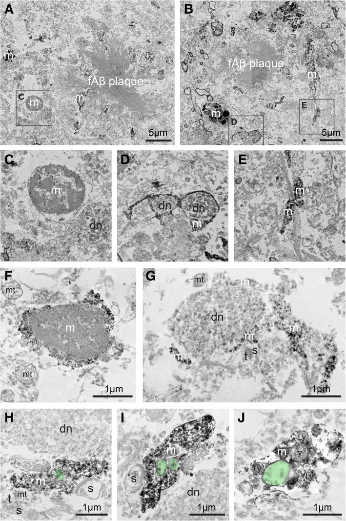
Alzheimer’s disease (AD) is the most common neurodegenerative disease, characterized by the deposition of extracellular fibrillar amyloid β (fΑβ) and the intracellular accumulation of neurofibrillary tangles. As AD progresses, Aβ drives a robust and prolonged inflammatory response via its recognition by microglia, the brain’s immune cells. Microglial reactivity to fAβ plaques may impair their normal surveillance duties, facilitating synaptic loss and neuronal death, as well as cognitive decline in AD. In the current study, we performed correlative light, transmission, and scanning electron microscopy to provide insights into microglial structural and functional heterogeneity. We analyzed microglial cell bodies and processes in areas containing fAβ plaques and neuronal dystrophy, dystrophy only, or appearing healthy, among the hippocampus CA1 of 14-month-old APPSwe-PS1Δe9 mice versus wild-type littermates. Our quantitative analysis revealed that microglial cell bodies in the AD model mice were larger and displayed ultrastructural signs of cellular stress, especially nearby plaques. Microglial cell bodies and processes were overall less phagocytic in AD model mice. However, they contained increased fibrillar materials and non-empty inclusions proximal to plaques. Microglial cell bodies and processes in AD model mice also displayed reduced association with extracellular space pockets that contained debris. In addition, microglial processes in healthy subregions of AD model mice encircled synaptic elements more often compared with plaque-associated processes. These observations in mice were qualitatively replicated in post-mortem hippocampal samples from two patients with AD (Braak stage 5). Together, our findings identify at the ultrastructural level distinct microglial transformations common to mouse and human in association with amyloid pathology.
Overview
The claims and statistics of this paper haven’t been checked yet.
Run the check — free, no account needed. It reads the open-access text and checks the sources it cites.
Integrity signals
Registry
Cites
Statistics
Figures
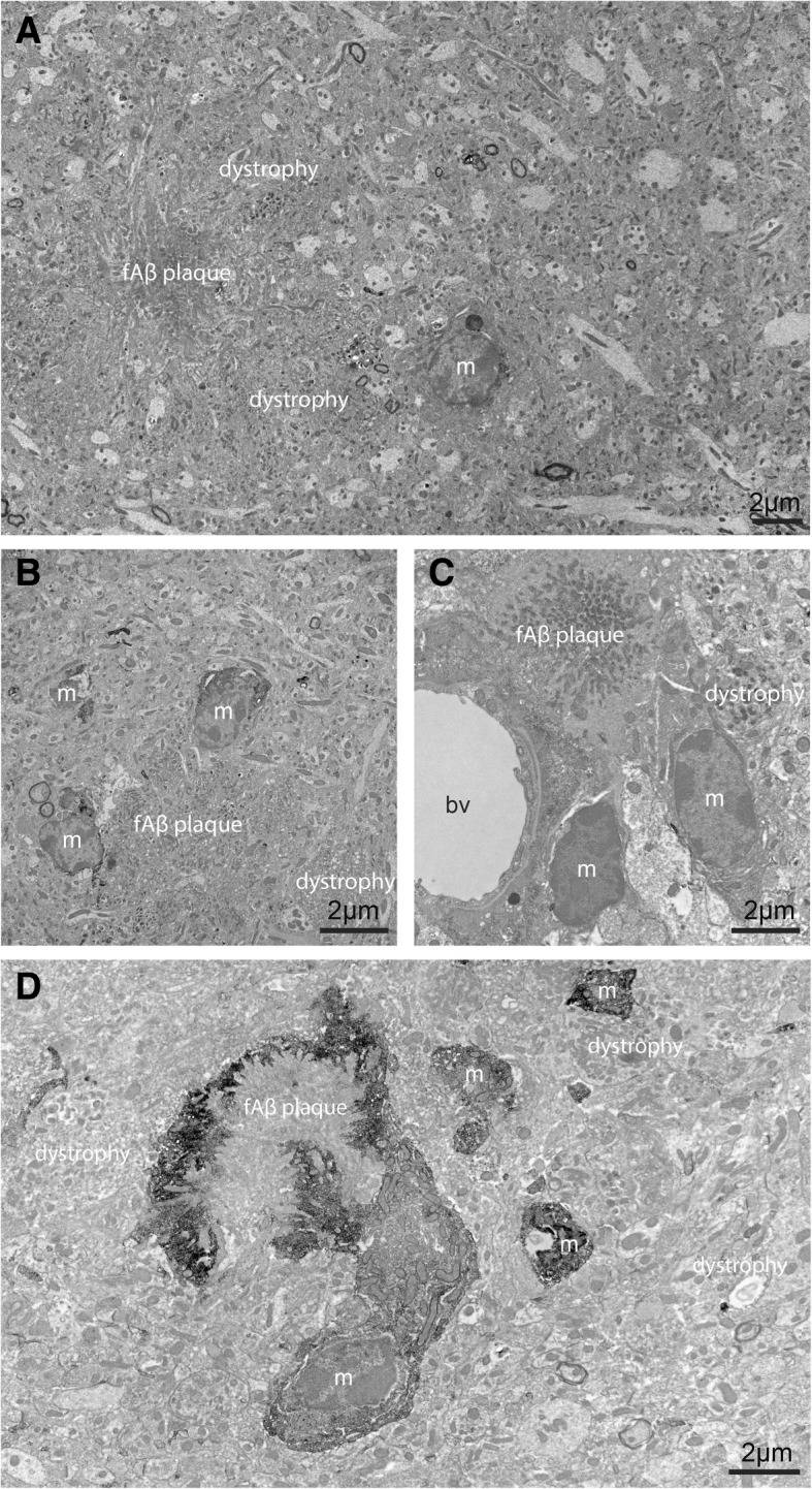
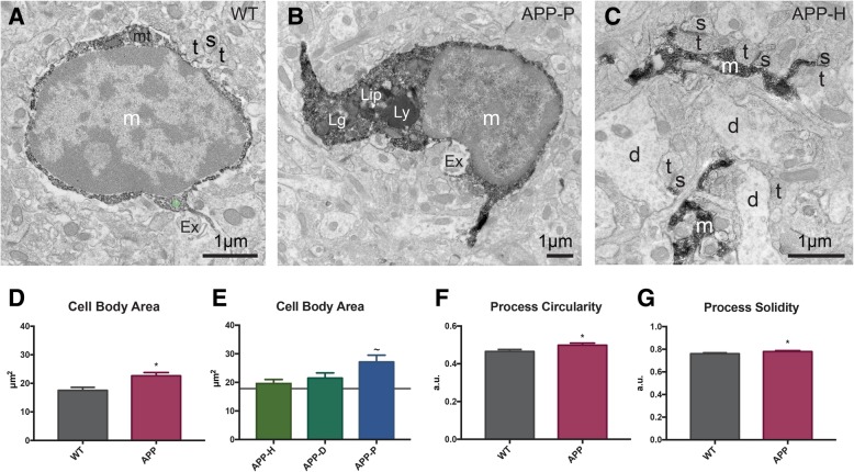
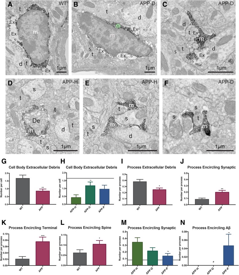
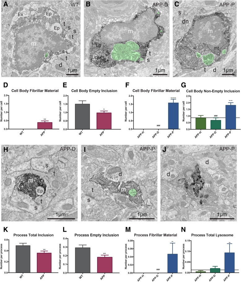
Verification
Claims the paper makes
The claims and statistics of this paper haven’t been checked yet.
Run the check — free, no account needed. It reads the open-access text and checks the sources it cites.
Claims other papers make about this one
Each is checked against this paper’s own text: does it say what the citing paper says it does?
The cited source says what the claim attributes to it.
In the citing paper
“In addition, in areas with limited plaque pathology, microglial processes encircled synaptic elements more often compared with plaque-associated processes [57].”
From Microglia specific deletion of miR-155 in Alzheimer’s disease mouse models reduces amyloid-β pathology but causes hyperexcitability and seizures (2023) · cites this paper as [57]
The cited source says what the claim attributes to it.
In the citing paper
“Previous studies have shown that cell bodies of microglia in AD model mice were larger and displayed ultrastructural signs of cellular stress, especially those nearby plaques and that these cells were overall less phagocytic [57].”
From Microglia specific deletion of miR-155 in Alzheimer’s disease mouse models reduces amyloid-β pathology but causes hyperexcitability and seizures (2023) · cites this paper as [57]
Self-check
Percentages, sums and reported differences re-computed from the paper’s own numbers.
The self-check needs the paper’s full text.
Citations
Cites
The Mouse Brain in Stereotaxic Coordinates
Paxinos, G. (2001) · Cited by 12,859
Sperling, R. A., Aisen, P. S., Beckett, L. A., Bennett, D. A., Craft, S., Fagan, A. M., Iwatsubo, T., Jack, C. R., Kaye, J., Montine, T. J., et al. (2011) · Alzheimer's & Dementia · doi:10.1016/j.jalz.2011.03.003 · Cited by 7,155
Resting microglial cells are highly dynamic surveillants of brain parenchyma in vivo
Nimmerjahn, A., Kirchhoff, F., & Helmchen, F. (2005) · Science · doi:10.1126/science.1110647 · Cited by 5,827
A Unique Microglia Type Associated with Restricting Development of Alzheimer’s Disease
Keren-Shaul, H., Spinrad, A., Weiner, A., Matcovitch-Natan, O., Dvir-Szternfeld, R., Ulland, T. K., David, E., Baruch, K., Lara-Astaiso, D., Toth, B., et al. (2017) · Cell · doi:10.1016/j.cell.2017.05.018 · Cited by 5,493 · cited 1×
“Microglial implication in the pathological process of AD is contextual [31, 32] and heterogeneous, as illustrated by recent single cell transcriptomic studies [33].”
BackgroundTerry, R. D., Masliah, E., Salmon, D. P., Butters, N., DeTeresa, R., Hill, R., Hansen, L. A., & Katzman, R. (1991) · Annals of Neurology · doi:10.1002/ana.410300410 · Cited by 4,397
Meta-Analysis of 74,046 Individuals Identifies 11 New Susceptibility Loci for Alzheimer’s Disease
(EADI), E. A. D. I., Lambert, J.-C., (GERAD), G. A. E. R. I. A. D., Alzheimer's Disease Genetic Consortium (ADGC), (CHARGE), C. F. H. A. A. R. I. G. E., Ibrahim-Verbaas, C. A., Harold, D., Naj, A. C., Sims, R., Bellenguez, C., et al. (2013) · Nature Genetics · doi:10.1038/ng.2802 · Cited by 4,336
Complement and microglia mediate early synapse loss in Alzheimer mouse models
Hong, S., Beja-Glasser, V. F., Nfonoyim, B. M., Frouin, A., Li, S., Ramakrishnan, S., Merry, K. M., Shi, Q., Rosenthal, A., Barres, B. A., et al. (2016) · Science · doi:10.1126/science.aad8373 · Cited by 3,343
Krasemann, S., Madore, C., Cialic, R., Baufeld, C., Calcagno, N., El Fatimy, R., Beckers, L., O’Loughlin, E., Xu, Y., Fanek, Z., et al. (2017) · Immunity · doi:10.1016/j.immuni.2017.08.008 · Cited by 2,979 · cited 1×
“Microglial implication in the pathological process of AD is contextual [31, 32] and heterogeneous, as illustrated by recent single cell transcriptomic studies [33].”
BackgroundVariant of TREM2 Associated with the Risk of Alzheimer's Disease
Jonsson, T., Stefansson, H., Steinberg, S., Jonsdottir, I., Jonsson, P. V., Snaedal, J., Bjornsson, S., Huttenlocher, J., Levey, A. I., Lah, J. J., et al. (2013) · New England Journal of Medicine · doi:10.1056/nejmoa1211103 · Cited by 2,558
Extensive Involvement of Autophagy in Alzheimer Disease: An Immuno-Electron Microscopy Study
Nixon, R. A., Wegiel, J., Kumar, A., Yu, W. H., Peterhoff, C., Cataldo, A., & Cuervo, A. M. (2005) · Journal of Neuropathology & Experimental Neurology · doi:10.1093/jnen/64.2.113 · Cited by 1,588
Research works printed but not linked: 4
- International AD. World Alzheimer Report 2016; 2016. p. 1–140.
- Alzheimer Association. 2018 Alzheimer’s disease facts and figures. Alzheimer’s Dement. 2018;2018:1–88.
- Ferreira ST, Lourenco MV, Oliveira MM, De Felice FG. Soluble amyloid-beta oligomers as synaptotoxins leading to cognitive impairment in Alzheimer’s disease. Front Cell Neurosci. 2015;9:9498.
- Solé-Domènech S, Cruz DL, Capetillo-Zarate E, Maxfield FR. Ageing research reviews Ageing Research Reviews. Elsevier B.V; 2016;32:89–103.
Cited by
Sorted by citations
Synaptic degeneration in Alzheimer disease
Tzioras, M., McGeachan, R. I., Durrant, C. S., & Spires-Jones, T. L. (2023) · Nature Reviews Neurology · doi:10.1038/s41582-022-00749-z · Cited by 504
Impact of TREM2R47H variant on tau pathology–induced gliosis and neurodegeneration
Gratuze, M., Leyns, C. E. G., Sauerbeck, A. D., St-Pierre, M.-K., Xiong, M., Kim, N., Serrano, J. R., Tremblay, M.-È., Kummer, T. T., Colonna, M., et al. (2020) · Journal of Clinical Investigation · doi:10.1172/jci138179 · Cited by 252
“(endoplasmic reticulum dilation, condensation of the nucleo- and cytoplasm, altered mitochondria) (67, 68) and have been seen previously in pathological conditions, such as AD (69).”
BackgroundSelf-citationTherapeutic B-cell depletion reverses progression of Alzheimer’s disease
Kim, K., Wang, X., Ragonnaud, E., Bodogai, M., Illouz, T., DeLuca, M., McDevitt, R. A., Gusev, F., Okun, E., Rogaev, E., et al. (2021) · Nature Communications · doi:10.1038/s41467-021-22479-4 · Cited by 206
Lombardi, M., Parolisi, R., Scaroni, F., Bonfanti, E., Gualerzi, A., Gabrielli, M., de Rosbo, N. K., Uccelli, A., Giussani, P., Viani, P., et al. (2019) · Acta Neuropathologica · doi:10.1007/s00401-019-02049-1 · Cited by 191
“Importantly, microglia exhibited condensed, electron-dense cytoplasm and nucleoplasm, resembling “dark” microglia, a recently described phenotype of reactive cells [7] [27], characterized by pronounced chromatin remodeling, which is indicative of altered gene expression.”
BackgroundSubramanian, J., Savage, J. C., & Tremblay, M. (2020) · Frontiers in Cellular Neuroscience · doi:10.3389/fncel.2020.592607 · Cited by 127
Self-citationPuntambekar, S. S., Moutinho, M., Lin, P. B.-C., Jadhav, V., Tumbleson-Brink, D., Balaji, A., Benito, M. A., Xu, G., Oblak, A., Lasagna-Reeves, C. A., et al. (2022) · Molecular Neurodegeneration · doi:10.1186/s13024-022-00545-9 · Cited by 113
“Taken as a whole, these data indicate that a) active elimination of neurons by neurodegenerative microglia may be dictated by the quality of intraneuronal pathological protein aggregates and b) microglial neurodegenerative responses modulated by CX3CR1 may be subject to region-dependent microglial heterogeneity along…”
BackgroundAPOE genotype and sex affect microglial interactions with plaques in Alzheimer’s disease mice
Stephen, T. L., Cacciottolo, M., Balu, D., Morgan, T. E., LaDu, M. J., Finch, C. E., & Pike, C. J. (2019) · Acta Neuropathologica Communications · doi:10.1186/s40478-019-0729-z · Cited by 115
Brain Ultrastructure: Putting the Pieces Together
Nahirney, P. C., & Tremblay, M. (2021) · Frontiers in Cell and Developmental Biology · doi:10.3389/fcell.2021.629503 · Cited by 111
Self-citationAugusto‐Oliveira, M., Arrifano, G. D. P., Delage, C. I., Tremblay, M., Lopez, M. E. C., & Verkhratsky, A. (2021) · Biological reviews/Biological reviews of the Cambridge Philosophical Society · doi:10.1111/brv.12797 · Cited by 114
Self-citationMicroglia control glutamatergic synapses in the adult mouse hippocampus
Basilico, B., Ferrucci, L., Ratano, P., Golia, M. T., Grimaldi, A., Rosito, M., Ferretti, V., Reverte, I., Sanchini, C., Marrone, M. C., et al. (2021) · Glia · doi:10.1002/glia.24101 · Cited by 104
Self-citationTzioras, M., Daniels, M. J. D., Davies, C., Baxter, P. S., King, D., McKay, S., Varga, B. V., Popovic, K., Hernandez, M., Stevenson, A. J., et al. (2023) · Cell Reports Medicine · doi:10.1016/j.xcrm.2023.101175 · Cited by 117
“These data show that microglia contain synaptic protein in human brain with more ingestion in AD, more near plaques, and more in APOE4 carriers.”
InfluentialSavage, J. C., St‐Pierre, M., Carrier, M., Hajj, H. E., Novak, S. W., Sánchez, M. G., Cicchetti, F., & Tremblay, M. (2020) · Journal of Neuroinflammation · doi:10.1186/s12974-020-01782-9 · Cited by 103
Self-citationA neurodegenerative cellular stress response linked to dark microglia and toxic lipid secretion
Flury, A. G., Aljayousi, L., Park, H.-J., Khakpour, M., Mechler, J., Aziz, S., McGrath, J. D., Deme, P., Sandberg, C., Ibáñez, F. G., et al. (2024) · Neuron · doi:10.1016/j.neuron.2024.11.018 · Cited by 102
Self-citationKrukowski, K., Nolan, A., Becker, M., Picard, K., Vernoux, N., Frias, E. S., Feng, X., Tremblay, M., & Rosi, S. (2021) · Brain Behavior and Immunity · doi:10.1016/j.bbi.2021.08.210 · Cited by 87
“In addition, in our qualitative analysis, neuronal cell bodies were distinguished by their pale nuclei and pale cytoplasm, heterochromatin pattern, as well as innervation from axon terminals (65, 68).”
BackgroundMethodInfluentialSelf-citationWalker, L. C. (2020) · Free Neuropathology · doi:10.17879/freeneuropathology-2020-3025 · Cited by 113
Bordeleau, M., Lacabanne, C., Fernández de Cossío, L., Vernoux, N., Savage, J. C., González-Ibáñez, F., & Tremblay, M.-È. (2020) · Journal of Neuroinflammation · doi:10.1186/s12974-020-01914-1 · Cited by 85
“Dilation of the endoplasmic reticulum and/or Golgi apparatus was noted when the distance between the cisternal membranes was 50 nm or greater [56, 57].”
BackgroundSelf-citationHashemiaghdam, A., & Mroczek, M. (2020) · Journal of Neuroimmunology · doi:10.1016/j.jneuroim.2020.577185 · Cited by 79
Microglia transcriptional states and their functional significance: Context drives diversity
Depp, C., Doman, J. L., Hingerl, M., Xia, J., & Stevens, B. (2025) · Immunity · doi:10.1016/j.immuni.2025.04.009 · Cited by 84
Different phenotypes of microglia in animal models of Alzheimer disease
Wei, Y., & Li, X. (2022) · Immunity & Ageing · doi:10.1186/s12979-022-00300-0 · Cited by 86
Picard, K., St‐Pierre, M., Vecchiarelli, H. A., Bordeleau, M., & Tremblay, M. (2021) · Neurochemistry International · doi:10.1016/j.neuint.2021.104987 · Cited by 79
Self-citationSt‐Pierre, M., Carrier, M., Ibáñez, F. G., Šimončičová, E., Wallman, M., Vallières, L., Parent, M., & Tremblay, M. (2022) · Journal of Neuroinflammation · doi:10.1186/s12974-022-02595-8 · Cited by 80
“In line with these findings, previous studies from our group identified a microglial state, the dark microglia, which are found nearby fibrillar Aβ in middle-age, 14-month-old APP-PS1 mice [mouse model of AD with a Swedish mutation in the APP and a humanized presenilin (PSEN1)] [64, 65].”
BackgroundMethodResultInfluentialSelf-citationShedding Light on the Dark Side of the Microglia
St‐Pierre, M., Šimončičová, E., Bögi, E., & Tremblay, M. (2020) · ASN NEURO · doi:10.1177/1759091420925335 · Cited by 66
“…observed microglia with a preserved, distinct heterochromatin pattern and a less-dark appearance that suggested the existence of an intermediate phenotype between the typical and DM (El Hajj et al., 2019), thus further supporting the idea that DM might be part of a heterogenous microglial spectrum.”
BackgroundResultInfluentialSelf-citationMicroglia gone awry: linking immunometabolism to neurodegeneration
Afridi, R., Lee, W.-H., & Suk, K. (2020) · Frontiers in Cellular Neuroscience · doi:10.3389/fncel.2020.00246 · Cited by 64
“The dark microglia are named after their dark appearance under an electronmicroscope (Bisht et al., 2016; El Hajj et al., 2019).”
BackgroundThe role of the immune system in driving neuroinflammation
Ní Chasaide, C., & Lynch, M. A. (2020) · Brain and Neuroscience Advances · doi:10.1177/2398212819901082 · Cited by 72
“Evidence suggests that these cells are hypermotile (Gyoneva et al., 2016) and less phagocytic, although they contain fibrillar material (El Hajj et al., 2019), so they can phagocytose, but not process, amyloid-β (Aβ).”
BackgroundMicroglial burden, activation and dystrophy patterns in frontotemporal lobar degeneration
Woollacott, I., Toomey, C. E., Strand, C., Courtney, R. J., Benson, B. C., Rohrer, J. D., & Lashley, T. (2020) · Journal of Neuroinflammation · doi:10.1186/s12974-020-01907-0 · Cited by 65
Bollinger, J. L., Dadosky, D. T., Flurer, J. K., Rainer, I. L., Woodburn, S. C., & Wohleb, E. S. (2022) · Neuropsychopharmacology · doi:10.1038/s41386-022-01519-7 · Cited by 55
Microglial Ultrastructure in the Hippocampus of a Lipopolysaccharide-Induced Sickness Mouse Model
Savage, J. C., St‐Pierre, M., Hui, C. W., & Tremblay, M. (2019) · Frontiers in Neuroscience · doi:10.3389/fnins.2019.01340 · Cited by 53
Self-citationBordeleau, M., Comin, C. H., de Cossío, L. F., Lacabanne, C., Freitas-Andrade, M., Ibáñez, F. G., Raman-Nair, J., Wakem, M., Chakravarty, M., Costa, L. D. F., et al. (2022) · Communications Biology · doi:10.1038/s42003-021-02947-9 · Cited by 53
Self-citationDysregulated CD200-CD200R signaling in early diabetes modulates microglia-mediated retinopathy
Pfeifer, C. W., Walsh, J. T., Santeford, A. C., Lin, J. B., Beatty, W. L., Terao, R., Liu, Y. A., Hase, K., Ruzycki, P. A., & Apte, R. S. (2023) · Proceedings of the National Academy of Sciences · doi:10.1073/pnas.2308214120 · Cited by 48
Sharma, M., Pal, P., & Gupta, S. K. (2024) · Neurochemistry International · doi:10.1016/j.neuint.2024.105761 · Cited by 49
McFarland, K. N., & Chakrabarty, P. (2022) · Neurotherapeutics · doi:10.1007/s13311-021-01179-3 · Cited by 48
Bordeleau, M., de Cossío, L. F., Lacabanne, C., Savage, J. C., Vernoux, N., Chakravarty, M. M., & Tremblay, M. (2021) · Brain Behavior & Immunity - Health · doi:10.1016/j.bbih.2021.100281 · Cited by 44
Self-citationTargeting innate immunity to protect and cure Alzheimer’s disease: opportunities and pitfalls
Cisbani, G., & Rivest, S. (2021) · Molecular Psychiatry · doi:10.1038/s41380-021-01083-4 · Cited by 44
“Of note, brains of subjects with AD showed an intensified immune response in both post-mortem studies [33] as well as in vivo [34] and microglia displayed ultrastructural modifications when associated with Aβ [35].”
BackgroundSelf-citationAloi, M. S., Prater, K. E., Sánchez, R. E. A., Beck, A., Pathan, J. L., Davidson, S., Wilson, A., Keene, C. D., de la Iglesia, H., Jayadev, S., et al. (2023) · Journal of Neuroinflammation · doi:10.1186/s12974-023-02745-6 · Cited by 43
“In addition, in areas with limited plaque pathology, microglial processes encircled synaptic elements more often compared with plaque-associated processes [57].”
BackgroundResultDe Felice, E., Gonçalves de Andrade, E., Golia, M. T., González Ibáñez, F., Khakpour, M., Di Castro, M. A., Garofalo, S., Di Pietro, E., Benatti, C., Brunello, N., et al. (2022) · Journal of Neuroinflammation · doi:10.1186/s12974-022-02655-z · Cited by 39
Self-citationde Andrade, E. G., Šimončičová, E., Carrier, M., Vecchiarelli, H. A., Robert, M.-È., & Tremblay, M. (2021) · Frontiers in Cellular Neuroscience · doi:10.3389/fncel.2021.647378 · Cited by 39
Self-citationLiang, F., Huang, T., Li, B., Zhao, Y., Zhang, X., & Xu, B. (2020) · Neuroreport · doi:10.1097/wnr.0000000000001429 · Cited by 38
McKee, C., Hoffos, M., Vecchiarelli, H. A., & Tremblay, M. (2023) · Frontiers in Pharmacology · doi:10.3389/fphar.2023.1125982 · Cited by 38
Self-citationLecours, C., St‐Pierre, M., Picard, K., Bordeleau, M., Bourque, M., Awogbindin, I. O., Benadjal, A., Ibáñez, F. G., Gagnon, D., Cantin, L., et al. (2020) · Brain Behavior and Immunity · doi:10.1016/j.bbi.2020.07.044 · Cited by 34
Self-citationIkzf1 as a novel regulator of microglial homeostasis in inflammation and neurodegeneration
Ballasch, I., García-García, E., Vila, C., Pérez-González, A., Sancho‐Balsells, A., Fernández, J., Soto, D., Puigdellívol, M., Gasull, X., Alberch, J., et al. (2023) · Brain Behavior and Immunity · doi:10.1016/j.bbi.2023.01.016 · Cited by 36
Digestive exophagy: Phagocyte digestion of objects too large for phagocytosis
Maxfield, F. R., Barbosa-Lorenzi, V. C., & Singh, R. K. (2019) · Traffic · doi:10.1111/tra.12712 · Cited by 33
Enlow, W., Bordeleau, M., Piret, J., Ibáñez, F. G., Uyar, O., Venable, M.-C., Goyette, N., Carbonneau, J., Tremblay, M., & Boivin, G. (2021) · Journal of Neuroinflammation · doi:10.1186/s12974-021-02221-z · Cited by 31
“It is suggested that microglia break down dead or dying neurons into pieces using exophagy before their phagocytic engulfment and digestion [46], hence allowing them to digest large debris and cells [49, 50].”
BackgroundSelf-citationVecchiarelli, H. A., & Tremblay, M. (2023) · Annual Review of Genetics · doi:10.1146/annurev-genet-022223-093643 · Cited by 32
Self-citationGonzález Ibáñez, F., Halvorson, T., Sharma, K., McKee, C. G., Carrier, M., Picard, K., Vernoux, N., Bisht, K., Deslauriers, J., Lalowski, M., et al. (2023) · Brain, Behavior, and Immunity · doi:10.1016/j.bbi.2023.09.006 · Cited by 28
Self-citationSt-Pierre, M.-K., Carrier, M., Lau, V., & Tremblay, M.-È. (2022) · Methods in Molecular Biology · doi:10.1007/978-1-0716-2409-8_3 · Cited by 29
Self-citationImaging the Neuroimmune Dynamics Across Space and Time
Carrier, M., Robert, M.-È., Ibáñez, F. G., Desjardins, M., & Tremblay, M. (2020) · Frontiers in Neuroscience · doi:10.3389/fnins.2020.00903 · Cited by 28
Self-citationHypothalamic Microglial Heterogeneity and Signature under High Fat Diet–Induced Inflammation
Mendes, N. F., Jara, C. P., Zanesco, A. M., & ARAUJO, E. P. D. (2021) · International Journal of Molecular Sciences · doi:10.3390/ijms22052256 · Cited by 28
St‐Pierre, M., VanderZwaag, J., Loewen, S. M., & Tremblay, M. (2022) · Frontiers in Cellular Neuroscience · doi:10.3389/fncel.2022.932572 · Cited by 27
Self-citationSt‐Pierre, M., Carrier, M., Ibáñez, F. G., Khakpour, M., Wallman, M., Parent, M., & Tremblay, M. (2023) · Journal of Neuroinflammation · doi:10.1186/s12974-023-02752-7 · Cited by 28
Self-citationKim, Y. S., Choi, S., Kim, K.-Y., Pelaez, J. M. N., Perkins, G. A., Choi, S., Kim, J., Nazarenkov, N., Rissman, R. A., Ju, W., et al. (2024) · Journal of Neuroinflammation · doi:10.1186/s12974-024-03214-4 · Cited by 24
“Dilated ER has been reported in several neurodegenerative diseases, including amyotrophic lateral sclerosis and AD [37].”
Microglia in Alzheimer’s Disease
Süß, P., & Schlachetzki, J. C. M. (2020) · Current Alzheimer Research · doi:10.2174/1567205017666200212155234 · Cited by 26
St‐Pierre, M., Ibáñez, F. G., Kroner, A., & Tremblay, M. (2023) · Journal of Neuroinflammation · doi:10.1186/s12974-023-02953-0 · Cited by 22
Self-citationStoiljković, M., Gutierrez, K. O., Kelley, C., Horváth, T. L., & Hajós, M. (2021) · Journal of Alzheimer s Disease · doi:10.3233/jad-210041 · Cited by 21
“…progression microglia in response to oligomeric and plaque A (cid:2) challenge tend to undergo substantial ultrastructural changes leading to aberrant activation involved in A (cid:2) -mediated neuronal dysfunctions and pathologic loss of synapses in cognitive-related brain regions [55, 56].”
Abo-Zaid, O. A., Sallam, M. W., & Ahmed, E. S. A. (2022) · The Journal of Prevention of Alzheimer s Disease · doi:10.14283/jpad.2022.26 · Cited by 24
“These plaques caused neuronal dystrophy, and finally neuronal death (34) that is represented by the increased number of dark neurons in the AD group.”
BackgroundBrain structure and structural basis of neurodegenerative diseases
Yang, J., Sui, S., & Liu, Z. (2022) · Biophysics Reports · doi:10.52601/bpr.2022.220013 · Cited by 20
Fundamental Neurochemistry Review: Lipids across microglial states
Traetta, M. E., Vecchiarelli, H. A., & Tremblay, M. (2024) · Journal of Neurochemistry · doi:10.1111/jnc.16259 · Cited by 21
Self-citationMultiscale ATUM-FIB Microscopy Enables Targeted Ultrastructural Analysis at Isotropic Resolution
Kislinger, G., Gnaegi, H., Kerschensteiner, M., Simons, M., Misgeld, T., & Schifferer, M. (2020) · iScience · doi:10.1016/j.isci.2020.101290 · Cited by 20
Vignon, A., Dudon, G., Oliva, G., Thirard, S., Alenda, U. G., Brugoux, A., Cazevieille, C., Imbert, J., Bellières, C., Lehmann, S., et al. (2025) · Journal of Hazardous Materials · doi:10.1016/j.jhazmat.2025.138640 · Cited by 22
The Dual Nature of Microglia in Alzheimer’s Disease: A Microglia-Neuron Crosstalk Perspective
Xie, Z., Meng, J., Wu, Z., Nakanishi, H., Hayashi, Y., Kong, W., Lan, F., Narengaowa, Yang, Q., Qing, H., et al. (2023) · The Neuroscientist · doi:10.1177/10738584211070273 · Cited by 19
Using focused ultrasound to modulate microglial structure and function
Grewal, S., de Andrade, E. G., Kofoed, R. H., Matthews, P. M., Aubert, I., Tremblay, M., & Morse, S. V. (2023) · Frontiers in Cellular Neuroscience · doi:10.3389/fncel.2023.1290628 · Cited by 19
Self-citationBobotis, B. C., Halvorson, T., Carrier, M., & Tremblay, M. (2024) · Frontiers in Cellular Neuroscience · doi:10.3389/fncel.2024.1317125 · Cited by 22
Self-citationWendt, S., Johnson, S., Weilinger, N. L., Groten, C., Sorrentino, S., Frew, J., Yang, L., Choi, H. B., Nygaard, H. B., & MacVicar, B. A. (2022) · Redox Biology · doi:10.1016/j.redox.2022.102448 · Cited by 20
“neuron-microglia interaction remains unknown [17].”
BackgroundPhenomic Microglia Diversity as a Druggable Target in the Hippocampus in Neurodegenerative Diseases
Lana, D., Magni, G., Landucci, E., Wenk, G. L., Pellegrini-Giampietro, D. E., & Giovannini, M. G. (2023) · International Journal of Molecular Sciences · doi:10.3390/ijms241813668 · Cited by 20
Bobotis, B. C., Khakpour, M., Braniff, O., de Andrade, E. G., Gargus, M., Allen, M., Carrier, M., Baillargeon, J., Rangachari, M., & Tremblay, M.-È. (2025) · Journal of Neuroinflammation · doi:10.1186/s12974-025-03341-6 · Cited by 17
Self-citationDecoeur, F., Picard, K., St‐Pierre, M., Greenhalgh, A. D., Delpech, J.-C., Séré, A., Layé, S., Tremblay, M., & Nadjar, A. (2022) · Frontiers in Cellular Neuroscience · doi:10.3389/fncel.2022.802411 · Cited by 16
Self-citationMicroglial Reactivity Correlates with Presynaptic Loss Independent of β‐Amyloid and Tau
Lan, G., Chen, X., Yang, J., Sun, P., Cai, Y. Q., Li, A., Zhu, Y., Liu, Z., Ma, S., Guo, T., et al. (2024) · Annals of Neurology · doi:10.1002/ana.26885 · Cited by 19
“One postmortem study 49 demonstrated that microglial processes contained synaptic elements in the hippocampus of AD patients, and the amyloidosis mice experienced greater synaptic engulfment by microglia compared to wild-type mice.”
Tolédano, A., Rodríguez‐Casado, A., Álvarez, M., & Toledano‐Díaz, A. (2024) · Brain Sciences · doi:10.3390/brainsci14111101 · Cited by 19
Pumo, A., & Legeay, S. (2024) · Brain Research · doi:10.1016/j.brainres.2024.148817 · Cited by 14
“Additionally, other microglial subsets that emerge during disease progression have been observed, which differ from the originally described DAMs, including plaques associated microglia and dark microglia (Hashemiaghdam and Mroczek, 2020; Bisht et al., 2016; El Hajj et al., 2019).”
Liang, F., Sun, F., He, B., & Wang, J. (2022) · Neuroscience · doi:10.1016/j.neuroscience.2022.03.043 · Cited by 12
Toledano‐Díaz, A., Álvarez, M., & Tolédano, A. (2022) · Journal of Central Nervous System Disease · doi:10.1177/11795735221128703 · Cited by 11
“Many authors consider that microglial cells accumulate in areas more strongly associated with amyloid deposits, even infiltrating amyloid plaques, although they also appear in less severely affected areas.(198,199) In”
BackgroundPromising molecular targets for pharmacological therapy of neurodegenerative pathologies
Неганова, М. Е., Aleksandrova, Y. R., Nebogatikov, V. O., Klochkov, S. G., & Ustyugov, A. A. (2020) · Acta Naturae · doi:10.32607/actanaturae.10925 · Cited by 11
The Role of Glia in Addiction: Dopamine as a Modulator of Glial Responses in Addiction
Jiménez-González, A., Acevedo, C. G., Ochoa-Aguilar, A., & Chavarría, A. (2021) · Cellular and Molecular Neurobiology · doi:10.1007/s10571-021-01105-3 · Cited by 10
Vecchiarelli, H. A., Bisht, K., Sharma, K. P., Novak, S. W., Traetta, M. E., Garcia‐Segura, M. E., St‐Pierre, M., Savage, J. C., Willis, C. M., Picard, K., et al. (2024) · bioRxiv (Cold Spring Harbor Laboratory) · doi:10.1101/2024.10.15.618087 · Cited by 10
Self-citationUltrastructural features of psychological stress resilience in the brain: a microglial perspective
Ibáñez, F. G., VanderZwaag, J., Deslauriers, J., & Tremblay, M. (2024) · Open Biology · doi:10.1098/rsob.240079 · Cited by 11
Self-citationMicroglia heterogeneity during neuroinflammation and neurodegeneration in the mouse retina
Rao, B., Liu, X., Xiao, J., Wu, X., He, F., Yang, Q., Zhao, W., Lin, X., & Zhang, J. (2024) · Brain Structure and Function · doi:10.1007/s00429-024-02875-4 · Cited by 12
“This is in agreement with previous studies in the CNS (Bisht K et al. 2016; El Hajj H et al. 2019).”
InfluentialCell type specific isolation of primary astrocytes and microglia from adult mouse spinal cord
Ahn, J. J., Islam, Y., & Miller, R. H. (2022) · Journal of Neuroscience Methods · doi:10.1016/j.jneumeth.2022.109599 · Cited by 8
Shulyatnikova, T. V., Tumanskiy, V. A., & Hayden, M. R. (2022) · International Journal of Molecular Sciences · doi:10.3390/ijms232214455 · Cited by 8
“5 μm in diameter, enclosing heterogeneous contents by a single membrane [44].”
BackgroundInfluentialInvestigation of microglial diversity in a LRRK2 G2019S mouse model of Parkinson's disease
Iovino, L., VanderZwaag, J., Kaur, G., Khakpour, M., Giusti, V., Donadon, M., Chiavegato, A., Lopes, L. T., Greggio, E., Tremblay, M., et al. (2024) · Neurobiology of Disease · doi:10.1016/j.nbd.2024.106481 · Cited by 7
Self-citationSt‐Pierre, M., Šimončičová, E., Carrier, M., & Tremblay, M. (2022) · Methods in molecular biology · doi:10.1007/978-1-0716-2655-9_4 · Cited by 5
Self-citationMicroglial Drivers of Alzheimer's Disease Pathology: An Evolution of Diverse Participating States
Kuhn, M. K., & Proctor, E. A. (2024) · Proteins Structure Function and Bioinformatics · doi:10.1002/prot.26723 · Cited by 5
“…neurons from Aβ oligomers that contribute to synapse disruption and cognitive deficits [169, 170], as a consequence of their interaction with Aβ and Aβ plaques, microglia display impaired surveillance activity and dampened homeostatic functions [171] and promote the spread of Aβ in the brain [172].”
Lau, V. Z., VanderZwaag, J., Murray, C. J., & Tremblay, M. (2025) · Journal of Neurochemistry · doi:10.1111/jnc.70086 · Cited by 6
Self-citationEvolving insights into the identity and function of dark microglia
Flury, A. G., St‐Pierre, M., Ayata, P., & Tremblay, M. (2025) · Trends in Neurosciences · doi:10.1016/j.tins.2025.11.002 · Cited by 7
Self-citationFelice, E. D., Bobotis, B. C., Rigillo, G., Khakpour, M., de Andrade, E. G., Benatti, C., Vilella, A., Tascedda, F., Limatola, C., Tremblay, M., et al. (2024) · Brain Behavior and Immunity · doi:10.1016/j.bbi.2024.11.034 · Cited by 5
Self-citationSomodji, E. A., & Gowrishankar, S. (2026) · npj Dementia · doi:10.1038/s44400-026-00086-8 · Cited by 6
“Indeed, a study that examined the ultrastructure of organelles within the main body and processes of microglia, noted that there were more non-empty inclusions within phagocytic vesicles of processes versus the main body 103 .”
St‐Pierre, M., Carrier, M., Ibáñez, F. G., Šimončičová, E., Wallman, M., Vallières, L., Parent, M., & Tremblay, M. (2022) · Research Square · doi:10.21203/rs.3.rs-1751270/v1 · Cited by 4
Self-citationEmerging Techniques for the Study of Microglia: Visualization, Depletion and Fate MappingPreprint
Bobotis, B. C., Halvorson, T., Carrier, M., & Tremblay, M. (2023) · Preprints.org · doi:10.20944/preprints202308.2190.v1 · Cited by 3
Self-citationXie, D., Zheng, Q., Lv, J., Zhang, Q., Cui, Z., Huang, S., Yu, W., Chen, B., Que, W., Fu, S., et al. (2025) · Advanced Science · doi:10.1002/advs.202510270 · Cited by 3
How Many Alzheimer–Perusini’s Atypical Forms Do We Still Have to Discover?
Donato, L., Mordà, D., Scimone, C., Alibrandi, S., D’Angelo, R., & Sidoti, A. (2023) · Biomedicines · doi:10.3390/biomedicines11072035 · Cited by 3
Biphasic Role of Microglia in Healthy and Diseased Brain
Kumar, M., Arora, P., & Sandhir, R. (2022) · Springer eBooks · doi:10.1007/978-981-16-8313-8_18 · Cited by 2
Ibáñez, F. G., Halvorson, T., Sharma, K. P., McKee, C., Carrier, M., Picard, K., Vernoux, N., Bisht, K., Deslauriers, J., Lalowski, M. M., et al. (2023) · bioRxiv (Cold Spring Harbor Laboratory) · doi:10.1101/2023.08.28.555135 · Cited by 2
Self-citationda Silva‐Araújo, E. R., Manhães-de-Castro, R., Toscano, A. E., Gouveia, H. J. C. B., de Sá, M. R., Júnior, G. B. A., da Silva, A. N., & Hernández, E. P. (2025) · Progress in Biophysics and Molecular Biology · doi:10.1016/j.pbiomolbio.2025.12.006 · Cited by 1
Caveolin-1 Regulates Ultrastructural Alterations of Astrocytes in Chronic Cerebral Hypoperfusion
Wu, M., Zhang, X., Wang, D., Shu, X., Chen, D., Zhao, Y., Lv, Q., Li, Y., Huang, Z., Gao, J., et al. (2025) · Inflammation · doi:10.1007/s10753-025-02431-0 · Cited by 1
Investigation of microglial diversity in a mouse model of Parkinson’s disease pathologyPreprint
Iovino, L., VanderZwaag, J., Kaur, G., Khakpour, P., Giusti, V., Chiavegato, A., Lopes, L. T., Greggio, E., Tremblay, M., & Civiero, L. (2023) · bioRxiv (Cold Spring Harbor Laboratory) · doi:10.1101/2023.11.23.567809 · Cited by 1
Self-citationEtebar, F. (2023) · Queensland University of Technology · doi:10.5204/thesis.eprints.244774
Kozyrev, N., Ibáñez, F. G., McKee, C., Traetta, M. E., Tellios, V., Nygard, K. L., Rylett, R. J., Tremblay, M., & Dekaban, G. A. (2025) · Research Square · doi:10.21203/rs.3.rs-6222217/v1
Self-citationWhite, A., Gavrilyuk, P., Gu, P., Moya, R. F., Thurston, R. H., Fickak, A., Rozema, N. B., Keshavaram, P., Vermilyea, S., Schlichte, R., et al. (2026) · Translational Neurodegeneration · doi:10.1186/s40035-026-00563-3
de Andrade, E. G., VanderZwaag, J., Kofoed, R. H., Carrier, M., Picard, K., Pekarik, K. H., Ibáñez, F. G., Khakpour, M., Hynynen, K., Aubert, I., et al. (2025) · bioRxiv (Cold Spring Harbor Laboratory) · doi:10.1101/2025.09.17.675183
“ER /Golgi apparatus dilation, associated with cellular stress, was identified when the distance between cisternal membranes was greater than 100 nm [17,19].”
Self-citationSherman, E., Qiu, K., Roberts, R., Shichman, L., Li, S., Sun, H., Ide, L., Tucker, A., Lee, S., Gniadzik, W., et al. (2025) · SSRN Electronic Journal · doi:10.1101/2025.10.17.683162 · Cited by 1
Enlow, W., Bordeleau, M., Piret, J., Ibáñez, F. G., Uyar, O., Venable, M.-C., Goyette, N., Carbonneau, J., Tremblay, M., & Boivin, G. (2021) · Figshare · doi:10.6084/m9.figshare.15179567
Self-citationAloi, M. S., Prater, K. E., Sánchez, R. E. A., Beck, A. I., Pathan, J. L., Davidson, S., Wilson, A. M., Keene, C. D., de la Iglesia, H. O., Jayadev, S., et al. (2024) · Figshare · doi:10.6084/m9.figshare.26573215
Metadata
- Authors
- El Hajj, H., Savage, J. C., Bisht, K., Parent, M., Vallières, L., Rivest, S., & Tremblay, M.-È.
- Affiliations
- Centre de recherche du CHU de Québec-Université Laval (CA)
- Université Laval (CA)
- Corresponding author
- Tremblay, Marie-Ève
- Venue
- Journal of Neuroinflammation
- Published
- 2019-04-16
- Volume
- 16
- Issue
- 1
- Pages
- 87-87
- ISSN
- 1742-2094 (ISSN-L)
- PMC
- PMC6469225
- OpenAlex
- W2946033659
- Cited by
- 119
- References
- 95
- Citations by year
- 2026: 7 · 2025: 11 · 2024: 23 · 2023: 16 · 2022: 22 · 2021: 18 · 2020: 18 · 2019: 4
- FWCI
- 6.82
- Type
- journal article
- Language
- en
- Open access
- gold
- doi.org
- Licence
- Locations
- Journal of Neuroinflammation (publishedVersion, cc-by)
- PubMed Central (publishedVersion, cc-by)
- PubMed (publishedVersion)
- DOAJ (DOAJ: Directory of Open Access Journals) (submittedVersion)
- Funders
- Fonds de recherche en santé du Québec
- Lebanese Ministry of Education and Higher Education
- Fondation du CHU de Québec
- The Banting Research Foundation
- The Scottish Rite Charitable Foundation of Canada
- NEURON-ERANET
- Topics
- Life Sciences › Neuroscience › Neurology › Neuroinflammation and Neurodegeneration Mechanisms
- Life Sciences › Neuroscience › Cellular and Molecular Neuroscience › Alzheimer's disease research and treatments
- Life Sciences › Neuroscience › Neurology › Neurological Disease Mechanisms and Treatments
- Keywords
- Alzheimer's disease, microglial heterogeneity, amyloid plaques, amyloid fibrils, APPswe/PS1ΔE9 mice, hippocampal CA1, microglial phagocytosis, neuronal dystrophy, Braak stages, neuroinflammation
- MeSH terms
- Aged, Aged, 80 and over, Alzheimer Disease, Animals, Hippocampus, Humans, Amyloid beta-Peptides, Microglia, Mice
About these checks
Registry, retraction, Cites and statistics results are computed, not judged, with one exception: a printed reference that no index lists is read once by a model before it counts as a citation error. Claims are extracted by a model and kept only when their quote appears verbatim in the text; each cited source is then checked, with a model’s help, for whether it says what the claim relies on. Those verdicts can be wrong — read the claim’s quote and the cited source before relying on one.
Statistics and arithmetic are recomputed, not judged.
Confidence bands are shown only for fields calibrated on our annotated test set. None is calibrated yet, so no band is shown.
What counts as a claim ›How we measure confidence ›How we check this ›
Are you an author, or do you see a problem with this page? Request a review
