

The DOI matches its Crossref record; 170 of the 172 references it cites were found in a registry or an index, and 2 are not checked yet.
Endo-lysosomal dysregulations and late-onset Alzheimer’s disease: impact of genetic risk factors
Open accessAbstract
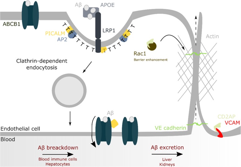
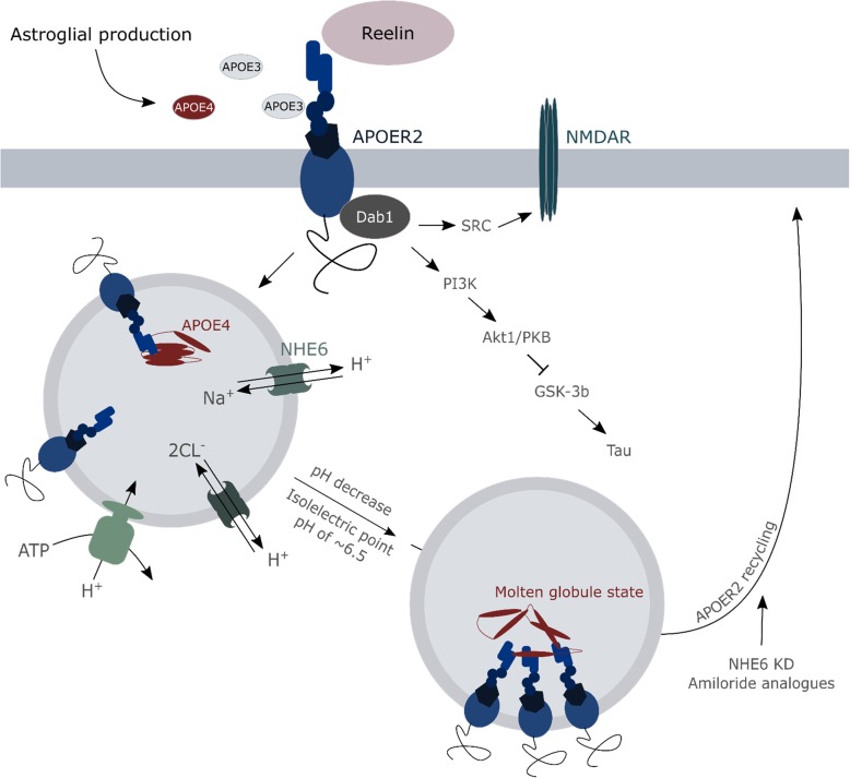
Increasing evidence supports that cellular dysregulations in the degradative routes contribute to the initiation and progression of neurodegenerative diseases, including Alzheimer's disease. Autophagy and endolysosomal homeostasis need to be maintained throughout life as they are major cellular mechanisms involved in both the production of toxic amyloid peptides and the clearance of misfolded or aggregated proteins. As such, alterations in endolysosomal and autophagic flux, as a measure of degradation activity in these routes or compartments, may directly impact as well on disease-related mechanisms such as amyloid-β clearance through the blood-brain-barrier and the interneuronal spreading of amyloid-β and/or Tau seeds, affecting synaptic function, plasticity and metabolism. The emerging of several genetic risk factors for late-onset Alzheimer's disease that are functionally related to endocytic transport regulation, including cholesterol metabolism and clearance, supports the notion that in particular the autophagy/lysosomal flux might become more vulnerable during ageing thereby contributing to disease onset. In this review we discuss our current knowledge of the risk genes APOE4, BIN1, CD2AP, PICALM, PLD3 and TREM2 and their impact on endolysosomal (dys)regulations in the light of late-onset Alzheimer's disease pathology.
Overview
The claims and statistics of this paper haven’t been checked yet.
Run the check — free, no account needed. It reads the open-access text and checks the sources it cites.
Integrity signals
Registry
Cites
Statistics
Figures
Verification
Claims the paper makes
The claims and statistics of this paper haven’t been checked yet.
Run the check — free, no account needed. It reads the open-access text and checks the sources it cites.
Claims other papers make about this one
Each is checked against this paper’s own text: does it say what the citing paper says it does?
The cited source says what the claim attributes to it.
In the citing paper
“multiple genetic risk alleles associated with AD are thought to be active in this pathway [34].”
From Microglia specific deletion of miR-155 in Alzheimer’s disease mouse models reduces amyloid-β pathology but causes hyperexcitability and seizures (2023) · cites this paper as [34]
The cited source says what the claim attributes to it.
In the citing paper
“Aβ results from the proteolysis of APP, a type I transmembrane protein targeted first at the plasma membrane, then rapidly endocytosed to endosomes to be metabolized to Aβ or subsequently sent to the lysosomal compartment for degradation (Wang X. et al., 2017; Van Acker et al., 2019).”
From Emerging Alternative Proteinases in APP Metabolism and Alzheimer’s Disease Pathogenesis: A Focus on MT1-MMP and MT5-MMP (2019) · cites this paper as Van Acker et al., 2019
Self-check
Percentages, sums and reported differences re-computed from the paper’s own numbers.
The self-check needs the paper’s full text.
Citations
Cites
Meta-Analysis of 74,046 Individuals Identifies 11 New Susceptibility Loci for Alzheimer’s Disease
(EADI), E. A. D. I., Lambert, J.-C., (GERAD), G. A. E. R. I. A. D., Alzheimer's Disease Genetic Consortium (ADGC), (CHARGE), C. F. H. A. A. R. I. G. E., Ibrahim-Verbaas, C. A., Harold, D., Naj, A. C., Sims, R., Bellenguez, C., et al. (2013) · Nature Genetics · doi:10.1038/ng.2802 · Cited by 4,336
Blood–brain barrier breakdown in Alzheimer disease and other neurodegenerative disorders
Sweeney, M. D., Sagare, A. P., & Zlokovic, B. V. (2018) · Nature Reviews Neurology · doi:10.1038/nrneurol.2017.188 · Cited by 3,076
TREM2 Variants in Alzheimer's Disease
Guerreiro, R., Wojtas, A., Bras, J., Carrasquillo, M., Rogaeva, E., Majounie, E., Cruchaga, C., Sassi, C., Kauwe, J. S. K., Younkin, S., et al. (2013) · New England Journal of Medicine · doi:10.1056/nejmoa1211851 · Cited by 3,020
Krasemann, S., Madore, C., Cialic, R., Baufeld, C., Calcagno, N., El Fatimy, R., Beckers, L., O’Loughlin, E., Xu, Y., Fanek, Z., et al. (2017) · Immunity · doi:10.1016/j.immuni.2017.08.008 · Cited by 2,979
Alzheimer Disease Genetics Consortium (ADGC), Kunkle, B. W., The European Alzheimer’s Disease Initiative (EADI), Cohorts for Heart and Aging Research in Genomic Epidemiology Consortium (CHARGE), Genetic and Environmental Risk in AD/Defining Genetic, P. A. E. R. F. A. D. C. (., Grenier-Boley, B., Sims, R., Bis, J. C., Damotte, V., Naj, A. C., et al. (2019) · Nature Genetics · doi:10.1038/s41588-019-0358-2 · Cited by 2,946
Variant of TREM2 Associated with the Risk of Alzheimer's Disease
Jonsson, T., Stefansson, H., Steinberg, S., Jonsdottir, I., Jonsson, P. V., Snaedal, J., Bjornsson, S., Huttenlocher, J., Levey, A. I., Lah, J. J., et al. (2013) · New England Journal of Medicine · doi:10.1056/nejmoa1211103 · Cited by 2,558
Jansen, I. E., Savage, J. E., Watanabe, K., Bryois, J., Williams, D. M., Steinberg, S., Sealock, J., Karlsson, I. K., Hägg, S., Athanasiu, L., et al. (2019) · Nature Genetics · doi:10.1038/s41588-018-0311-9 · Cited by 2,277
The cholinergic system in the pathophysiology and treatment of Alzheimer’s disease
Hampel, H., Mesulam, M.-M., Cuello, A. C., Farlow, M. R., Giacobini, E., Grossberg, G. T., Khachaturian, A. S., Vergallo, A., Cavedo, E., Snyder, P. J., et al. (2018) · Brain · doi:10.1093/brain/awy132 · Cited by 1,918
Depletion of microglia and inhibition of exosome synthesis halt tau propagation
Asai, H., Ikezu, S., Tsunoda, S., Medalla, M., Luebke, J., Haydar, T., Wolozin, B., Butovsky, O., Kügler, S., & Ikezu, T. (2015) · Nature Neuroscience · doi:10.1038/nn.4132 · Cited by 1,591
Brain Infarction and the Clinical Expression of Alzheimer Disease<subtitle>The Nun Study</subtitle>
Snowdon, D. A. (1997) · JAMA: The Journal of the American Medical Association · doi:10.1001/jama.1997.03540340047031 · Cited by 1,590
Cited by
Sorted by citations
Items 101–200 of 226
Astrocytes Excessively Engulf Synapses in a Mouse Model of Alzheimer’s Disease
Li, L., Lü, S., Zhu, J., Yu, X., Hou, S., Huang, Y., Niu, X., Du, X., & Liu, R. (2024) · International Journal of Molecular Sciences · doi:10.3390/ijms25021160 · Cited by 17
Mitochondrial dysfunction, cause or consequence in neurodegenerative diseases?
Acker, Z. P. V., Leroy, T., & Annaert, W. G. (2024) · BioEssays · doi:10.1002/bies.202400023 · Cited by 20
Self-citationDefective Lysosomal Lipid Catabolism as a Common Pathogenic Mechanism for Dementia
Lee, J. Y., Marian, O. C., & Don, A. S. (2021) · NeuroMolecular Medicine · doi:10.1007/s12017-021-08644-4 · Cited by 18
Pollack, S. J., Dakkak, D., Guo, T., Chennell, G., Gomez-Suaga, P., Noble, W., Jimenez-Sanchez, M., & Hanger, D. P. (2024) · Cellular and Molecular Life Sciences · doi:10.1007/s00018-024-05337-6 · Cited by 14
Alpha adaptins show isoform‐specific association with neurofibrillary tangles in Alzheimer's disease
Srinivasan, S., Gál, J., Bachstetter, A. D., & Nelson, P. T. (2021) · Neuropathology and Applied Neurobiology · doi:10.1111/nan.12776 · Cited by 15
Njoku, G. C., & Kanmogne, G. D. (2025) · International Journal of Molecular Sciences · doi:10.3390/ijms26189069 · Cited by 15
Krokidis, M. G., Exarchos, T. P., & Vlamos, P. (2021) · Mathematical Biosciences & Engineering · doi:10.3934/mbe.2021094 · Cited by 14
Krishna, G., Santhoshkumar, R., Sivakumar, P. T., Alladi, S., Mahadevan, A., Dahale, A. B., Arshad, F., & Subramanian, S. (2022) · Journal of Alzheimer s Disease · doi:10.3233/jad-220829 · Cited by 14
Kim, Y., Lee, Y., Choo, M., Yun, N., Cho, J. W., & Oh, Y. J. (2023) · Journal of Biological Chemistry · doi:10.1016/j.jbc.2023.105479 · Cited by 14
A Lipid-Raft Theory of Alzheimer's Disease
Rappoport, A. (2024) · Annual Review of Biochemistry · doi:10.1146/annurev-biochem-052024-115455 · Cited by 15
Zhou, X., Kedia, S., Meng, R., & Gerstein, M. (2024) · PLoS ONE · doi:10.1371/journal.pone.0312848 · Cited by 14
LAMP2 deficiency attenuates the neurodegeneration markers induced by HSV-1 infection
Kristen, H., Sastre, I., Aljama, S., Fuentes, M., Recuero, M., Frank, A., Martín, Á. G., Sánchez‐Juan, P., Lage, C., Bullido, M. J., et al. (2021) · Neurochemistry International · doi:10.1016/j.neuint.2021.105032 · Cited by 14
Potential Role for Herpesviruses in Alzheimer’s Disease
Duggan, M. R., Torkzaban, B., Ahooyi, T. M., & Khalili, K. (2020) · Journal of Alzheimer's Disease · doi:10.3233/jad-200814 · Cited by 13
Isoform- and cell-state-specific APOE homeostasis and function
Lindner, K., & Gavin, A.-C. (2024) · Neural Regeneration Research · doi:10.4103/nrr.nrr-d-23-01470 · Cited by 13
Mishra, S., Jayadev, S., & Young, J. E. (2024) · Philosophical Transactions of the Royal Society B Biological Sciences · doi:10.1098/rstb.2022.0389 · Cited by 13
Schultheis, N., Becker, R., Berhanu, G., Kapral, A., Roseman, M., Shah, S., Connell, A., & Selleck, S. B. (2023) · Frontiers in Genetics · doi:10.3389/fgene.2022.1012706 · Cited by 13
Shultz, A., Vincze, A., Yau, T. T., Poirier, E. L., Demanche, H., & Rauch, J. N. (2025) · Neurobiology of Disease · doi:10.1016/j.nbd.2025.107058 · Cited by 13
Apolipoprotein E intersects with amyloid-β within neurons
Konings, S. C., Nyberg, E., Martinsson, I., Torres-Garcia, L., Klementieva, O. L., Almeida, C. G., & Gouras, G. K. (2023) · Life Science Alliance · doi:10.26508/lsa.202201887 · Cited by 12
Role of TREM2 in immune and neurological diseases: Structure, function, and implications
Yin, P., Su, Z., Shu, X., Dong, Z., & Tian, Y. (2024) · International Immunopharmacology · doi:10.1016/j.intimp.2024.113286 · Cited by 14
Differences in Recycling of Apolipoprotein E3 and E4—LDL Receptor Complexes—A Mechanistic Hypothesis
Kim, M., & Bezprozvanny, I. (2021) · International Journal of Molecular Sciences · doi:10.3390/ijms22095030 · Cited by 11
Crosstalk between the Rho and Rab family of small GTPases in neurodegenerative disorders
Akhtar, S. N., Bunner, W., Brennan, E., Lu, Q., & Szatmari, E. M. (2023) · Frontiers in Cellular Neuroscience · doi:10.3389/fncel.2023.1084769 · Cited by 13
Kapadia, A., Theil, S., Opitz, S., Villacampa, N., Beckert, H., Schoch, S., Heneka, M. T., Kumar, S., & Walter, J. (2023) · Autophagy · doi:10.1080/15548627.2023.2252300 · Cited by 13
α-Linolenic acid modulates phagocytosis of extracellular Tau and induces microglial migrationPreprint
Desale, S. E., & Chinnathambi, S. (2020) · bioRxiv (Cold Spring Harbor Laboratory) · doi:10.1101/2020.04.15.042143 · Cited by 12
Heiby, J. C., & Ori, A. (2022) · Current Opinion in Systems Biology · doi:10.1016/j.coisb.2022.100416 · Cited by 11
Wirth, S., Schlößer, A., Beiersdorfer, A., Schweizer, M., Woo, M. S., Friese, M. A., Lohr, C., & Grochowska, K. M. (2024) · Glia · doi:10.1002/glia.24539 · Cited by 12
Ünal, Ö., Akgun-Unal, N., & Baltacı, A. K. (2025) · Biogerontology · doi:10.1007/s10522-025-10254-7 · Cited by 11
BIN1K358R suppresses glial response to plaques in mouse model of Alzheimer's disease
Garcia‐Agudo, L. F., Shi, Z., Smith, I. F., Kramár, E. A., Tran, K., Kawauchi, S., Wang, S., Collins, S., Walker, A., Shi, K., et al. (2024) · Alzheimer s & Dementia · doi:10.1002/alz.13767 · Cited by 12
Meraz‐Ríos, M. A., Rochín-Hernández, L. J., Ramírez-Reyes, L., Figueroa‐Corona, M. D. P., Sánchez-González, V., Orozco-Barajas, M., & Jiménez-Acosta, M. A. (2023) · International Journal of Molecular Sciences · doi:10.3390/ijms241612606 · Cited by 11
Temporal Quantitative Profiling of Newly Synthesized Proteins during Aβ Accumulation
Ma, Y., McClatchy, D. B., Martínez‐Bartolomé, S., Bamberger, C., & Yates, J. R. (2020) · Journal of Proteome Research · doi:10.1021/acs.jproteome.0c00645 · Cited by 10
Dysregulation of neuroproteasomes by ApoE isoforms drives endogenous Tau aggregationPreprint
Paradise, V., Sabu, M., Bafia, J., Sharif, N., Nguyen, C., Konrad-Vicario, K., Mukim, R. D., Wang, X., Corjuc, B., Fu, J. M., et al. (2022) · bioRxiv (Cold Spring Harbor Laboratory) · doi:10.1101/2022.11.29.518293 · Cited by 10
What does current genAI actually mean for student learning?
Dinsmore, D. L., & Fryer, L. K. (2025) · Learning and Individual Differences · doi:10.1016/j.lindif.2025.102834 · Cited by 10
Zhan, L., Li, J., Jew, B., & Sul, J. H. (2021) · PLoS Genetics · doi:10.1371/journal.pgen.1009772 · Cited by 9
Jadhav, V., Stair, J. G., Eck, R. J., Smukowski, S. N., Currey, H. N., García‐Toscano, L., Hincks, J. C., Latimer, C. S., Valdmanis, P. N., Kraemer, B. C., et al. (2024) · Neurobiology of Disease · doi:10.1016/j.nbd.2024.106441 · Cited by 9
Genome‐wide association analysis identifies APOE as a mitophagy modifier in Lewy body disease
Hou, X., Heckman, M. G., Fiesel, F. C., Koga, S., Soto‐Beasley, A. I., Watzlawik, J. O., Zhao, J., Valentino, R. R., Johnson, P. W., White, L. J., et al. (2025) · Alzheimer s & Dementia · doi:10.1002/alz.70198 · Cited by 9
Patient-derived iPSCs, a reliable in vitro model for the investigation of Alzheimer’s disease
Asiamah, E. A., Guo, R., Kong, D., Feng, B., He, J., Zhang, W., Liu, X., Du, X., Ma, Z., Liu, B., et al. (2021) · Reviews in the Neurosciences · doi:10.1515/revneuro-2020-0065 · Cited by 9
Hao, M., Chu, J., Zhang, T., Yin, T., Gu, Y., Liang, W., Ji, W., Zhuang, J., Liu, Y., Gao, J., et al. (2024) · Neural Regeneration Research · doi:10.4103/nrr.nrr-d-23-01736 · Cited by 8
Whole‐genome sequencing study in Koreans identifies novel loci for Alzheimer's disease
Kang, M.-I., Farrell, J. J., Zhu, C., Park, H., Kang, S., Seo, E. H., Choi, K. Y., Jun, G., Won, S., Gim, J., et al. (2024) · Alzheimer s & Dementia · doi:10.1002/alz.14128 · Cited by 8
Shoff, T. A., Orman, B. L. V., Onwudiwe, V. C., Genereux, J. C., & Julian, R. R. (2025) · ACS Chemical Neuroscience · doi:10.1021/acschemneuro.4c00721 · Cited by 8
Diverse signaling mechanisms and heterogeneity of astrocyte reactivity in Alzheimer's disease
Qin, H., Zhou, L., Haque, F. T., Martín-Jiménez, C., Trang, A., Benveniste, E. N., & Wang, Q. (2023) · Journal of Neurochemistry · doi:10.1111/jnc.16002 · Cited by 10
Marshall, K. E., Mengham, K., Spink, M. C., Vania, L., Pollard, H. J., Darrow, M. C., Duke, E., Harkiolaki, M., & Serpell, L. C. (2024) · Structure · doi:10.1016/j.str.2024.02.010 · Cited by 8
Arnst, N., Mairal, P. B., García‐González, L., Arnaud, L., Greetham, L., Nivet, E., Rivera, S., & Dityatev, A. E. (2021) · Cells · doi:10.3390/cells10071705 · Cited by 8
Romanò, R., Fiore, V. S. D., Ruotolo, G., Mazzoni, M., Rosati, J. D., Conforti, F. L., & Bucci, C. (2025) · International Journal of Molecular Sciences · doi:10.3390/ijms26072867 · Cited by 7
SIRT-1 Activity Sustains Cholesterol Synthesis in the Brain
Paulazo, M. A., & Sodero, A. O. (2021) · Neuroscience · doi:10.1016/j.neuroscience.2021.09.017 · Cited by 8
Girolimetti, G., Calcagnile, M., & Bucci, C. (2025) · International Journal of Molecular Sciences · doi:10.3390/ijms26209858 · Cited by 7
Afghah, Z., Khan, N., Datta, G., Halcrow, P. W., Geiger, J. D., & Chen, X. (2024) · International Journal of Molecular Sciences · doi:10.3390/ijms25116200 · Cited by 6
Balak, C. D., Schlachetzki, J. C. M., Lana, A. J., West, E., Hong, C., DuGal, J., Zhou, Y., Li, B., Spann, N. J., Saisan, P. A., et al. (2026) · Immunity · doi:10.1016/j.immuni.2026.07.008 · Cited by 7
Huo, Y., Gao, Y., Zheng, Q., Zhao, D., Guo, T., Zhang, S., Zeng, Y., Cheng, Y., Gu, H., Zhang, L., et al. (2020) · Frontiers in Cell and Developmental Biology · doi:10.3389/fcell.2020.595357 · Cited by 6
Fahimi, A., Noroozi, M., & Salehi, A. (2021) · Handbook of clinical neurology · doi:10.1016/b978-0-12-819975-6.00011-x · Cited by 6
Hou, X., Heckman, M. G., Fiesel, F. C., Koga, S., Soto‐Beasley, A. I., Watzlawik, J. O., Zhao, J., Valentino, R. R., Johnson, P. W., White, L. J., et al. (2023) · medRxiv · doi:10.1101/2023.10.16.23297100 · Cited by 6
Sena, P. P., Friedrich, L., Villarreal, A. E., Fath, F., Sopco, L., Hernández‐Guillamón, M., Pereira, M. L. S., Britton, G. B., Weber, J. J., & Schmidt, T. (2025) · Frontiers in Molecular Neuroscience · doi:10.3389/fnmol.2025.1681079 · Cited by 6
Biological classification of memory clinic patients
Mastenbroek, S. E., Collij, L. E., Anijärv, T. E., Rittmo, J. Ö., Young, A. L., Strandberg, O. T., Smith, R. A., Spotorno, N., Palmqvist, S., Mattsson, N., et al. (2025) · Brain · doi:10.1093/brain/awaf411 · Cited by 7
Liu, Y., Initiative, A. D. N., Wang, X.-J., Tan, L., Tan, C., & Xu, W. (2023) · Journal of Alzheimer s Disease · doi:10.3233/jad-230516 · Cited by 5
Mutation of TRPML1 Channel and Pathogenesis of Neurodegeneration in Haimeria
Gao, J., Li, H., Lv, H., & Cheng, X. (2023) · Molecular Neurobiology · doi:10.1007/s12035-023-03874-y · Cited by 5
SLC38A9 is directly involved in Tat-induced endolysosome dysfunction and senescence in astrocytes
Rezagholizadeh, N., Datta, G., Hasler, W. A., Nguon, E. C., Smokey, E. V., Khan, N., & Chen, X. (2025) · Life Science Alliance · doi:10.26508/lsa.202503231 · Cited by 5
Liu, Q. F., Choi, H., Son, T., Kim, Y.-M., Kanmani, S., Chin, Y., Kim, S.-N., Kim, K. K., Kim, K., & Koo, B. (2022) · Current Alzheimer Research · doi:10.2174/1567205019666220413082130 · Cited by 5
miR-155-5p promotes hepatic steatosis via PICALM-mediated autophagy in aging hepatocytes
Chen, X., Lu, T., Zheng, Y., Lin, Z., Liu, C., Yuan, D., & Yuan, C. (2024) · Archives of Gerontology and Geriatrics · doi:10.1016/j.archger.2024.105327 · Cited by 7
Desale, S. E., & Chinnathambi, S. (2020) · doi:10.21203/rs.3.rs-22830/v1 · Cited by 5
Protective ApoE variants support neuronal function by extracting peroxidated lipids
Ralhan, I., Do, A. D., Bae, J.-Y., Feringa, F. M., Cai, W., Chang, J., Chik, K., Lee, N. Y., Gerry, C., van der Kant, R., et al. (2025) · bioRxiv · doi:10.1101/2025.03.05.641722 · Cited by 4
Chen, Y., Chen, L., Liu, C., Huang, C., Liu, B., Zhou, H., Liu, X., Liang, J., Zhao, T., Zhao, L., et al. (2025) · Progress in Materials Science · doi:10.1016/j.pmatsci.2025.101644 · Cited by 4
Johnson, E. A., Nowar, R., Viola, K. L., Huang, W., Zhou, S., Bicca, M. A., Zhu, W., Kranz, D. L., Klein, W. L., & Silverman, R. B. (2025) · Proceedings of the National Academy of Sciences · doi:10.1073/pnas.2402117122 · Cited by 5
Amyloid‐β plaques may be reduced in advanced stages of cerebral amyloid angiopathy in the elderly
Okamoto, K., Amari, M., Fukuda, T., Suzuki, K., & Takatama, M. (2020) · Neuropathology · doi:10.1111/neup.12662 · Cited by 4
The role of membrane trafficking and retromer complex in Parkinson's and Alzheimer's disease
Abdul‐Rahman, T., Ghosh, S., Kalmanovich, J. B., Awuah, W. A., Zivcevska, M., Khalifa, S., Bassey, E. E., Ali, N. A., Ferreira, M. M. D. S., Umar, T. P., et al. (2023) · Journal of Neuroscience Research · doi:10.1002/jnr.25261 · Cited by 4
NHE6-Depletion Corrects ApoE4-Mediated Synaptic Impairments and Reduces Amyloid Plaque LoadPreprint
Pohlkamp, T., Xian, X., Wong, C. H. Y., Durakoglugil, M. S., Werthmann, G. C., Saido, T., Evers, B. M., White, C. L., Connor, J., Hammer, R. E., et al. (2021) · bioRxiv (Cold Spring Harbor Laboratory) · doi:10.1101/2021.03.22.436385 · Cited by 4
Rose, S. E., Prater, K. E., Mamde, S., Smith, I., Lin, Y. F., Cochoit, A., Reid, A. N., Johnson, C. S. C., Qiu, W., Strohbehn, S. D., et al. (2025) · bioRxiv (Cold Spring Harbor Laboratory) · doi:10.1101/2025.03.16.643481 · Cited by 4
INPP5D/SHIP1 regulates endo-lysosomal function and cargo-selective phagocytosis in human microglia
Terzioglu, G., Karp, E. S., Heuer, S. E., Chadarevian, J. P., Ahsan, F. M., Haage, V. C., Davtyan, H., Fogo, G. M., Curle, A. J., Ashour, N., et al. (2025) · doi:10.1101/2025.10.27.684632 · Cited by 3
Hudgins, A. D., Zhou, S., Arey, R. N., Rosenfeld, M. G., Murphy, C. T., & Suh, Y. (2024) · Neuron · doi:10.1016/j.neuron.2024.04.009 · Cited by 4
Yuan, F., Tang, Y., Zheng, F., & Xie, Q. (2024) · Journal of Innate Immunity · doi:10.1159/000541542 · Cited by 3
Belapurkar, V., Swamy, H. S. M., Singh, N., Kedia, S., Setty, S. R. G., Jose, M., & Nair, D. K. (2023) · Cellular and Molecular Life Sciences · doi:10.1007/s00018-023-04939-w · Cited by 4
Quantitative analysis of proteostasis networks: are there applications for dementia?
McClatchy, D. B. (2024) · Expert Review of Proteomics · doi:10.1080/14789450.2024.2320807 · Cited by 3
Early-Onset Alzheimer’s disease and amyloid precursor protein gene mutations
Varte, V., Ralte, L., & Kumar, N. S. (2025) · Discover Neuroscience · doi:10.1186/s13064-025-00203-y · Cited by 7
SARS-CoV-2 spike triggers TLR7-dependent endolysosome dysfunction and senescence in human astrocytes
Hasler, W. A., McKay, E., Datta, G., Johnson, S., Rezagholizadeh, N., & Chen, X. (2025) · Journal of Neuroinflammation · doi:10.1186/s12974-025-03586-1 · Cited by 2
Goniotaki, D., Hausherr, M., Lynham, S., Ale, A., Chennell, G., Marcotti, S., Marcus, K., Noble, W. J., Hanger, D. P., Fraser, G. M., et al. (2025) · Alzheimer s & Dementia · doi:10.1002/alz.70977 · Cited by 2
HOPS disruption impairs APP trafficking and processing, promoting exosomal secretion of APP-CTFs
Draper, D., George, A. E., Veenendaal, T., van Dijk, S., Soltani, E., Sanzà, P., Verweij, F. J., Klumperman, J., & Farı́as, G. G. (2026) · Neurobiology of Disease · doi:10.1016/j.nbd.2026.107269 · Cited by 2
A lipid-raft theory of Alzheimer's disease
Rappoport, A. (2025) · Elsevier eBooks · doi:10.1016/b978-0-44-345008-2.00017-5 · Cited by 2
Autophagic processes in early- and late-onset Alzheimer's disease
Xie, C., Aman, Y., Frank, J., Lagartos-Donate, M. J., Gudmundsrud, R., Čechová, K., Shi, L., Vyhnálek, M., & Fang, E. F. (2021) · Elsevier eBooks · doi:10.1016/b978-0-12-822003-0.00012-7 · Cited by 2
Chaudhary, S., Kumar, P., & Kaushik, M. (2025) · International Journal of Biological Macromolecules · doi:10.1016/j.ijbiomac.2025.142896 · Cited by 2
Hyaluronidases as Targets for the Treatment of Neurological Diseases
Sherman, L. S., Sorg, B. A., Matsumoto, S. G., Su, W., Banine, F., & Back, S. A. (2025) · Proteoglycan Research · doi:10.1002/pgr2.70036 · Cited by 2
High-fat diet impairs intermediate-term memory by autophagic-lysosomal dysfunction in Drosophila
Tong, Y. A., Jiang, M., Onuki, K., Itoh, M., & Tonoki, A. (2025) · PLoS Genetics · doi:10.1371/journal.pgen.1011818 · Cited by 1
An E3 ligase and autophagy adaptor regulates tau proteostasis through nested phase separation
(2024) · Nature Cell Biology · doi:10.1038/s41556-024-01466-z · Cited by 1
Biomarkers for Alzheimer's Disease
Panwar, D., Sharma, P., Sharma, S., Malik, M., & Kaur, J. (2024) · Advances in bioinformatics and biomedical engineering book series · doi:10.4018/979-8-3693-6442-0.ch003 · Cited by 1
An autophagy adaptor TRIAD3A promotes tau fibrillation by phase separation
Zhou, J., Chuang, Y. ‘., Redding-Ochoa, J., Platero, A. J., Barrett, A. H., Troncoso, J. C., Worley, P. F., & Zhang, W. (2024) · Alzheimer s & Dementia · doi:10.1002/alz.095036 · Cited by 1
Hu, J.-Z., Gao, Y., Song, X.-N., Wang, D., Du, X.-Z., Wang, X., Hu, X., & Liu, S. (2025) · Scientific Reports · doi:10.1038/s41598-025-10283-9 · Cited by 1
Exploring the Protein–Metabolite Interplay to Discover Novel Drug Targets for Alzheimer's Disease
Yin, K., Gu, X., Jiang, Z., Su, W., Duan, Q., He, S., Cao, B., Chen, Q., Zhang, Q., & Chen, Y. (2025) · The Journal of Gene Medicine · doi:10.1002/jgm.70035 · Cited by 1
Hou, X., Richardson, T., Heckman, M. G., Fiesel, F. C., White, L. J., Koga, S., Ross, O. A., Dickson, D. W., & Springer, W. (2025) · Acta Neuropathologica · doi:10.1007/s00401-025-02964-6 · Cited by 2
Xi, R., Zheng, X., Zhao, J., & Fu, Y. (2026) · Journal of Neuroinflammation · doi:10.1186/s12974-026-03819-x · Cited by 1
Kawachi, Y., Koçak, G., Korolchuk, V. I., Kataura, T., & Sarkar, S. (2026) · Frontiers in Neuroscience · doi:10.3389/fnins.2026.1857866 · Cited by 2
Technological advances expand our knowledge of lysosomal dysfunction in neurodegeneration
Chen, C., Chen, C., & Sidransky, E. (2022) · Neural Regeneration Research · doi:10.4103/1673-5374.346490 · Cited by 1
Genome Editing Technology for the Study and Correction of Neurodegenerative Diseases
Grivennikov, I. A., & Tarantul, V. Z. (2021) · Neurochemical Journal · doi:10.1134/s181971242104005x · Cited by 1
Crosstalk Between Autophagy and Nutrigenomics in Neurodegenerative Diseases
Russell, A. E., Badr, A., Rai, A., Carafice, C., Rai, A., Daily, K. P., Whitham, O. D., Amer, A. O., Brown, C. M., & Dakhlallah, D. A. (2022) · Nutritional neurosciences · doi:10.1007/978-981-16-9205-5_13 · Cited by 1
Miyahara, H., Dai, J., Li, Y., Cui, X., Takeuchi, H., Hachiya, N., Kametani, F., Yazaki, M., Mori, M., & Higuchi, K. (2022) · Amyloid · doi:10.1080/13506129.2022.2153667 · Cited by 1
α- Linolenic Acid Modulates Phagocytosis of Extracellular Tau and Induces Microglial MigrationPreprint
Desale, S. E., & Chinnathambi, S. (2020) · doi:10.21203/rs.3.rs-30891/v1 · Cited by 1
Phospholipase D3 regulates lysosomal morphology, biogenesis, and function in neuronsPreprint
Wang, Y., Carvajal‐Tapia, C., Jain, S., Wang, E., Rieschick, P., Hepowit, N. L., Appelbaum, N., Prusky, A., Anghelina, T., Kulsomphob, A., et al. (2024) · bioRxiv (Cold Spring Harbor Laboratory) · doi:10.1101/2024.09.26.615214 · Cited by 2
APP-CTFβ/C99 Oligomers Drive Synaptic Vesicle Tethering through C-Terminal InteractionsPreprint
Kapadia, A., Cijffers, E., Van, S., & Hafner, A. (2026) · SSRN Electronic Journal · doi:10.2139/ssrn.7162192 · Cited by 1
Chan, D., Spires‐Jones, T. L., Provenzano, F. A., Glennon, E. B. C., & Duff, K. (2025) · Oxford University Press eBooks · doi:10.1093/med/9780190065324.003.0020
PF-04691502, a PI3K/mTOR Dual Inhibitor, Ameliorates AD-like Pathology in a Mouse Model of AD
Lanza, M., Basilotta, R., Caccamo, A., Casili, G., Repici, A., Oddo, S., & Esposito, E. (2025) · Cells · doi:10.3390/cells14181474
Aloi, M. S., Prater, K. E., Sánchez, R. E. A., Beck, A. I., Pathan, J. L., Davidson, S., Wilson, A. M., Keene, C. D., de la Iglesia, H. O., Jayadev, S., et al. (2024) · Figshare · doi:10.6084/m9.figshare.26573215
Delvenne, A., Vandendriessche, C., Gobom, J., Burgelman, M., Dujardin, P., Nolf, C. D., Tijms, B. M., Teunissen, C. E., Schindler, S. E., Verhey, F. R. J., et al. (2024) · Figshare · doi:10.6084/m9.figshare.26750357
Delvenne, A., Vandendriessche, C., Gobom, J., Burgelman, M., Dujardin, P., Nolf, C. D., Tijms, B. M., Teunissen, C. E., Schindler, S. E., Verhey, F. R. J., et al. (2024) · Figshare · doi:10.6084/m9.figshare.26750360
Aloi, M. S., Prater, K. E., Sánchez, R. E. A., Beck, A. I., Pathan, J. L., Davidson, S., Wilson, A. M., Keene, C. D., de la Iglesia, H. O., Jayadev, S., et al. (2024) · Figshare · doi:10.6084/m9.figshare.26573209
Nordengen, K., Kirsebom, B.-E., Richter, G., Pålhaugen, L., Gísladóttir, B., Siafarikas, N., Nakling, A. E., Rongve, A., Bråthen, G., Grøntvedt, G. R., et al. (2024) · Figshare · doi:10.6084/m9.figshare.26653528
Metadata
- Authors
- Van Acker, Z. P., Bretou, M., & Annaert, W.
- Affiliations
- VIB-KU Leuven Center for Brain & Disease Research (BE)
- Corresponding author
- Annaert, Wim
- Venue
- Molecular Neurodegeneration
- Published
- 2019-06-03
- Volume
- 14
- Issue
- 1
- Pages
- 20-20
- ISSN
- 1750-1326 (ISSN-L)
- PMC
- PMC6547588
- OpenAlex
- W2948656839
- Cited by
- 222
- References
- 171
- Citations by year
- 2026: 10 · 2025: 31 · 2024: 53 · 2023: 26 · 2022: 26 · 2021: 40 · 2020: 31 · 2019: 5
- FWCI
- 3.00
- Type
- journal article
- Language
- en
- Open access
- gold
- doi.org
- Locations
- Molecular Neurodegeneration (publishedVersion, cc-by)
- PubMed Central (publishedVersion, cc-by)
- PubMed (publishedVersion)
- Lirias (KU Leuven) (submittedVersion, other-oa)
- DOAJ (DOAJ: Directory of Open Access Journals) (submittedVersion, cc-by-sa)
- Funders
- Onderzoeksraad, KU Leuven (C16/15/073)
- FWO (S006617N, G078117N, G05601N)
- Hercules (AKUL/11/30, AKUL/13/39)
- SAO (2917/033, S#20180020)
- Alzheimer Association (AARF-18-564825)
- Topics
- Life Sciences › Neuroscience › Cellular and Molecular Neuroscience › Alzheimer's disease research and treatments
- Life Sciences › Biochemistry, Genetics and Molecular Biology › Cell Biology › Autophagy in Disease and Therapy
- Life Sciences › Biochemistry, Genetics and Molecular Biology › Cell Biology › Cellular transport and secretion
- Keywords
- late-onset Alzheimer's disease, endolysosomal dysfunction, autophagy, autophagic flux, lysosomal homeostasis, genetic risk factors, APOE ε4, BIN1, CD2AP, PICALM, PLD3, TREM2, amyloid-beta clearance, blood-brain barrier, cholesterol metabolism, endocytic transport, aging, neurodegeneration
- MeSH terms
- Alzheimer Disease, Animals, Autophagy, Humans, Lysosomes, Membrane Glycoproteins, Risk Factors, Amyloid beta-Protein Precursor
About these checks
Registry, retraction, Cites and statistics results are computed, not judged, with one exception: a printed reference that no index lists is read once by a model before it counts as a citation error. Claims are extracted by a model and kept only when their quote appears verbatim in the text; each cited source is then checked, with a model’s help, for whether it says what the claim relies on. Those verdicts can be wrong — read the claim’s quote and the cited source before relying on one.
Statistics and arithmetic are recomputed, not judged.
Confidence bands are shown only for fields calibrated on our annotated test set. None is calibrated yet, so no band is shown.
What counts as a claim ›How we measure confidence ›How we check this ›
Are you an author, or do you see a problem with this page? Request a review
