

The DOI matches its Crossref record; 55 of the 55 references it cites were found in a registry or an index.
CX3CR1 deficiency aggravates amyloid driven neuronal pathology and cognitive decline in Alzheimer’s disease
Open accessAbstract
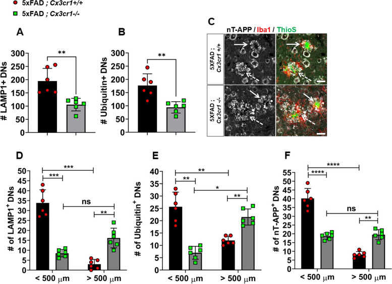
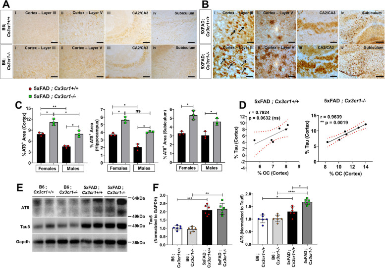
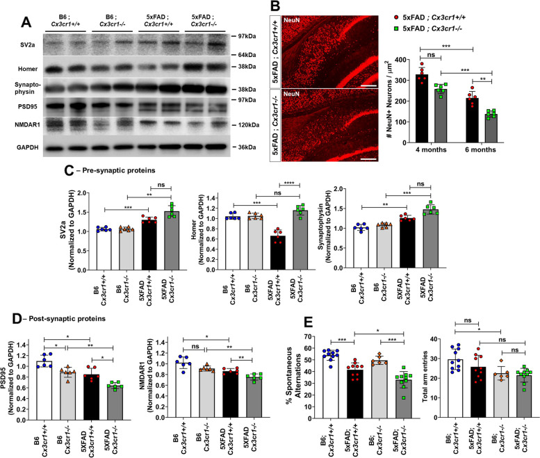
Background: Despite its identification as a key checkpoint regulator of microglial activation in Alzheimer’s disease, the overarching role of CX3CR1 signaling in modulating mechanisms of Aβ driven neurodegeneration, including accumulation of hyperphosphorylated tau is not well understood. Methodology: Accumulation of soluble and insoluble Aβ species, microglial activation, synaptic dysregulation, and neurodegeneration is investigated in 4- and 6-month old 5xFAD;Cx3cr1+/+ and 5xFAD;Cx3cr1−/− mice using immunohistochemistry, western blotting, transcriptomic and quantitative real time PCR analyses of purified microglia. Flow cytometry based, in-vivo Aβ uptake assays are used for characterization of the effects of CX3CR1-signaling on microglial phagocytosis and lysosomal acidification as indicators of clearance of methoxy-X-04+ fibrillar Aβ. Lastly, we use Y-maze testing to analyze the effects of Cx3cr1 deficiency on working memory. Results: Disease progression in 5xFAD;Cx3cr1−/− mice is characterized by increased deposition of filamentous plaques that display defective microglial plaque engagement. Microglial Aβ phagocytosis and lysosomal acidification in 5xFAD;Cx3cr1−/− mice is impaired in-vivo. Interestingly, Cx3cr1 deficiency results in heighted accumulation of neurotoxic, oligomeric Aβ, along with severe neuritic dystrophy, preferential loss of post-synaptic densities, exacerbated tau pathology, neuronal loss and cognitive impairment. Transcriptomic analyses using cortical RNA, coupled with qRT-PCR using purified microglia from 6 month-old mice indicate dysregulated TGFβ-signaling and heightened ROS metabolism in 5xFAD;Cx3cr1−/− mice. Lastly, microglia in 6 month-old 5xFAD;Cx3cr1−/− mice express a ‘degenerative’ phenotype characterized by increased levels of Ccl2, Ccl5, Il-1β, Pten and Cybb along with reduced Tnf, Il-6 and Tgfβ1 mRNA. Conclusions: Cx3cr1 deficiency impairs microglial uptake and degradation of fibrillar Aβ, thereby triggering increased accumulation of neurotoxic Aβ species. Furthermore, loss of Cx3cr1 results in microglial dysfunction typified by dampened TGFβ-signaling, increased oxidative stress responses and dysregulated pro-inflammatory activation. Our results indicate that Aβ-driven microglial dysfunction in Cx3cr1−/− mice aggravates tau hyperphosphorylation, neurodegeneration, synaptic dysregulation and impairs working memory.
Overview
The claims and statistics of this paper haven’t been checked yet.
Run the check — free, no account needed. It reads the open-access text and checks the sources it cites.
Integrity signals
Registry
Cites
Statistics
Figures
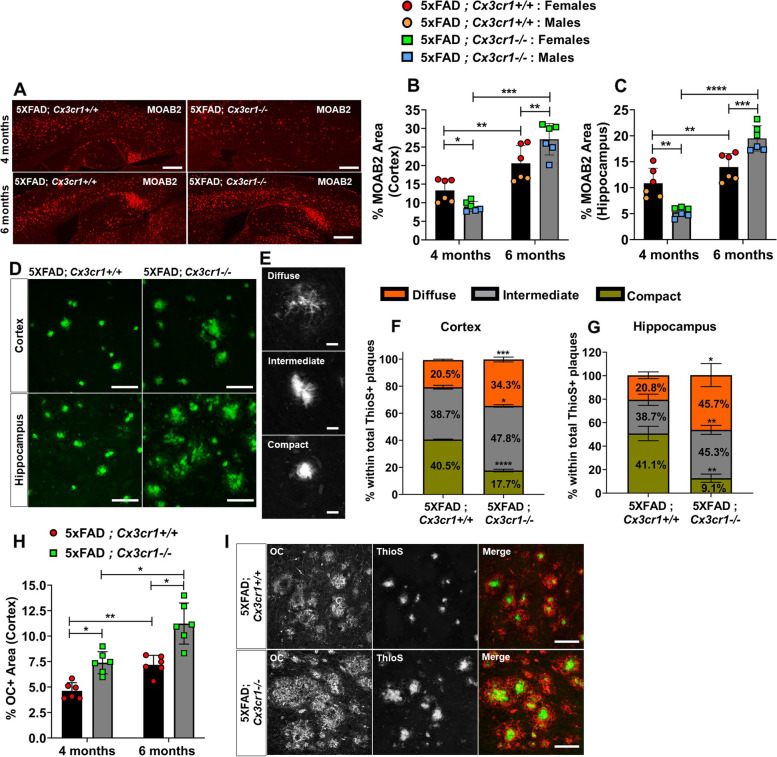
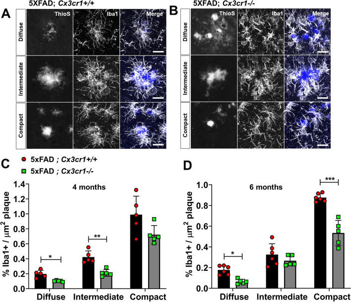
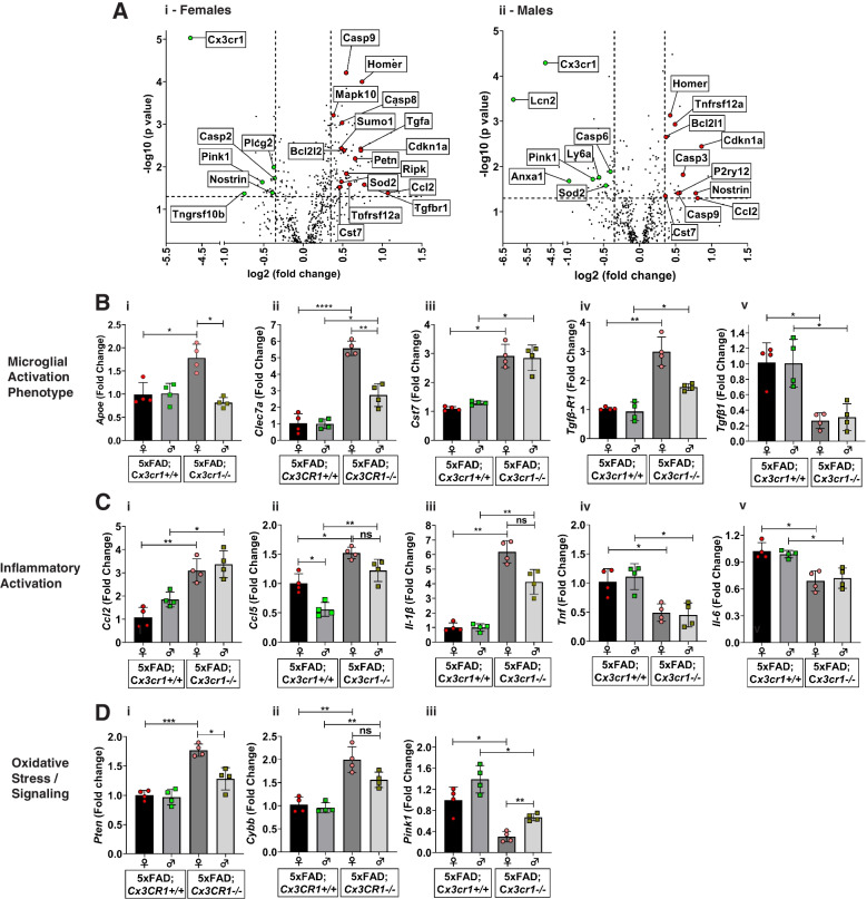
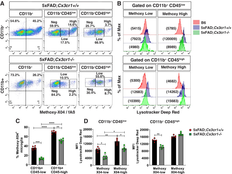
Verification
Claims the paper makes
The claims and statistics of this paper haven’t been checked yet.
Run the check — free, no account needed. It reads the open-access text and checks the sources it cites.
Claims other papers make about this one
Each is checked against this paper’s own text: does it say what the citing paper says it does?
The source backs part of the claim, not all of it — or backs it for a different population, condition or comparison.
In the citing paper
“Earlier studies on other microglial AD risk genes reported opposing effects on AD-related pathologies using various animal models at different ages (8–14).”
From The effect of Abi3 locus deletion on the progression of Alzheimer’s disease-related pathologies (2023) · cites this paper as [12]
Experimental — accuracy not yet measuredThe cited source says what the claim attributes to it.
In the citing paper
“These studies have led to the notion that microglia can have differential effects on AD pathology depending on the disease stage.”
From The effect of Abi3 locus deletion on the progression of Alzheimer’s disease-related pathologies (2023) · cites this paper as [12]
In the citing paper
“Deletion of CX3CR1 or depression of the CX3CL1/CX3CR1 axis reduces Aβ deposition [130–132] but exacerbates tau pathology, such as increased phosphorylation and aggregation of tau, and this is associated with worsened behavioral and cognitive impairments [132–137] (Fig. 1).”
From Tau and neuroinflammation in Alzheimer’s disease: interplay mechanisms and clinical translation (2023) · cites this paper as [132]
Self-check
Percentages, sums and reported differences re-computed from the paper’s own numbers.
The self-check needs the paper’s full text.
Citations
Cites
A Unique Microglia Type Associated with Restricting Development of Alzheimer’s Disease
Keren-Shaul, H., Spinrad, A., Weiner, A., Matcovitch-Natan, O., Dvir-Szternfeld, R., Ulland, T. K., David, E., Baruch, K., Lara-Astaiso, D., Toth, B., et al. (2017) · Cell · doi:10.1016/j.cell.2017.05.018 · Cited by 5,493
Synaptic Pruning by Microglia Is Necessary for Normal Brain Development
Paolicelli, R. C., Bolasco, G., Pagani, F., Maggi, L., Scianni, M., Panzanelli, P., Giustetto, M., Ferreira, T. A., Guiducci, E., Dumas, L., et al. (2011) · Science · doi:10.1126/science.1202529 · Cited by 4,244
Krasemann, S., Madore, C., Cialic, R., Baufeld, C., Calcagno, N., El Fatimy, R., Beckers, L., O’Loughlin, E., Xu, Y., Fanek, Z., et al. (2017) · Immunity · doi:10.1016/j.immuni.2017.08.008 · Cited by 2,979 · cited 13×
“In contrast, activation of TREM2-APOE signaling in AD results in a subset of microglia that display a ‘neurodegenerative phenotype’, characterized by suppression of homeostatic genes like Cx3cr1 and P2ry12 and upregulation of pro-inflammatory markers like Clec7a, Ccl5, Ccl2, IL1b, and Nos2.”
BackgroundInfluentialHammond, T. R., Dufort, C., Dissing-Olesen, L., Giera, S., Young, A., Wysoker, A., Walker, A. J., Segel, M., Nemesh, J., Saunders, A., et al. (2018) · doi:10.1101/406140 · Cited by 2,225
Alzheimer’s Disease Risk Genes and Mechanisms of Disease Pathogenesis
Karch, C. M., & Goate, A. M. (2015) · Biological Psychiatry · doi:10.1016/j.biopsych.2014.05.006 · Cited by 1,357 · cited 1×
“Lastly, large scale GWAS studies identifying single-nucleotide polymorphisms (SNPs) proximal to microglial-enriched, innate immune genes like CR1, CD33, MEF2C, HLA-DRB5-DRB1, PTK2b and TREM2 that significantly increase the risk of AD [5] have further underscored the critical role of microglia in neurodegeneration and…”
BackgroundZhan, Y., Paolicelli, R. C., Sforazzini, F., Weinhard, L., Bolasco, G., Pagani, F., Vyssotski, A. L., Bifone, A., Gozzi, A., Ragozzino, D., et al. (2014) · Nature Neuroscience · doi:10.1038/nn.3641 · Cited by 1,236 · cited 2×
“Cx3cr1 deficiency is associated with a transient deficit in microglial abundance during early post-natal development [11, 12].”
BackgroundMicroglial brain region-dependent diversity and selective regional sensitivities to aging
Grabert, K., Michoel, T., Karavolos, M. H., Clohisey, S., Baillie, J. K., Stevens, M. P., Freeman, T. C., Summers, K. M., & McColl, B. W. (2016) · Nature Neuroscience · doi:10.1038/nn.4222 · Cited by 1,151
Zhou, Y., Song, W. M., Andhey, P. S., Swain, A., Levy, T., Miller, K. R., Poliani, P. L., Cominelli, M., Grover, S., Gilfillan, S., et al. (2020) · Nature Medicine · doi:10.1038/s41591-019-0695-9 · Cited by 1,150 · cited 6×
“In contrast, activation of TREM2-APOE signaling in AD results in a subset of microglia that display a ‘neurodegenerative phenotype’, characterized by suppression of homeostatic genes like Cx3cr1 and P2ry12 and upregulation of pro-inflammatory markers like Clec7a, Ccl5, Ccl2, IL1b, and Nos2.”
BackgroundInfluentialYuan, P., Condello, C., Keene, C. D., Wang, Y., Bird, T. D., Paul, S. M., Luo, W., Colonna, M., Baddeley, D., & Grutzendler, J. (2016) · Neuron · doi:10.1016/j.neuron.2016.05.003 · Cited by 817 · cited 9×
“In contrast, activation of TREM2-APOE signaling in AD results in a subset of microglia that display a ‘neurodegenerative phenotype’, characterized by suppression of homeostatic genes like Cx3cr1 and P2ry12 and upregulation of pro-inflammatory markers like Clec7a, Ccl5, Ccl2, IL1b, and Nos2.”
BackgroundInfluentialStructural Classification of Toxic Amyloid Oligomers
Glabe, C. G. (2008) · Journal of Biological Chemistry · doi:10.1074/jbc.r800016200 · Cited by 783
Cited by
Sorted by citations
Items 101–113 of 113
Analysis of GRK2 aggregation in the pathology of Alzheimer disease in animal models
Alla, J. A., Perhal, A. F., Fu, X., Langer, A., el Faramawy, Y., & Quitterer, U. (2026) · Cell Reports Medicine · doi:10.1016/j.xcrm.2026.102707
Wei, H., Wang, B., Yang, J., & Liao, L. (2026) · Frontiers in Bioscience-Landmark · doi:10.31083/fbl45499
Microglia detection and phagocytosis of dying neurons is regulated by CX3CR1Preprint
Barasa, M. N., Pietramale, A. N., & Hill, R. (2026) · bioRxiv (Cold Spring Harbor Laboratory) · doi:10.64898/2026.04.02.716180
Zhang, B., Zhao, R., Li, X.-Y., Wei, Z.-H., Wang, D.-M., & Lu, M.-H. (2026) · Experimental Neurology · doi:10.1016/j.expneurol.2026.115845
Microglia-centered cellular crosstalk in neurodegeneration
Huang, H., & Mass, E. (2026) · Advances in immunology · doi:10.1016/bs.ai.2026.06.001
Smith, B. A., Klimko, C. P., Rodriguez, C. I., Rill, N. O., Davies, M. L., Dankmeyer, J. L., Hunter, M., Braun, C. T., Hernandez-Viezcas, Y., Xander, C. J., et al. (2026) · Frontiers in Microbiology · doi:10.3389/fmicb.2026.1865125
“…previous reports, the data contradictorily suggest simultaneous activation and impairment of microglia migration and uptake as well as an indication of pathological neuroinflammation (Yang et al., 2022; Izume et al., 2024; Qian et al., 2024; Gómez Morillas et al., 2021; Puntambekar et al., 2022).”
Neuroinflammation and Tauopathies
Zeenny, M.-N., Salame, I., Maatouk, L., & Akoury, E. (2026) · Molecular Biology Reports · doi:10.1007/s11033-026-12253-4
Chemokines in Alzheimer's Disease: Early Defence, Late Damage and the Impact of Sex and Infection
Aldana, B. I., & Freude, K. (2026) · Basic & Clinical Pharmacology & Toxicology · doi:10.1111/bcpt.70273
Mo, L., ZHU, X., Wang, N., Song, J., Cai, J., & Qian, B. (2026) · Journal of Environmental Sciences · doi:10.1016/j.jes.2026.105611
The human retina in Alzheimer’s disease: Pathology, mechanisms, and biomarkers
Müller, L. P. D. S., Majumdar, S., & Recio, B. S. (2026) · Ageing Research Reviews · doi:10.1016/j.arr.2026.103275
Chu, M., Tan, M., Gan, X., Cui, S., & Shi, L. (2026) · Molecular Neurobiology · doi:10.1007/s12035-026-06091-5
Lv, J., Xia, Z., Wang, X., Zhao, P., & Wu, Z. (2026) · Journal of Neuroinflammation · doi:10.1186/s12974-026-03997-8
“…phagocytic activity is tightly controlled by surface receptors that sense microenviron-mental cues, among which the CX3C chemokine ligand 1 (CX3CL1, also known as fractalkine)/CX3C motif chemokine receptor 1 (CX3CR1) axis plays a key role in maintaining appropriate phagocytic function [30, 31].”
Microgliopathy in poststroke depression: exploring mechanisms and therapeutic strategies
Yang, C., Wu, S., Cao, H., Yin, Y., Zhang, Y., Zhang, L., Chen, R., & Tao, X. (2026) · Neuroscience · doi:10.1016/j.neuroscience.2026.08.046
Metadata
- Authors
- Puntambekar, S. S., Moutinho, M., Lin, P. B.-C., Jadhav, V., Tumbleson-Brink, D., Balaji, A., Benito, M. A., Xu, G., Oblak, A., Lasagna-Reeves, C. A., Landreth, G. E., & Lamb, B. T.
- Affiliations
- Indiana University Indianapolis (US)
- Indiana University School of Medicine (US)
- University of Washington (US)
- Indiana Clinical and Translational Sciences Institute (US)
- Corresponding author
- Puntambekar, Shweta S.
- Lamb, Bruce T.
- ORCID
- Venue
- Molecular Neurodegeneration
- Published
- 2022-06-28
- Volume
- 17
- Issue
- 1
- Pages
- 47-47
- ISSN
- 1750-1326 (ISSN-L)
- PMC
- PMC9241248
- OpenAlex
- W4283643624
- Cited by
- 113
- References
- 55
- Citations by year
- 2026: 26 · 2025: 33 · 2024: 23 · 2023: 26 · 2022: 5
- FWCI
- 13.45
- Type
- journal article
- Language
- en
- Open access
- gold
- doi.org
- Locations
- Molecular Neurodegeneration (publishedVersion, cc-by)
- PubMed Central (publishedVersion, cc-by)
- PubMed (publishedVersion)
- Purdue University Indianapolis (Indiana University) (submittedVersion, cc-by)
- DOAJ (DOAJ: Directory of Open Access Journals) (submittedVersion, cc-by-sa)
- IUScholarWorks (Indiana University) (submittedVersion, cc-by)
- Funders
- National Institutes of Health (NIH RF1AG051495, 1RF1AG069425-01)
- Topics
- Life Sciences › Neuroscience › Cellular and Molecular Neuroscience › Alzheimer's disease research and treatments
- Life Sciences › Neuroscience › Neurology › Neuroinflammation and Neurodegeneration Mechanisms
- Life Sciences › Neuroscience › Neurology › Neurological Disease Mechanisms and Treatments
- Keywords
- CX3CR1, Alzheimer's disease, amyloid-beta, microglial dysfunction, 5xFAD mice, tau pathology, neurodegeneration, cognitive decline, TGF-beta signaling, oxidative stress, microglial phagocytosis, lysosomal acidification, microglial polarization, synaptic loss, working memory, neuroinflammation, amyloid plaques, amyloid-beta oligomers
- MeSH terms
- CX3C Chemokine Receptor 1, Alzheimer Disease, Amyloidosis, Animals, Neurons, Transforming Growth Factor beta, Mice, Plaque, Amyloid, Amyloidogenic Proteins, Cognitive Dysfunction
About these checks
Registry, retraction, Cites and statistics results are computed, not judged, with one exception: a printed reference that no index lists is read once by a model before it counts as a citation error. Claims are extracted by a model and kept only when their quote appears verbatim in the text; each cited source is then checked, with a model’s help, for whether it says what the claim relies on. Those verdicts can be wrong — read the claim’s quote and the cited source before relying on one.
Statistics and arithmetic are recomputed, not judged.
Confidence bands are shown only for fields calibrated on our annotated test set. None is calibrated yet, so no band is shown.
What counts as a claim ›How we measure confidence ›How we check this ›
Are you an author, or do you see a problem with this page? Request a review
