

The DOI matches its Crossref record; 55 of the 55 references it cites were found in a registry or an index.
CX3CR1 deficiency aggravates amyloid driven neuronal pathology and cognitive decline in Alzheimer’s disease
Open accessAbstract
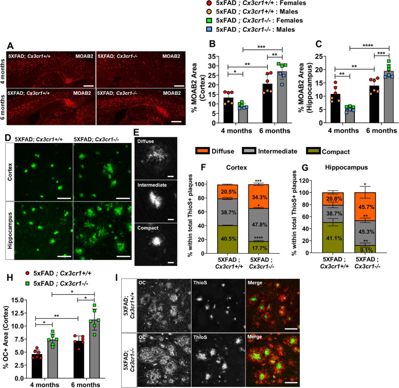
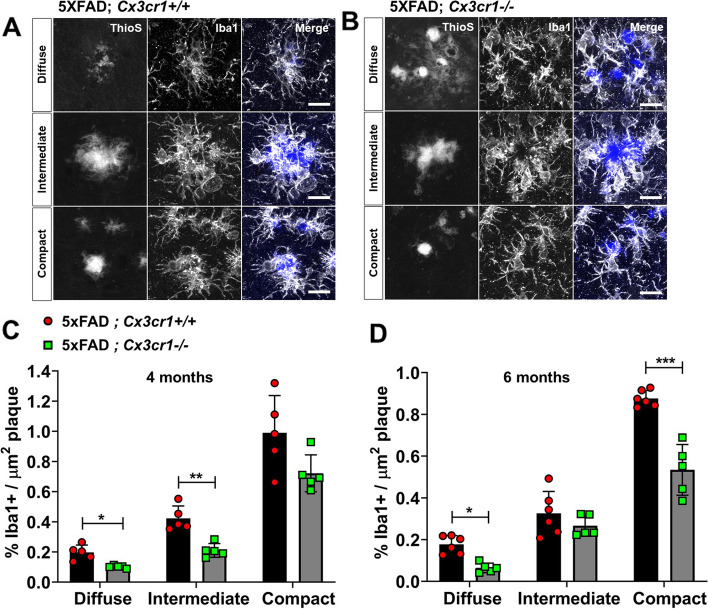
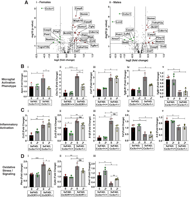
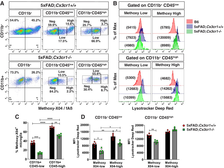
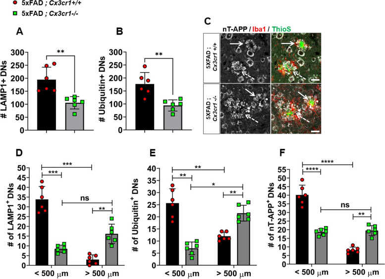
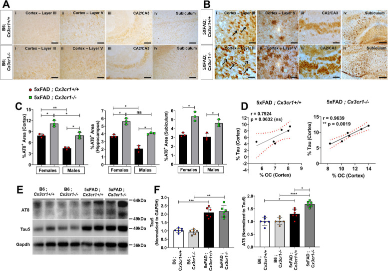
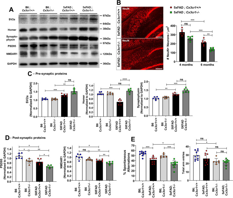
Background: Despite its identification as a key checkpoint regulator of microglial activation in Alzheimer’s disease, the overarching role of CX3CR1 signaling in modulating mechanisms of Aβ driven neurodegeneration, including accumulation of hyperphosphorylated tau is not well understood. Methodology: Accumulation of soluble and insoluble Aβ species, microglial activation, synaptic dysregulation, and neurodegeneration is investigated in 4- and 6-month old 5xFAD;Cx3cr1+/+ and 5xFAD;Cx3cr1−/− mice using immunohistochemistry, western blotting, transcriptomic and quantitative real time PCR analyses of purified microglia. Flow cytometry based, in-vivo Aβ uptake assays are used for characterization of the effects of CX3CR1-signaling on microglial phagocytosis and lysosomal acidification as indicators of clearance of methoxy-X-04+ fibrillar Aβ. Lastly, we use Y-maze testing to analyze the effects of Cx3cr1 deficiency on working memory. Results: Disease progression in 5xFAD;Cx3cr1−/− mice is characterized by increased deposition of filamentous plaques that display defective microglial plaque engagement. Microglial Aβ phagocytosis and lysosomal acidification in 5xFAD;Cx3cr1−/− mice is impaired in-vivo. Interestingly, Cx3cr1 deficiency results in heighted accumulation of neurotoxic, oligomeric Aβ, along with severe neuritic dystrophy, preferential loss of post-synaptic densities, exacerbated tau pathology, neuronal loss and cognitive impairment. Transcriptomic analyses using cortical RNA, coupled with qRT-PCR using purified microglia from 6 month-old mice indicate dysregulated TGFβ-signaling and heightened ROS metabolism in 5xFAD;Cx3cr1−/− mice. Lastly, microglia in 6 month-old 5xFAD;Cx3cr1−/− mice express a ‘degenerative’ phenotype characterized by increased levels of Ccl2, Ccl5, Il-1β, Pten and Cybb along with reduced Tnf, Il-6 and Tgfβ1 mRNA. Conclusions: Cx3cr1 deficiency impairs microglial uptake and degradation of fibrillar Aβ, thereby triggering increased accumulation of neurotoxic Aβ species. Furthermore, loss of Cx3cr1 results in microglial dysfunction typified by dampened TGFβ-signaling, increased oxidative stress responses and dysregulated pro-inflammatory activation. Our results indicate that Aβ-driven microglial dysfunction in Cx3cr1−/− mice aggravates tau hyperphosphorylation, neurodegeneration, synaptic dysregulation and impairs working memory.
Overview
The claims and statistics of this paper haven’t been checked yet.
Run the check — free, no account needed. It reads the open-access text and checks the sources it cites.
Integrity signals
Registry
Cites
Statistics
Figures
Verification
Claims the paper makes
The claims and statistics of this paper haven’t been checked yet.
Run the check — free, no account needed. It reads the open-access text and checks the sources it cites.
Claims other papers make about this one
Each is checked against this paper’s own text: does it say what the citing paper says it does?
The source backs part of the claim, not all of it — or backs it for a different population, condition or comparison.
In the citing paper
“Earlier studies on other microglial AD risk genes reported opposing effects on AD-related pathologies using various animal models at different ages (8–14).”
From The effect of Abi3 locus deletion on the progression of Alzheimer’s disease-related pathologies (2023) · cites this paper as [12]
Experimental — accuracy not yet measuredThe cited source says what the claim attributes to it.
In the citing paper
“These studies have led to the notion that microglia can have differential effects on AD pathology depending on the disease stage.”
From The effect of Abi3 locus deletion on the progression of Alzheimer’s disease-related pathologies (2023) · cites this paper as [12]
In the citing paper
“Deletion of CX3CR1 or depression of the CX3CL1/CX3CR1 axis reduces Aβ deposition [130–132] but exacerbates tau pathology, such as increased phosphorylation and aggregation of tau, and this is associated with worsened behavioral and cognitive impairments [132–137] (Fig. 1).”
From Tau and neuroinflammation in Alzheimer’s disease: interplay mechanisms and clinical translation (2023) · cites this paper as [132]
Self-check
Percentages, sums and reported differences re-computed from the paper’s own numbers.
The self-check needs the paper’s full text.
Citations
Cites
A Unique Microglia Type Associated with Restricting Development of Alzheimer’s Disease
Keren-Shaul, H., Spinrad, A., Weiner, A., Matcovitch-Natan, O., Dvir-Szternfeld, R., Ulland, T. K., David, E., Baruch, K., Lara-Astaiso, D., Toth, B., et al. (2017) · Cell · doi:10.1016/j.cell.2017.05.018 · Cited by 5,493
Synaptic Pruning by Microglia Is Necessary for Normal Brain Development
Paolicelli, R. C., Bolasco, G., Pagani, F., Maggi, L., Scianni, M., Panzanelli, P., Giustetto, M., Ferreira, T. A., Guiducci, E., Dumas, L., et al. (2011) · Science · doi:10.1126/science.1202529 · Cited by 4,244
Krasemann, S., Madore, C., Cialic, R., Baufeld, C., Calcagno, N., El Fatimy, R., Beckers, L., O’Loughlin, E., Xu, Y., Fanek, Z., et al. (2017) · Immunity · doi:10.1016/j.immuni.2017.08.008 · Cited by 2,979 · cited 13×
“In contrast, activation of TREM2-APOE signaling in AD results in a subset of microglia that display a ‘neurodegenerative phenotype’, characterized by suppression of homeostatic genes like Cx3cr1 and P2ry12 and upregulation of pro-inflammatory markers like Clec7a, Ccl5, Ccl2, IL1b, and Nos2.”
BackgroundInfluentialHammond, T. R., Dufort, C., Dissing-Olesen, L., Giera, S., Young, A., Wysoker, A., Walker, A. J., Segel, M., Nemesh, J., Saunders, A., et al. (2018) · doi:10.1101/406140 · Cited by 2,225
Alzheimer’s Disease Risk Genes and Mechanisms of Disease Pathogenesis
Karch, C. M., & Goate, A. M. (2015) · Biological Psychiatry · doi:10.1016/j.biopsych.2014.05.006 · Cited by 1,357 · cited 1×
“Lastly, large scale GWAS studies identifying single-nucleotide polymorphisms (SNPs) proximal to microglial-enriched, innate immune genes like CR1, CD33, MEF2C, HLA-DRB5-DRB1, PTK2b and TREM2 that significantly increase the risk of AD [5] have further underscored the critical role of microglia in neurodegeneration and…”
BackgroundZhan, Y., Paolicelli, R. C., Sforazzini, F., Weinhard, L., Bolasco, G., Pagani, F., Vyssotski, A. L., Bifone, A., Gozzi, A., Ragozzino, D., et al. (2014) · Nature Neuroscience · doi:10.1038/nn.3641 · Cited by 1,236 · cited 2×
“Cx3cr1 deficiency is associated with a transient deficit in microglial abundance during early post-natal development [11, 12].”
BackgroundMicroglial brain region-dependent diversity and selective regional sensitivities to aging
Grabert, K., Michoel, T., Karavolos, M. H., Clohisey, S., Baillie, J. K., Stevens, M. P., Freeman, T. C., Summers, K. M., & McColl, B. W. (2016) · Nature Neuroscience · doi:10.1038/nn.4222 · Cited by 1,151
Zhou, Y., Song, W. M., Andhey, P. S., Swain, A., Levy, T., Miller, K. R., Poliani, P. L., Cominelli, M., Grover, S., Gilfillan, S., et al. (2020) · Nature Medicine · doi:10.1038/s41591-019-0695-9 · Cited by 1,150 · cited 6×
“In contrast, activation of TREM2-APOE signaling in AD results in a subset of microglia that display a ‘neurodegenerative phenotype’, characterized by suppression of homeostatic genes like Cx3cr1 and P2ry12 and upregulation of pro-inflammatory markers like Clec7a, Ccl5, Ccl2, IL1b, and Nos2.”
BackgroundInfluentialYuan, P., Condello, C., Keene, C. D., Wang, Y., Bird, T. D., Paul, S. M., Luo, W., Colonna, M., Baddeley, D., & Grutzendler, J. (2016) · Neuron · doi:10.1016/j.neuron.2016.05.003 · Cited by 817 · cited 9×
“In contrast, activation of TREM2-APOE signaling in AD results in a subset of microglia that display a ‘neurodegenerative phenotype’, characterized by suppression of homeostatic genes like Cx3cr1 and P2ry12 and upregulation of pro-inflammatory markers like Clec7a, Ccl5, Ccl2, IL1b, and Nos2.”
BackgroundInfluentialStructural Classification of Toxic Amyloid Oligomers
Glabe, C. G. (2008) · Journal of Biological Chemistry · doi:10.1074/jbc.r800016200 · Cited by 783
Cited by
Sorted by citations
Tau and neuroinflammation in Alzheimer’s disease: interplay mechanisms and clinical translation
Chen, Y., & Yu, Y. (2023) · Journal of Neuroinflammation · doi:10.1186/s12974-023-02853-3 · Cited by 516
Microglia and Astrocytes in Alzheimer’s Disease: Significance and Summary of Recent Advances
Deng, Q., Wu, C., Parker, E., Liu, T. C.-Y., Duan, R., & Yang, L. (2023) · Aging and disease · doi:10.14336/ad.2023.0907 · Cited by 102
“Cx3cr1 deficiency impairs the uptake of and degradation of fibrillar Aβ by microglia, resulting in increased accumulation of neurotoxic Aβ species [212].”
BackgroundOxygen metabolism abnormality and Alzheimer's disease: An update
Liu, G., Yang, C., Wang, X., Chen, X., Wang, Y., & Le, W. (2023) · Redox Biology · doi:10.1016/j.redox.2023.102955 · Cited by 87
Wu, X., Gao, Y.-Z., Shi, C., Tong, J.-H., Ma, D., Shen, J.-C., Yang, J., & Ji, M. (2023) · Neuropharmacology · doi:10.1016/j.neuropharm.2023.109646 · Cited by 77
Brain high-throughput multi-omics data reveal molecular heterogeneity in Alzheimer’s disease
Eteleeb, A. M., Novotny, B. C., Tarraga, C. S., Sohn, C., Dhungel, E., Brase, L., Nallapu, A., Buss, J., Farias, F., Bergmann, K., et al. (2024) · PLOS Biology · doi:10.1371/journal.pbio.3002607 · Cited by 64
Neuroinflammation in retinitis pigmentosa: Therapies targeting the innate immune system
Zhao, L., Hou, C., & Yan, N. (2022) · Frontiers in Immunology · doi:10.3389/fimmu.2022.1059947 · Cited by 65
Interaction between autophagy and the NLRP3 inflammasome in Alzheimer’s and Parkinson’s disease
Lu, R., Zhang, L., & Yang, X. (2022) · Frontiers in Aging Neuroscience · doi:10.3389/fnagi.2022.1018848 · Cited by 58
The Role and Pathogenesis of Tau Protein in Alzheimer’s Disease
Hong, X., Huang, L., Lei, F., Tian, L., Luo, Y., Zeng, M., & Wang, Z. (2025) · Biomolecules · doi:10.3390/biom15060824 · Cited by 67
“These factors degrade the extracellular matrix surrounding synapses and disrupt the interaction between synapses and glial cells, ultimately contributing to synaptic structural damage and functional impairment [111–113].”
Alzheimer’s disease pathogenesis: standing at the crossroad of lipid metabolism and immune response
Wang, Z., Zhang, L., & Qin, C. (2025) · Molecular Neurodegeneration · doi:10.1186/s13024-025-00857-6 · Cited by 60
Fractalkine/CX3CR1-Dependent Modulation of Synaptic and Network Plasticity in Health and Disease
Camacho-Hernández, N. P., & Peña‐Ortega, F. (2023) · Neural Plasticity · doi:10.1155/2023/4637073 · Cited by 52
“For instance, CX3CR1 deficiency induces a reduction in Aβ accumulation in AD transgenic mice [115–117] due to the increased phagocytic activity of microglia [116, 117]; although, opposite effects have been recently reported [107].”
BackgroundAlzheimer’s genes in microglia: a risk worth investigating
Sudwarts, A., & Thinakaran, G. (2023) · Molecular Neurodegeneration · doi:10.1186/s13024-023-00679-4 · Cited by 52
“Interestingly, although Cx3cr1 ablation in the 5XFAD model resulted in fewer Aβ deposits at 4 months of age, there was an acceleration of Aβ deposition by 6 months, concomitant with dysregulation of microglial activation, impaired phagocytic function, and tau phosphorylation [111].”
BackgroundTGF-β Signaling in Microglia: A Key Regulator of Development, Homeostasis and Reactivity
Li, L., Sun, B., Harris, O. A., & Luo, J. (2024) · Biomedicines · doi:10.3390/biomedicines12112468 · Cited by 38
“The loss of the fractalkine receptor (CX3CR1), a homeostatic gene in microglia, in 5xFAD transgenic mice, led to microglial dysregulation and heightened reactivity, resulting in impaired A β phagocytosis and clearance, along with aberrant TGF-β signaling [74].”
New Pathways Identify Novel Drug Targets for the Prevention and Treatment of Alzheimer’s Disease
Penke, B., Szűcs, M., & Bogár, F. (2023) · International Journal of Molecular Sciences · doi:10.3390/ijms24065383 · Cited by 39
“Activation of the CX3CR1 (C-X3-C Motif Chemokine Receptor 1) gene for improved MG phagocytotic activity [176].”
BackgroundTondo, G., Marchi, F. D., Bonardi, F., Menegon, F., Verrini, G., Aprile, D., Anselmi, M., Mazzini, L., & Comi, C. (2024) · Journal of Clinical Medicine · doi:10.3390/jcm13113098 · Cited by 45
Microglial phagocytosis in Alzheimer disease
Brown, G. C., St George-Hyslop, P., Paolicelli, R. C., & Lemke, G. (2026) · Nature Reviews Neurology · doi:10.1038/s41582-025-01162-y · Cited by 36
CX3CL1/CX3CR1 signal mediates M1-type microglia and accelerates high-altitude-induced forgetting
Wang, X., Xie, Y., Niu, Y., Wan, B., Lu, Y., Luo, Q., & Zhu, L. (2023) · Frontiers in Cellular Neuroscience · doi:10.3389/fncel.2023.1189348 · Cited by 32
“In a mouse model of Alzheimer’s disease, CX3CR1 deficiency was reported to trigger microglia dysfunction, and exacerbate neurotoxicity and phosphorylation of tau, leading to neuronal synaptic dysregulation and the impairment of memory function (Puntambekar et al., 2022).”
BackgroundZhang, M., Han, X., Yan, L., Fu, Y., Kou, H., Shang, C., Wang, J., Liu, H., Jiang, C., Wang, J., et al. (2024) · CNS Neuroscience & Therapeutics · doi:10.1111/cns.14781 · Cited by 32
“Furthermore, the association between chemokines and various neurological disorders, including multiple sclerosis, Parkinson's disease, Huntington's disease, and Alzheimer's disease, 61–65 has been extensively reported.”
BackgroundZhu, Y., Li, L., Jin, X., Li, Z., Wang, C., Teng, L., Li, Y., Zhang, Y., & Wang, D. (2023) · International Journal of Biological Macromolecules · doi:10.1016/j.ijbiomac.2023.128819 · Cited by 29
Maurya, S. K., Gupta, S., & Mishra, R. (2023) · Frontiers in Molecular Neuroscience · doi:10.3389/fnmol.2022.1072046 · Cited by 26
Li, T., Tan, X., Tian, L., Jia, C., Cheng, C., Chen, X., Wei, M., Wang, Y., Hu, Y., Jia, Q., et al. (2023) · Journal of Neuroinflammation · doi:10.1186/s12974-023-02956-x · Cited by 29
“However, DAM becomes dysregulated at later stages and accelerates neurodegeneration [29, 30].”
BackgroundLavekar, S. S., Harkin, J., Hernandez, M., Gomes, C., Patil, S., Huang, K.-C., Puntambekar, S. S., Lamb, B. T., & Meyer, J. S. (2023) · Scientific Reports · doi:10.1038/s41598-023-40382-4 · Cited by 24
Self-citationXie, X., Hao, J.-J., Shi, J.-Z., Zhou, Y., Liu, P., Wang, F., Zheng, X.-M., Yu, X., Wang, C., Yan, Y., et al. (2023) · International Immunopharmacology · doi:10.1016/j.intimp.2023.109994 · Cited by 23
Tanshinone IIA alleviates atherosclerosis in LDLR−/− mice by regulating efferocytosis of macrophages
Wang, J., Zhang, Y., Feng, X., Du, M., Li, S., Chang, X., & Liu, P. (2023) · Frontiers in Pharmacology · doi:10.3389/fphar.2023.1233709 · Cited by 23
The effect of Abi3 locus deletion on the progression of Alzheimer’s disease-related pathologies
Karahan, H., Smith, D. C., Kim, B., McCord, B., Mantor, J., John, S. K., Al-Amin, M. M., Dabin, L. C., & Kim, J. (2023) · Frontiers in Immunology · doi:10.3389/fimmu.2023.1102530 · Cited by 20
Jury-Garfe, N., Redding-Ochoa, J., You, Y., Martínez, P., Karahan, H., Chimal-Juárez, E., Johnson, T. S., Zhang, J., Resnick, S., Kim, J., et al. (2024) · Acta Neuropathologica · doi:10.1007/s00401-024-02775-1 · Cited by 22
Self-citationAdvancements and Challenges in Antiamyloid Therapy for Alzheimer’s Disease: A Comprehensive Review
Beshir, S. A., Hussain, N., Menon, V. B., Haddad, A. H. I. A., Zeer, R. A. K. A., & Elnour, A. A. (2024) · International Journal of Alzheimer s Disease · doi:10.1155/2024/2052142 · Cited by 32
Ma, Y., Liu, N., Wang, Y., Zhang, A., Zhu, Z., Zhang, Z., Li, Y., Jian, G., Fu, G., Dong, M., et al. (2023) · EClinicalMedicine · doi:10.1016/j.eclinm.2023.101987 · Cited by 24
Microglial Transforming Growth Factor-β Signaling in Alzheimer’s Disease
Vidovic, N., & Spittau, B. (2024) · International Journal of Molecular Sciences · doi:10.3390/ijms25063090 · Cited by 22
“In addition, impaired A β phagocytosis and clearance were observed as well as aberrant TGF β signaling [137].”
BackgroundTang, S., Zhang, Y., Botchway, B. O. A., Wang, X., Huang, M., & Liu, X. (2024) · Molecular Neurobiology · doi:10.1007/s12035-024-04498-6 · Cited by 23
Role of chemokines in aging and age-related diseases
Chaudhary, J. K., Danga, A. K., Kumari, A., Bhardwaj, A., & Rath, P. C. (2024) · Mechanisms of Ageing and Development · doi:10.1016/j.mad.2024.112009 · Cited by 24
Chen, D., Jiang, H., Sun, L., Nurzat, Y., Qin, H., Zhao, Z., Liu, D., Zheng, S., Wang, L., Fu, Y., et al. (2025) · Journal of Nanobiotechnology · doi:10.1186/s12951-025-03730-2 · Cited by 19
“I Statistical analysis of the fluorescence intensity of CD206, n = 5, * p < 0.05 [48]), and Cx3cr1 (maintaining microglial homeostasis; reduced expression indicates protective phenotype shift [49]).”
InfluentialSola-Sevilla, N., Mesa-Lombardo, A., Aleixo, M., Expósito, S., Diaz-Perdigón, T., Azqueta, A., Zamani, F., Suzuki, T., Maioli, S., Eroli, F., et al. (2023) · Journal of Neuroimmune Pharmacology · doi:10.1007/s11481-023-10084-9 · Cited by 19
“Specifically, in AD, they have been proposed to have a key role on Aβ phagocytosis and, therefore, on AD pathology progression (Park et al. 2019; Singh et al. 2022; Puntambekar et al. 2022).”
BackgroundSirkis, D. W., Solsberg, C. W., Johnson, T. P., Bonham, L. W., Sturm, V. E., Lee, S. E., Rankin, K. P., Rosen, H. J., Boxer, A. L., Seeley, W. W., et al. (2023) · Genome Medicine · doi:10.1186/s13073-023-01205-3 · Cited by 19
Le, L., Feidler, A. M., Rodríguez, L., Cealie, M. Y., Plunk, E. C., Li, H., Kara-Pabani, K., Lamantia, C. E., O’Banion, M. K., & Majewska, A. K. (2023) · Brain Behavior and Immunity · doi:10.1016/j.bbi.2025.04.022 · Cited by 19
CX3CL1 Regulation of Gliosis in Neuroinflammatory and Neuroprotective Processes
Gutiérrez, I. L., Martín‐Hernández, D., MacDowell, K. S., García‐Bueno, B., Caso, J. R., Leza, J. C., & Madrigal, J. L. M. (2025) · International Journal of Molecular Sciences · doi:10.3390/ijms26030959 · Cited by 20
“Furthermore, more recent analyses have concluded that the suppression of CX3CR1 in 5xFAD mice increases the deposition of A β , causes neuritic dystrophy, and exacerbates tau pathology, neuronal loss, and cognitive impairment [50].”
Chemokines in patients with Alzheimer's disease: A meta-analysis
Wang, H., Zong, Y., Zhu, L., Wang, W., & Han, Y. (2023) · Frontiers in Aging Neuroscience · doi:10.3389/fnagi.2023.1047810 · Cited by 19
“Meanwhile, CXC3CR1 deficiency increased the Aβ level, and aggravated tau pathology, inducing cognitive decline (Puntambekar et al., 2022).”
BackgroundPLCG2 modulates TREM2 expression and signaling in response to Alzheimer's disease pathology
Messenger, E. J., Baar, S. A., Bedford, L. M., Tsai, A. P., Lin, P. B., Ferguson, C. A., Xu, G., Wallace, A., Landreth, G. E., Lamb, B. T., et al. (2025) · Alzheimer's & Dementia · doi:10.1002/alz.70231 · Cited by 16
Self-citationGlial Cells in the Early Stages of Neurodegeneration: Pathogenesis and Therapeutic Targets
Ahremenko, E. A., Andreev, A., Apushkin, D., & Korkotian, E. (2025) · International Journal of Molecular Sciences · doi:10.3390/ijms262411995 · Cited by 20
“In 5xFAD mice, Puntambekar et al. [113] demonstrated that CX3CR1 deficiency impairs clearance of fibrillar A β , leading to accumulation of neurotoxic A β oligomers, neuronal loss, cognitive deficits, and activation of a neurodegenerative microglial phenotype [114,115].”
Mechanisms and routes of G-protein coupled receptor-mediated Tau degradation in Alzheimer’s disease
Ananthanarayana, V., Chidambaram, H., Govindaraj, P., Vijayalakshmi, K., & Chinnathambi, S. (2025) · Cytokine & Growth Factor Reviews · doi:10.1016/j.cytogfr.2025.07.003 · Cited by 14
Kourti, M., & Metaxas, A. (2024) · Neurobiology of Disease · doi:10.1016/j.nbd.2024.106427 · Cited by 17
Fazzina, M., Bergonzoni, M., Massenzio, F., Monti, B., Frabetti, F., & Casadei, R. (2024) · Scientific Reports · doi:10.1038/s41598-024-52415-7 · Cited by 12
Chirico, F. D., Poeta, E., Babini, G., Piccolino, I., Monti, B., & Massenzio, F. (2022) · Frontiers in Cellular Neuroscience · doi:10.3389/fncel.2022.1038721 · Cited by 11
“Upon inflammatory stimuli, microglia undergo activation and release of pro-inflammatory mediators responsible for triggering chronic neuroinflammation widely described in neurodegenerative diseases (Zhang et al., 2017; Massenzio et al., 2018; Puntambekar et al., 2022).”
BackgroundZhou, J., Ye, W., Chen, L., Li, J., Zhou, Y., Bai, C., & Luo, L. (2024) · Brain Research Bulletin · doi:10.1016/j.brainresbull.2024.110939 · Cited by 11
Zhan, L., Qiu, M., Zheng, J., Lai, M., Lin, K., Dai, J., Sun, W., & Xu, E. (2024) · Cell Communication and Signaling · doi:10.1186/s12964-024-01830-4 · Cited by 11
“…role for FKN/CX3CR1 has been supported in studies of amyotrophic lateral sclerosis [14], Parkinson’s disease [15–17] and multiple sclerosis in some studies [18], whereas a neurotoxic role for FKN/CX3CR1 has been demonstrated in Alzheimer’s disease [19, 20] and stroke [21] in another.”
BackgroundSingle-Cell RNA-seq reveals transcriptomic modulation of Alzheimer’s disease by activated protein C
Fatmi, M. K., Wang, H., Slotabec, L. A., Wen, C., Seale, B. C., Zhao, B., & Li, J. (2024) · Aging · doi:10.18632/aging.205624 · Cited by 10
“Deficiency of Cx3cr1 has shown increased Aβ pathology as well as cognitive decline [28], 5xFAD mice did not show a decrease in Cx3cr1 expression.”
BackgroundLin, Y.-Y., Chang, W.-H., Hsieh, S.-L. E., & Cheng, I. H.-J. (2024) · Journal of Neuroinflammation · doi:10.1186/s12974-024-03253-x · Cited by 10
“In the AD mouse model, enhanced microglial phagocytosis reduces the Aβ burden and alleviates memory declines [20–23], while suppressed microglial phagocytosis aggra-vates the Aβ pathology [20, 24].”
BackgroundWang, F., Wang, J., Luo, N., Luo, Y., Gao, Z., Ye, C., Jiang, M., Shen, Z., Xiao, J., Xue, P., et al. (2025) · Ecotoxicology and Environmental Safety · doi:10.1016/j.ecoenv.2025.117736 · Cited by 13
An, Y., Yu, Y., Liu, H., Xi, Y., Pi, M., Xu, R. C., Huang, Y., Li, S., & Gu, X. (2024) · Experimental Neurology · doi:10.1016/j.expneurol.2024.115114 · Cited by 8
Yang, L. G., Zou, Y., Fan, J., Yin, P., Qin, H., Li, Z., Wu, F., Li, X., Teng, H., Zhang, Y., et al. (2025) · Pharmaceuticals · doi:10.3390/ph18060816 · Cited by 7
Wang, S., Wang, J., Hu, Z., Wu, L., & Huang, L. (2026) · Experimental Neurology · doi:10.1016/j.expneurol.2026.115663 · Cited by 7
Zeng, H.-X., Qin, S.-J., Andersson, J., Li, S.-P., Zeng, Q.-G., Li, J.-H., Wu, Q.-Z., Meng, W.-J., Oudin, A., Kanninen, K. M., et al. (2024) · Environmental Pollution · doi:10.1016/j.envpol.2024.125440 · Cited by 8
Liu, J., Sun, Z., Liu, X., Chiu, K., Ma, L., & Wang, J. (2025) · Frontiers in Aging · doi:10.3389/fragi.2025.1549848 · Cited by 6
CX3CR1: a potential microglia-specific PET imaging target in Alzheimer’s and Parkinson’s diseases
Yang, H., Wang, Y., Xu, Y., & Wang, C. (2025) · Frontiers in Pharmacology · doi:10.3389/fphar.2025.1678159 · Cited by 6
Evolving insights into the identity and function of dark microglia
Flury, A. G., St‐Pierre, M., Ayata, P., & Tremblay, M. (2025) · Trends in Neurosciences · doi:10.1016/j.tins.2025.11.002 · Cited by 7
Widjaya, M. A., Liu, C.-H., Lee, S., & Cheng, W. (2023) · Journal of Molecular Neuroscience · doi:10.1007/s12031-023-02152-9 · Cited by 5
“It is involved in AD process, leading to Aβ aggregation and cognitive decline (Puntambekar et al. 2022).”
BackgroundCX3CL1-CX3CR1 pathway mediates hyperthermia-induced microglial processes retraction
Song, R., Liu, C., Peng, M., Qian, Z., Wei, M., Yu, F., Yao, M., Li, X., & Feng, B. (2025) · Brain Behavior and Immunity · doi:10.1016/j.bbi.2025.03.007 · Cited by 5
He, Q., Zhou, T., & He, Q. (2025) · Brain Sciences · doi:10.3390/brainsci15070759 · Cited by 5
“…overexpression in microglia [85,86] Early phase: Continues repair [4] Long-term: Pathological synaptic pruning [88–90], chronic neuroinflammation [51,91,92], A β acceleration [93,94] It is now well established that the CX3CR1 signaling axis plays a stage-dependent dual role following stroke [8,36].”
InfluentialOkhovat, M., Layman, C. E., Davis, B. A., Pederson, A., O’Niel, A., Holden, S. J., Kessler, K., Acharya, S. N., Wheeler, K. J., Nevonen, K. A., et al. (2025) · Scientific Reports · doi:10.1038/s41598-025-28244-7 · Cited by 6
“Prior studies in AD mouse models have shown that CX3CR1 deficiency exacerbates Aβ pathology, tau hyperphosphorylation, neuritic dystrophy, synaptic loss, neurodegeneration, and cognitive impairment 22,23 .”
ZHAO, J.-F., JIANG, Y.-R., GUO, T.-L., JIAO, Y.-Q., & WANG, X. (2024) · Biocell · doi:10.32604/biocell.2024.050120 · Cited by 4
Andreo‐Lopez, J., Nuñez‐Diaz, C., Huynh, K. D., Nguyen, M. M. T., Cunha, C. D., Cantero‐Molina, F., Campos‐Moreno, C., Zimbone, S., Bellia, F., Giuffrida, M. L., et al. (2025) · Aging Cell · doi:10.1111/acel.70094 · Cited by 3
Wyatt‐Johnson, S. K., Desai, J. M., Wireman, A., Eipers, P. G., Morrow, C. D., Vornhagen, J., & Brutkiewicz, R. R. (2025) · Alzheimer s & Dementia · doi:10.1002/alz.70493 · Cited by 3
Barczánfalvi, G., Nyári, T., Tolnai, J., Tiszlavicz, L., Gulyás, B., & Gulya, K. (2025) · International Journal of Molecular Sciences · doi:10.3390/ijms26157134 · Cited by 2
“While 7 of these parameters have been previously examined in the context of amyloid pathology [11,14,105,106,112,113], the remaining 12 have primarily been applied in general structural–morphometric studies of biological tissues, offering novel perspectives on the microstructural organization of…”
Wen, W., Chen, J., Xiang, J., Zhang, S., Liu, J., Wang, J., Wang, P., & Xu, S. (2025) · Chinese Journal of Natural Medicines · doi:10.1016/s1875-5364(25)60927-7 · Cited by 3
Xu, Y., Peng, Y., Zhi, F., Yang, Y., Li, J., Fan, L., Xia, Y., & Shao, N. (2025) · Brain Behavior and Immunity · doi:10.1016/j.bbi.2025.106149 · Cited by 3
Ye, T., Lv, X., Li, Q., Liao, X., Zhan, L., Dai, C., Xiang, K., Lin, X., Sun, J., & Liu, J. (2026) · Brain Behavior and Immunity · doi:10.1016/j.bbi.2026.106769 · Cited by 3
Morrison, V. E., Houpert, M., Trapani, J. B., Brockman, A. A., Kingsley, P. J., Katdare, K. A., Layden, H. M., Nguena‐Jones, G., Trevisan, A., Maguire‐Zeiss, K. A., et al. (2023) · Cell Reports · doi:10.1016/j.celrep.2023.113423 · Cited by 4
“5,8 23–25 Overall, phagocytosis appears to be a lens through which microglia see their environment and make phenotypic changes based on the specific needs of that environment.”
The meal Maketh the Microglia: Why studying microglial phagocytosis is critical to stroke research
Morrison, V. E., & Bix, G. J. (2023) · Neurochemistry International · doi:10.1016/j.neuint.2023.105488 · Cited by 3
Jury-Garfe, N., You, Y.-W., Martínez, P., Redding-Ochoa, J., Karahan, H., Johnson, T., Zhang, J., Kim, J., Troncoso, J., & Lasagna-Reeves, C. A. (2023) · bioRxiv · doi:10.1101/2023.07.27.550884 · Cited by 2
Self-citationMicroglial Regulation of Neural Networks in Neuropsychiatric Disorders
Cao, Z.-L., Zhu, L., Wang, H., & Zhu, L. (2025) · The Neuroscientist · doi:10.1177/10738584251316558 · Cited by 2
Gong, X., Chen, Y., Chen, C., Chen, C., & Zhang, X. (2025) · Food Bioscience · doi:10.1016/j.fbio.2025.107147 · Cited by 2
Kaur, H., Saikia, B., Choudhary, G. P., Prajapat, M., Ghosh, K., Ghosh, S., Mondal, P., Prakash, A., & Medhi, B. (2025) · Metabolic Brain Disease · doi:10.1007/s11011-025-01665-z · Cited by 2
“Relative quantification was determined using the comparative Ct (2 −ΔΔCt ) method, where the ΔCt represents the difference between the Ct values of the target genes and the internal reference gene (Puntambekar et al. 2022).”
Septyaningtrias, D. E., Sholikah, T. A., Suhda, S., Sumiwi, Y. A. A., & Susilowati, R. (2025) · Journal of Neuroimmune Pharmacology · doi:10.1007/s11481-025-10245-y · Cited by 2
“…other studies suggest that CX3CR1 downregulation may have detrimental consequences, including increased microglial activation (Bhaskar et al. 2010 ), impaired tau clearance (Bolós et al. 2017), and increased aggregation of tau and amyloid beta (Bhaskar et al. 2010; Puntambekar et al. 2022 ).”
Inflammatory Mediators of Alzheimer’s Disease Characterized in a Mouse Model (APP/PS1)
Jorda, A., Alvarez-Gamez, K., Campo-Palacio, I., Campos-Campos, J., Colmena, C., Singh, S. K., Miralles, M. J. C., Aldasoro, C., Aldasoro, M., & Vallés, S. L. (2026) · NeuroSci · doi:10.3390/neurosci7010023 · Cited by 2
“CCR2 deficiency exacerbates cognitive impairment and β -amyloid deposition in animal models of Alzheimer’s disease [13,14].”
Therapeutic potential of exerkines in neurodegenerative and mental disorders: a narrative review
Yang, S., Sang, H.-W., Kim, S., Cho, E., Choi, Y., Kang, D., Jang, Y. C., Park, D.-H., Kwak, H., & Yook, J. S. (2026) · Frontiers in Physiology · doi:10.3389/fphys.2026.1793043 · Cited by 2
Fu, H., Yang, J., & Zhang, M. Y. (2026) · Frontiers in Aging Neuroscience · doi:10.3389/fnagi.2026.1838306 · Cited by 2
“…CX3CL1, expressed by neurons, signals through CX3CR1 on microglia to maintain their homeostatic state; loss of this signaling in AD models results in impaired microglial plaque engagement, skewing toward a neurodegenerative phenotype, and aggravated cognitive decline (Puntambekar et al., 2022).”
Wu, X., Wu, B., Kang, J., Chen, L., Deng, Y., Chen, S.-D., Dong, Q., Feng, J., Cheng, W., & Yu, J. (2024) · Molecular Psychiatry · doi:10.1038/s41380-024-02717-z · Cited by 3
Protection of dauricine and daurisoline on PC12 cells damaged by glutamate or Aβ25-35
Ma, X., Dai, H., Liu, S.-Y., Liu, X., Zhang, J., & Meng, X. (2025) · Brain Research · doi:10.1016/j.brainres.2025.149609 · Cited by 2
Chinnathambi, S., Rangappa, N., Velmurugan, G., & Chandrashekar, M. (2025) · Advances in protein chemistry and structural biology · doi:10.1016/bs.apcsb.2025.10.018 · Cited by 1
Association of blood-based DNA methylation of lncRNAs with Alzheimer’s disease diagnosis
Tian, G., Zheng, D., Zhang, N., Deng, W., Xu, J., Huang, C., Chen, Q., Hu, S., Li, X., Wang, H., et al. (2025) · Clinical Epigenetics · doi:10.1186/s13148-025-02015-7 · Cited by 2
“These results were consistent with the protective roles of TREM2 [31] and CX3CR1 [32] and the risk associations of BACE1 [33] and MAPT [34] with AD pathophysiology.”
Li, R., Zhang, J., Wang, Q., Fan, J.-Q., & Lin, B. (2026) · Experimental & Molecular Medicine · doi:10.1038/s12276-025-01618-7 · Cited by 1
Xu, Z., Chen, M., Liang, F., Song, S., Lei, J., Huang, X., Hu, D., Tang, L., Xu, P., & Lu, L. Z.-B. (2026) · Journal of Translational Medicine · doi:10.1186/s12967-025-07621-w · Cited by 1
“Concurrently, the significant downregulation of anti-inflammatory markers CX3CR1 and P2RY12 [27, 28] ( p 0.001) in the TIM-3 ⁺ subpopulation further exacerbates neuroinflammation and impairs microglial homeostatic maintenance.”
InfluentialMicroglial Senescence in Neurodegenerative Diseases: Mechanisms and Therapeutic Perspectives
Liu, Y., Hu, Z., Xiao, Y., Han, X., Cheng, X., & Ye, T. (2026) · ACS Chemical Neuroscience · doi:10.1021/acschemneuro.6c00016 · Cited by 1
CX3CR1-mediated immune networks in sepsis: implications for precision therapy
Tang, Y., Lu, J. H., Liu, Y., Yu, Z., Chen, H., Liu, L., Chao, J., Xie, J. F., & Qiu, H. P. (2026) · Cell Death Discovery · doi:10.1038/s41420-026-03102-1 · Cited by 1
Wei, R., Zhang, X., Zhang, J., Chang, Z., Wang, K., Chen, J., Feng, Q., & Li, N. (2026) · Small · doi:10.1002/smll.202509328 · Cited by 1
Bang-feng, L., Chen, X., Xie, R., Mo, Y.-F., Wu, Y.-Z., Meng, Y., Han, X.-L., Chen, M., & Peng, Y. (2026) · Frontiers in Immunology · doi:10.3389/fimmu.2026.1818802 · Cited by 2
Peripheral biomarkers of neuroinflammation in Alzheimer’s disease and vascular dementia
Ouyang, X., Chia, R. S. L., Chai, Y. L., Chong, Sim, M. A., Jo, D., Arumugam, T. V., Sethi, G., Dawe, G. S., Chen, C. P., et al. (2026) · Biochemical Pharmacology · doi:10.1016/j.bcp.2026.118077 · Cited by 1
Zheng, X., Zhang, X., Zhang, Y., Chen, C., & Ji, E. (2024) · Frontiers in Psychiatry · doi:10.3389/fpsyt.2024.1392437 · Cited by 1
Majewska, A. K., Le, L., Feidler, A. M., Li, H., Kara-Pabani, K., Lamantia, C. E., & O’Banion, M. K. (2024) · Research Square · doi:10.21203/rs.3.rs-3976896/v1 · Cited by 1
Tutrow, K. D., Harkin, J., Varghese, L., Hernandez, M., Huang, K.-C., Fang, Y., Wilcox, P., Bedford, L. M., Lin, T., Zhang, C., et al. (2025) · bioRxiv (Cold Spring Harbor Laboratory) · doi:10.1101/2025.07.01.662163 · Cited by 1
Self-citationSirkis, D. W., Solsberg, C. W., Johnson, T. P., Bonham, L. W., Sturm, V. E., Lee, S. E., Rankin, K. P., Rosen, H. J., Boxer, A. L., Seeley, W. W., et al. (2022) · bioRxiv (Cold Spring Harbor Laboratory) · doi:10.1101/2022.10.28.514304
Brain cross-omics integration in Alzheimer’s diseasePreprint
Eteleeb, A. M., Novotny, B. C., Soriano‐Tárraga, C., Sohn, C., Dhungel, E., Brase, L., Nallapu, A., Buss, J., Farias, F., Bergmann, K., et al. (2022) · medRxiv · doi:10.1101/2022.12.10.22283295
Zhu, Y., Li, L., Jin, X., Li, Z., Wang, C., Zhang, Y., & Wang, D. (2023) · SSRN Electronic Journal · doi:10.2139/ssrn.4531462
Microglia - a vital marker in early Alzheimer’s disease
Ma, L. (2024) · E3S Web of Conferences · doi:10.1051/e3sconf/202455305035
Neuro-inflammatory Responses in Alzheimer’s v/s Parkinson’s Diseases
Amrutha, K., & Singh, S. (2024) · BENTHAM SCIENCE PUBLISHERS eBooks · doi:10.2174/9789815238754124010005
Gupta, S. K., Gupta, A., Kansal, D., Kumar, D., & Patil, V. M. (2025) · Springer eBooks · doi:10.1007/978-981-96-6831-1_8
RetractedChinnathambi, S., Rangappa, N., & Chandrashekar, M. (2025) · Advances in protein chemistry and structural biology · doi:10.1016/bs.apcsb.2025.10.023
Ananathanarayana, V., Upadhyay, R., Illanad, G. H., Krishnan, D., Bochageri, N. P., Sevanan, M., Bagewadi, Z., Sivasamy, R., & Chinnathambi, S. (2025) · Advances in immunology · doi:10.1016/bs.ai.2025.11.002
GPR55 deficiency exacerbates cognitive impairments and Alzheimer's disease-like pathology in mice
Fan, B., Liang, Y., Zhi, T., Wu, L., Wu, Y., Yang, Y., Xie, Z., & Wu, X. (2025) · Neurochemistry International · doi:10.1016/j.neuint.2025.106105
Li, T., Tan, X., Tian, L., Jia, C., Cheng, C., Chen, X., Wei, M., Wang, Y., Hu, Y., Jia, Q., et al. (2023) · Figshare · doi:10.6084/m9.figshare.24603498
Hossain, M. K., & Kim, H. (2026) · Tissue and Cell · doi:10.1016/j.tice.2026.103443
Metadata
- Authors
- Puntambekar, S. S., Moutinho, M., Lin, P. B.-C., Jadhav, V., Tumbleson-Brink, D., Balaji, A., Benito, M. A., Xu, G., Oblak, A., Lasagna-Reeves, C. A., Landreth, G. E., & Lamb, B. T.
- Affiliations
- Indiana University Indianapolis (US)
- Indiana University School of Medicine (US)
- University of Washington (US)
- Indiana Clinical and Translational Sciences Institute (US)
- Corresponding author
- Puntambekar, Shweta S.
- Lamb, Bruce T.
- ORCID
- Venue
- Molecular Neurodegeneration
- Published
- 2022-06-28
- Volume
- 17
- Issue
- 1
- Pages
- 47-47
- ISSN
- 1750-1326 (ISSN-L)
- PMC
- PMC9241248
- OpenAlex
- W4283643624
- Cited by
- 113
- References
- 55
- Citations by year
- 2026: 26 · 2025: 33 · 2024: 23 · 2023: 26 · 2022: 5
- FWCI
- 13.45
- Type
- journal article
- Language
- en
- Open access
- gold
- doi.org
- Locations
- Molecular Neurodegeneration (publishedVersion, cc-by)
- PubMed Central (publishedVersion, cc-by)
- PubMed (publishedVersion)
- Purdue University Indianapolis (Indiana University) (submittedVersion, cc-by)
- DOAJ (DOAJ: Directory of Open Access Journals) (submittedVersion, cc-by-sa)
- IUScholarWorks (Indiana University) (submittedVersion, cc-by)
- Funders
- National Institutes of Health (NIH RF1AG051495, 1RF1AG069425-01)
- Topics
- Life Sciences › Neuroscience › Cellular and Molecular Neuroscience › Alzheimer's disease research and treatments
- Life Sciences › Neuroscience › Neurology › Neuroinflammation and Neurodegeneration Mechanisms
- Life Sciences › Neuroscience › Neurology › Neurological Disease Mechanisms and Treatments
- Keywords
- CX3CR1, Alzheimer's disease, amyloid-beta, microglial dysfunction, 5xFAD mice, tau pathology, neurodegeneration, cognitive decline, TGF-beta signaling, oxidative stress, microglial phagocytosis, lysosomal acidification, microglial polarization, synaptic loss, working memory, neuroinflammation, amyloid plaques, amyloid-beta oligomers
- MeSH terms
- CX3C Chemokine Receptor 1, Alzheimer Disease, Amyloidosis, Animals, Neurons, Transforming Growth Factor beta, Mice, Plaque, Amyloid, Amyloidogenic Proteins, Cognitive Dysfunction
About these checks
Registry, retraction, Cites and statistics results are computed, not judged, with one exception: a printed reference that no index lists is read once by a model before it counts as a citation error. Claims are extracted by a model and kept only when their quote appears verbatim in the text; each cited source is then checked, with a model’s help, for whether it says what the claim relies on. Those verdicts can be wrong — read the claim’s quote and the cited source before relying on one.
Statistics and arithmetic are recomputed, not judged.
Confidence bands are shown only for fields calibrated on our annotated test set. None is calibrated yet, so no band is shown.
What counts as a claim ›How we measure confidence ›How we check this ›
Are you an author, or do you see a problem with this page? Request a review
