

The DOI matches its Crossref record; 55 of the 55 references it cites were found in a registry or an index.
CX3CR1 deficiency aggravates amyloid driven neuronal pathology and cognitive decline in Alzheimer’s disease
Open accessAbstract
Background: Despite its identification as a key checkpoint regulator of microglial activation in Alzheimer’s disease, the overarching role of CX3CR1 signaling in modulating mechanisms of Aβ driven neurodegeneration, including accumulation of hyperphosphorylated tau is not well understood. Methodology: Accumulation of soluble and insoluble Aβ species, microglial activation, synaptic dysregulation, and neurodegeneration is investigated in 4- and 6-month old 5xFAD;Cx3cr1+/+ and 5xFAD;Cx3cr1−/− mice using immunohistochemistry, western blotting, transcriptomic and quantitative real time PCR analyses of purified microglia. Flow cytometry based, in-vivo Aβ uptake assays are used for characterization of the effects of CX3CR1-signaling on microglial phagocytosis and lysosomal acidification as indicators of clearance of methoxy-X-04+ fibrillar Aβ. Lastly, we use Y-maze testing to analyze the effects of Cx3cr1 deficiency on working memory. Results: Disease progression in 5xFAD;Cx3cr1−/− mice is characterized by increased deposition of filamentous plaques that display defective microglial plaque engagement. Microglial Aβ phagocytosis and lysosomal acidification in 5xFAD;Cx3cr1−/− mice is impaired in-vivo. Interestingly, Cx3cr1 deficiency results in heighted accumulation of neurotoxic, oligomeric Aβ, along with severe neuritic dystrophy, preferential loss of post-synaptic densities, exacerbated tau pathology, neuronal loss and cognitive impairment. Transcriptomic analyses using cortical RNA, coupled with qRT-PCR using purified microglia from 6 month-old mice indicate dysregulated TGFβ-signaling and heightened ROS metabolism in 5xFAD;Cx3cr1−/− mice. Lastly, microglia in 6 month-old 5xFAD;Cx3cr1−/− mice express a ‘degenerative’ phenotype characterized by increased levels of Ccl2, Ccl5, Il-1β, Pten and Cybb along with reduced Tnf, Il-6 and Tgfβ1 mRNA. Conclusions: Cx3cr1 deficiency impairs microglial uptake and degradation of fibrillar Aβ, thereby triggering increased accumulation of neurotoxic Aβ species. Furthermore, loss of Cx3cr1 results in microglial dysfunction typified by dampened TGFβ-signaling, increased oxidative stress responses and dysregulated pro-inflammatory activation. Our results indicate that Aβ-driven microglial dysfunction in Cx3cr1−/− mice aggravates tau hyperphosphorylation, neurodegeneration, synaptic dysregulation and impairs working memory.
Overview
The claims and statistics of this paper haven’t been checked yet.
Run the check — free, no account needed. It reads the open-access text and checks the sources it cites.
Integrity signals
Registry
Cites
Statistics
Figures
Verification
Claims the paper makes
The claims and statistics of this paper haven’t been checked yet.
Run the check — free, no account needed. It reads the open-access text and checks the sources it cites.
Claims other papers make about this one
Each is checked against this paper’s own text: does it say what the citing paper says it does?
The source backs part of the claim, not all of it — or backs it for a different population, condition or comparison.
In the citing paper
“Earlier studies on other microglial AD risk genes reported opposing effects on AD-related pathologies using various animal models at different ages (8–14).”
From The effect of Abi3 locus deletion on the progression of Alzheimer’s disease-related pathologies (2023) · cites this paper as [12]
Experimental — accuracy not yet measuredThe cited source says what the claim attributes to it.
In the citing paper
“These studies have led to the notion that microglia can have differential effects on AD pathology depending on the disease stage.”
From The effect of Abi3 locus deletion on the progression of Alzheimer’s disease-related pathologies (2023) · cites this paper as [12]
In the citing paper
“Deletion of CX3CR1 or depression of the CX3CL1/CX3CR1 axis reduces Aβ deposition [130–132] but exacerbates tau pathology, such as increased phosphorylation and aggregation of tau, and this is associated with worsened behavioral and cognitive impairments [132–137] (Fig. 1).”
From Tau and neuroinflammation in Alzheimer’s disease: interplay mechanisms and clinical translation (2023) · cites this paper as [132]
Self-check
Percentages, sums and reported differences re-computed from the paper’s own numbers.
The self-check needs the paper’s full text.
Citations
Cites
A Unique Microglia Type Associated with Restricting Development of Alzheimer’s Disease
Keren-Shaul, H., Spinrad, A., Weiner, A., Matcovitch-Natan, O., Dvir-Szternfeld, R., Ulland, T. K., David, E., Baruch, K., Lara-Astaiso, D., Toth, B., et al. (2017) · Cell · doi:10.1016/j.cell.2017.05.018 · Cited by 5,493
Synaptic Pruning by Microglia Is Necessary for Normal Brain Development
Paolicelli, R. C., Bolasco, G., Pagani, F., Maggi, L., Scianni, M., Panzanelli, P., Giustetto, M., Ferreira, T. A., Guiducci, E., Dumas, L., et al. (2011) · Science · doi:10.1126/science.1202529 · Cited by 4,244
Krasemann, S., Madore, C., Cialic, R., Baufeld, C., Calcagno, N., El Fatimy, R., Beckers, L., O’Loughlin, E., Xu, Y., Fanek, Z., et al. (2017) · Immunity · doi:10.1016/j.immuni.2017.08.008 · Cited by 2,979 · cited 13×
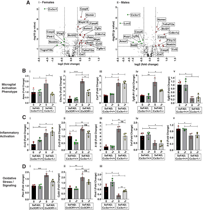
“In contrast, activation of TREM2-APOE signaling in AD results in a subset of microglia that display a ‘neurodegenerative phenotype’, characterized by suppression of homeostatic genes like Cx3cr1 and P2ry12 and upregulation of pro-inflammatory markers like Clec7a, Ccl5, Ccl2, IL1b, and Nos2.”
BackgroundInfluentialHammond, T. R., Dufort, C., Dissing-Olesen, L., Giera, S., Young, A., Wysoker, A., Walker, A. J., Segel, M., Nemesh, J., Saunders, A., et al. (2018) · doi:10.1101/406140 · Cited by 2,225
Alzheimer’s Disease Risk Genes and Mechanisms of Disease Pathogenesis
Karch, C. M., & Goate, A. M. (2015) · Biological Psychiatry · doi:10.1016/j.biopsych.2014.05.006 · Cited by 1,357 · cited 1×
“Lastly, large scale GWAS studies identifying single-nucleotide polymorphisms (SNPs) proximal to microglial-enriched, innate immune genes like CR1, CD33, MEF2C, HLA-DRB5-DRB1, PTK2b and TREM2 that significantly increase the risk of AD [5] have further underscored the critical role of microglia in neurodegeneration and…”
BackgroundZhan, Y., Paolicelli, R. C., Sforazzini, F., Weinhard, L., Bolasco, G., Pagani, F., Vyssotski, A. L., Bifone, A., Gozzi, A., Ragozzino, D., et al. (2014) · Nature Neuroscience · doi:10.1038/nn.3641 · Cited by 1,236 · cited 2×
“Cx3cr1 deficiency is associated with a transient deficit in microglial abundance during early post-natal development [11, 12].”
BackgroundMicroglial brain region-dependent diversity and selective regional sensitivities to aging
Grabert, K., Michoel, T., Karavolos, M. H., Clohisey, S., Baillie, J. K., Stevens, M. P., Freeman, T. C., Summers, K. M., & McColl, B. W. (2016) · Nature Neuroscience · doi:10.1038/nn.4222 · Cited by 1,151
Zhou, Y., Song, W. M., Andhey, P. S., Swain, A., Levy, T., Miller, K. R., Poliani, P. L., Cominelli, M., Grover, S., Gilfillan, S., et al. (2020) · Nature Medicine · doi:10.1038/s41591-019-0695-9 · Cited by 1,150 · cited 6×
“In contrast, activation of TREM2-APOE signaling in AD results in a subset of microglia that display a ‘neurodegenerative phenotype’, characterized by suppression of homeostatic genes like Cx3cr1 and P2ry12 and upregulation of pro-inflammatory markers like Clec7a, Ccl5, Ccl2, IL1b, and Nos2.”
BackgroundInfluentialYuan, P., Condello, C., Keene, C. D., Wang, Y., Bird, T. D., Paul, S. M., Luo, W., Colonna, M., Baddeley, D., & Grutzendler, J. (2016) · Neuron · doi:10.1016/j.neuron.2016.05.003 · Cited by 817 · cited 9×
“In contrast, activation of TREM2-APOE signaling in AD results in a subset of microglia that display a ‘neurodegenerative phenotype’, characterized by suppression of homeostatic genes like Cx3cr1 and P2ry12 and upregulation of pro-inflammatory markers like Clec7a, Ccl5, Ccl2, IL1b, and Nos2.”
BackgroundInfluentialStructural Classification of Toxic Amyloid Oligomers
Glabe, C. G. (2008) · Journal of Biological Chemistry · doi:10.1074/jbc.r800016200 · Cited by 783
He, Z., Guo, J. L., McBride, J. D., Narasimhan, S., Kim, H., Changolkar, L., Zhang, B., Gathagan, R. J., Yue, C., Dengler, C., et al. (2018) · Nature Medicine · doi:10.1038/nm.4443 · Cited by 645 · cited 1×
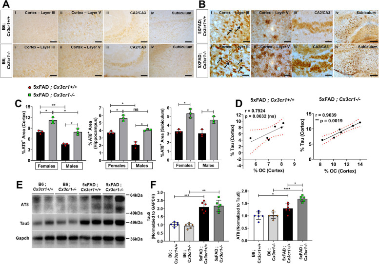
“Studies using stereotaxic injections of pathological human-AD tau into 5xFAD and APP-KI mice have shown that seeding and spread of pTau is affected by plaque burdens, where increased Aβ loads exacerbate MAPT pathology and lead to worsening of memory deficits [47].”
BackgroundRegulation of Tau Pathology by the Microglial Fractalkine Receptor
Bhaskar, K., Konerth, M., Kokiko-Cochran, O. N., Cardona, A., Ransohoff, R. M., & Lamb, B. T. (2010) · Neuron · doi:10.1016/j.neuron.2010.08.023 · Cited by 639 · cited 4×
“Taken together with published studies showing that increased pTau pathology in hTau;Cx3cr1−/− mice is driven by microglial IL-1β [23, 24], our data suggests that Cx3cr1 deficiency may drive pTau accumulation in 5xFAD mice via microglial TREM2-IL1β signaling.”
BackgroundInfluentialSelf-citationAmyloid oligomers: formation and toxicity of Aβ oligomers
Sakono, M., & Zako, T. (2010) · FEBS Journal · doi:10.1111/j.1742-4658.2010.07568.x · Cited by 633
Microglial dysfunction in brain aging and Alzheimer's disease
Mosher, K. I., & Wyss-Coray, T. (2014) · Biochemical Pharmacology · doi:10.1016/j.bcp.2014.01.008 · Cited by 618
Combadière, C., Feumi, C., Raoul, W., Keller, N., Rodéro, M., Pézard, A., Lavalette, S., Houssier, M., Jonet, L., Picard, E., et al. (2007) · Journal of Clinical Investigation · doi:10.1172/jci31692 · Cited by 618
Maphis, N., Xu, G., Kokiko-Cochran, O. N., Jiang, S., Cardona, A., Ransohoff, R. M., Lamb, B. T., & Bhaskar, K. (2015) · Brain · doi:10.1093/brain/awv081 · Cited by 595 · cited 3×
“Taken together with published studies showing that increased pTau pathology in hTau;Cx3cr1−/− mice is driven by microglial IL-1β [23, 24], our data suggests that Cx3cr1 deficiency may drive pTau accumulation in 5xFAD mice via microglial TREM2-IL1β signaling.”
BackgroundSelf-citationMicroglial Cx3cr1 knockout prevents neuron loss in a mouse model of Alzheimer's disease
Fuhrmann, M., Bittner, T., Jung, C. K. E., Burgold, S., Page, R. M., Mitteregger, G., Haass, C., LaFerla, F. M., Kretzschmar, H., & Herms, J. (2010) · Nature Neuroscience · doi:10.1038/nn.2511 · Cited by 562 · cited 1×
“have demonstrated that the loss of Cx3cr1 reduces neuronal loss in cortical layer III in the 5xTg mouse model of AD [51].”
BackgroundLee, S., Varvel, N. H., Konerth, M. E., Xu, G., Cardona, A. E., Ransohoff, R. M., & Lamb, B. T. (2010) · The American Journal of Pathology · doi:10.2353/ajpath.2010.100265 · Cited by 522 · cited 9×
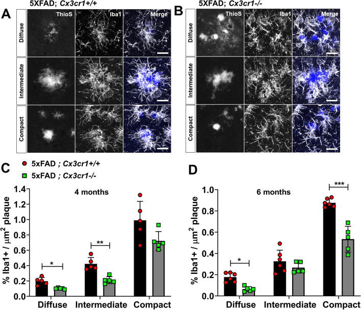
“Interestingly, 4 month-old APPPS1;Cx3cr1−/− mice display impaired microglial plaque engulfment with altered activation of plaqueassociated microglia as evidenced by reduced CD68 and CD45 immunoreactivity [25].”
BackgroundMethodResultInfluentialSelf-citationBidirectional Microglia–Neuron Communication in Health and Disease
Szepesi, Z., Manouchehrian, O., Bachiller, S., & Deierborg, T. (2018) · Frontiers in Cellular Neuroscience · doi:10.3389/fncel.2018.00323 · Cited by 459 · cited 1×
“Signaling via CX3CR1 and its neuronal ligand CX3CL1 represents one of multiple important neuro-glial communication axes that maintain microglial homeostasis [9, 10].”
BackgroundDistinct amyloid-β and tau-associated microglia profiles in Alzheimer’s disease
Gerrits, E., Brouwer, N., Kooistra, S. M., Woodbury, M. E., Vermeiren, Y., Lambourne, M., Mulder, J., Kummer, M., Möller, T., Biber, K., et al. (2021) · Acta Neuropathologica · doi:10.1007/s00401-021-02263-w · Cited by 441
Molecular Mechanisms of Amyloid Oligomers Toxicity
Kayed, R., & Lasagna‐Reeves, C. A. (2012) · Journal of Alzheimer s Disease · doi:10.3233/jad-2012-129001 · Cited by 440
Self-citationDark microglia: A new phenotype predominantly associated with pathological states
Bisht, K., Sharma, K. P., Lecours, C., Gabriela Sánchez, M., El Hajj, H., Milior, G., Olmos‐Alonso, A., Gómez‐Nicola, D., Luheshi, G., Vallières, L., et al. (2016) · Glia · doi:10.1002/glia.22966 · Cited by 421 · cited 1×
“These cells display signs of oxidative stress, including an electron-dense cytoplasm and nucleoplasm, cytoplasmic fragmentation, dilation of Golgi- and ER membranes and mitochondrial alterations [42].”
BackgroundThe amyloid cascade hypothesis: are we poised for success or failure?
Karran, E., & De Strooper, B. (2016) · Journal of Neurochemistry · doi:10.1111/jnc.13632 · Cited by 402 · cited 1×
“These underlying signaling pathways are postulated to lay the framework for cognitive impairment seen in AD [1].”
BackgroundNeuron–glia crosstalk in health and disease: fractalkine and CX 3 CR1 take centre stage
Sheridan, G. K., & Murphy, K. J. (2013) · Open Biology · doi:10.1098/rsob.130181 · Cited by 358
Pan, X.-D., Zhu, Y.-G., Lin, N., Zhang, J., Ye, Q.-Y., Huang, H.-P., & Chen, X.-C. (2011) · Molecular Neurodegeneration · doi:10.1186/1750-1326-6-45 · Cited by 292
Impact of TREM2R47H variant on tau pathology–induced gliosis and neurodegeneration
Gratuze, M., Leyns, C. E. G., Sauerbeck, A. D., St-Pierre, M.-K., Xiong, M., Kim, N., Serrano, J. R., Tremblay, M.-È., Kummer, T. T., Colonna, M., et al. (2020) · Journal of Clinical Investigation · doi:10.1172/jci138179 · Cited by 252 · cited 1×
“Studies using the PS19 mouse model of tauopathy have demonstrated that TREM2 signaling aggravates NFT pathology, associated with pro-inflammatory, Il-1β+ microglia [40].”
BackgroundAPOE and TREM2 regulate amyloid-responsive microglia in Alzheimer’s disease
Nguyen, A. T., Wang, K., Hu, G., Wang, X., Miao, Z., Azevedo, J. A., Suh, E., Van Deerlin, V. M., Choi, D., Roeder, K., et al. (2020) · Acta Neuropathologica · doi:10.1007/s00401-020-02200-3 · Cited by 241 · cited 7×
“In contrast, activation of TREM2-APOE signaling in AD results in a subset of microglia that display a ‘neurodegenerative phenotype’, characterized by suppression of homeostatic genes like Cx3cr1 and P2ry12 and upregulation of pro-inflammatory markers like Clec7a, Ccl5, Ccl2, IL1b, and Nos2.”
BackgroundInfluentialModulating neurotoxicity through CX3CL1/CX3CR1 signaling
Limatola, C., & Ransohoff, R. M. (2014) · Frontiers in Cellular Neuroscience · doi:10.3389/fncel.2014.00229 · Cited by 210 · cited 1×
“However, the downstream effects of attenuated CX3CR1 on the microglial phenotype and subsequent mechanisms that impact neurodegenerative pathology in AD remain elusive, in part due to the lack of comprehensive, transgenic animal models [14, 39].”
BackgroundQuaternary Structure Defines a Large Class of Amyloid-β Oligomers Neutralized by Sequestration
Liu, P., Reed, M. N., Kotilinek, L., Grant, M. K. O., Forster, C. L., Qiang, W., Shapiro, S. L., Reichl, J. H., Chiang, A. C. A., Jankowsky, J. L., et al. (2015) · Cell Reports · doi:10.1016/j.celrep.2015.05.021 · Cited by 148 · cited 1×
“Interestingly, studies in the ArcTau, Tg2576 and hAPPJ20 mouse models of AD have shown that soluble, OC+ oAβ, rather than insoluble Aβ plaques facilitate pTau pathology and cognitive deficits [37, 48].”
BackgroundNi, H., Wang, Y., An, K., Liu, Q., Xu, L., Zhu, C., Deng, H., He, Q., Wang, T., Xu, M., et al. (2019) · Journal of Neuroinflammation · doi:10.1186/s12974-018-1391-2 · Cited by 139
Neuroinflammation and fractalkine signaling in Alzheimer’s disease
Finneran, D. J., & Nash, K. R. (2019) · Journal of Neuroinflammation · doi:10.1186/s12974-019-1412-9 · Cited by 132 · cited 1×
“However, the downstream effects of attenuated CX3CR1 on the microglial phenotype and subsequent mechanisms that impact neurodegenerative pathology in AD remain elusive, in part due to the lack of comprehensive, transgenic animal models [14, 39].”
BackgroundSoluble Aβ Promotes Wild-Type Tau Pathology In Vivo
Chabrier, M. A., Blurton-Jones, M., Agazaryan, A. A., Nerhus, J. L., Martinez-Coria, H., & LaFerla, F. M. (2012) · The Journal of Neuroscience · doi:10.1523/jneurosci.0172-12.2012 · Cited by 123 · cited 2×
“Interestingly, studies in the ArcTau, Tg2576 and hAPPJ20 mouse models of AD have shown that soluble, OC+ oAβ, rather than insoluble Aβ plaques facilitate pTau pathology and cognitive deficits [37, 48].”
BackgroundOpposing Effects of Membrane-Anchored CX3CL1 on Amyloid and Tau Pathologies via the p38 MAPK Pathway
Lee, S., Xu, G., Jay, T. R., Bhatta, S., Kim, K.-W., Jung, S., Landreth, G. E., Ransohoff, R. M., & Lamb, B. T. (2014) · The Journal of Neuroscience · doi:10.1523/jneurosci.0853-14.2014 · Cited by 123 · cited 1×
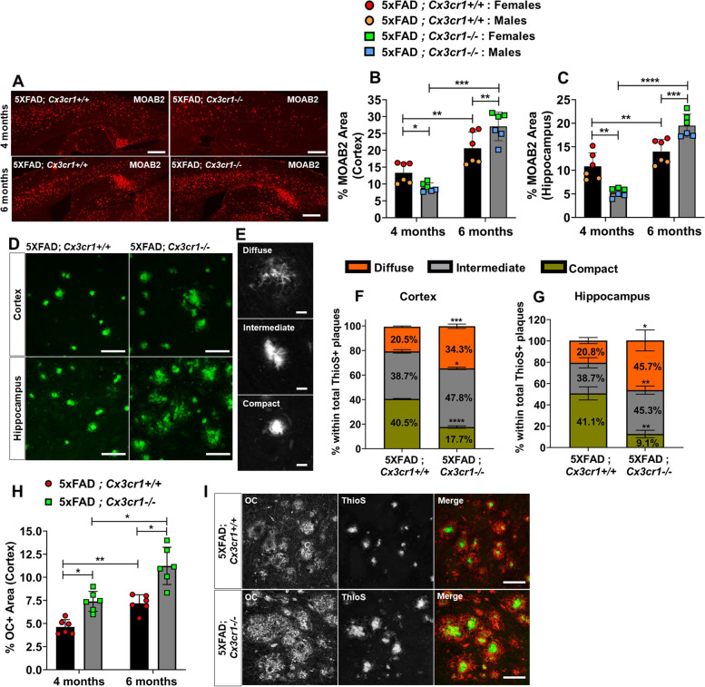
“As seen in our previous studies using APPPS1 mice [25, 26], loss of Cx3cr1 resulted in significantly reduced MOAB2+ plaque load in the cortex and hippocampus in 4-month-old 5xFAD mice (Fig.”
ResultSelf-citationUltrastructural evidence of microglial heterogeneity in Alzheimer’s disease amyloid pathology
El Hajj, H., Savage, J. C., Bisht, K., Parent, M., Vallières, L., Rivest, S., & Tremblay, M.-È. (2019) · Journal of Neuroinflammation · doi:10.1186/s12974-019-1473-9 · Cited by 119 · cited 1×
“Taken as a whole, these data indicate that a) active elimination of neurons by neurodegenerative microglia may be dictated by the quality of intraneuronal pathological protein aggregates and b) microglial neurodegenerative responses modulated by CX3CR1 may be subject to region-dependent microglial heterogeneity along…”
BackgroundReceptors, Ion Channels, and Signaling Mechanisms Underlying Microglial Dynamics
Madry, C., & Attwell, D. (2015) · Journal of Biological Chemistry · doi:10.1074/jbc.r115.637157 · Cited by 109 · cited 1×
“Signaling via CX3CR1 and its neuronal ligand CX3CL1 represents one of multiple important neuro-glial communication axes that maintain microglial homeostasis [9, 10].”
BackgroundPomilio, C., Gorojod, R. M., Riudavets, M., Vinuesa, A., Presa, J., Gregosa, A., Bentivegna, M., Alaimo, A., Alcon, S. P., Sevlever, G., et al. (2020) · GeroScience · doi:10.1007/s11357-020-00161-9 · Cited by 109 · cited 1×
“Studies have indicated that prolonged exposure to toxic Aβ peptides downregulates expression of phagocytic receptors such as CD47, CD36 and RAGE [45], impairs microglial autophagy and induces lysosomal dysfunction in AD [46], an observation corroborated invivo by significant alteration of endocytosis/phagocytosis and…”
BackgroundPossible Mechanisms of Tau Spread and Toxicity in Alzheimer’s Disease
Zhang, H., Cao, Y., Ma, L., Wei, Y., & Li, H. (2021) · Frontiers in Cell and Developmental Biology · doi:10.3389/fcell.2021.707268 · Cited by 109 · cited 1×
“We hypothesize that this increase is reflective of the release of tau from degenerating / dying neurons into the interstitial space, which in-turn potentiates the seeding and spreading of neurotoxic tau [50].”
BackgroundFractalkine Over Expression Suppresses α-Synuclein-mediated Neurodegeneration
Nash, K. R., Moran, P., Finneran, D. J., Hudson, C., Robinson, J., Morgan, D., & Bickford, P. C. (2014) · Molecular Therapy · doi:10.1038/mt.2014.175 · Cited by 107 · cited 1×
“Indeed, disruption of Cx3cr1 increases pro-inflammatory microglial signaling that correlates with heightened loss of dopaminergic neurons in the substantia nigra in murine models of Parkinson’s disease [16, 17].”
BackgroundCx3cr1- deficient microglia exhibit a premature aging transcriptome
Gyoneva, S., Hosur, R., Gosselin, D., Zhang, B., Ouyang, Z., Cotleur, A. C., Peterson, M., Allaire, N., Challa, R., Cullen, P., et al. (2019) · Life Science Alliance · doi:10.26508/lsa.201900453 · Cited by 103 · cited 1×
“Similarly, sc-RNA seq studies that have demonstrated that transcriptomic differences observed in FACS purified, CD11b+CD45+ microglia from 2 month-old B6;Cx3cr1−/− mice are not evident in aged microglia from 12 and 24 month-old Cx3cr1 deficient mice as compared to cells isolated from agematched B6;Cx3cr1+/+ animals [3…”
BackgroundCastro‐Sánchez, S., García‐Yagüe, Á. J., López‐Royo, T., Casarejos, M. J., Lanciego, J. L., & Lastres‐Becker, I. (2018) · Glia · doi:10.1002/glia.23338 · Cited by 89
Xu, J., Zhang, P., Huang, Y., Zhou, Y., Hou, Y., Bekris, L. M., Lathia, J. D., Chiang, C., Li, L., Pieper, A. A., et al. (2021) · Genome Research · doi:10.1101/gr.272484.120 · Cited by 83 · cited 1×
“inflammatory signaling in AD [2, 3], and molecular networks between these unique diseaseassociated glial clusters can reciprocally shape their activation profiles [4].”
Backgroundδ-secretase in neurodegenerative diseases: mechanisms, regulators and therapeutic opportunities
Zhang, Z., Tian, Y., & Ye, K. (2020) · Translational Neurodegeneration · doi:10.1186/s40035-019-0179-3 · Cited by 81
CX3CR1 Is a Modifying Gene of Survival and Progression in Amyotrophic Lateral Sclerosis
López-López, A., Gámez, J., Syriani, E., Morales, M., Salvadó, M., Rodríguez, M. J., Mahy, N., & Vidal-Taboada, J. M. (2014) · PLoS ONE · doi:10.1371/journal.pone.0096528 · Cited by 74 · cited 1×
“TREM2, APOE, PLCγ2), diseasemodifying SNPs around microglial genes, particularly the loss-of-function CX3CR1-V249I variant, have been associated with increased neuronal loss and disease severity in macular degeneration, ALS and AD [7, 8, 38].”
BackgroundEstrada, L. D., Oliveira-Cruz, L., & Cabrera, D. (2018) · Current Protein and Peptide Science · doi:10.2174/1389203719666171129094937 · Cited by 59 · cited 1×
“3Biv, Bv), signaling components that have been implicated in a protective microglial phenotype [35].”
BackgroundFan, Q., He, W., Gayen, M., Benoit, M. R., Luo, X., Hu, X., & Yan, R. (2020) · The Journal of Neuroscience · doi:10.1523/jneurosci.1333-19.2019 · Cited by 58 · cited 2×
“Lastly, overexpression of CX3CL1 in the PS19 model of tauopathy, reduces synaptic and neuronal loss and improves memory and cognition [41].”
BackgroundZhang, J., Liu, Y., Liu, X., Li, S., Cheng, C., Chen, S., & Le, W. (2018) · Translational Neurodegeneration · doi:10.1186/s40035-018-0138-4 · Cited by 57 · cited 1×
“Similar results have been reported in the SOD1-G93A model of ALS, where Cx3cr1 deficiency is associated with increased SOD1 aggregation, neuronal loss and inflammatory microglial activation with impaired autophagy-lysosomal degradation pathways and autophagosome maturation [18, 19].”
ResultThe intracellular domain of CX3CL1 regulates adult neurogenesis and Alzheimer’s amyloid pathology
Fan, Q., Gayen, M., Singh, N., Gao, F., He, W., Hu, X., Tsai, L., & Yan, R. (2019) · The Journal of Experimental Medicine · doi:10.1084/jem.20182238 · Cited by 51 · cited 1×
“5xFAD;CX3CL1-ct mice show decreased plaque loads and reduced neuronal loss [27, 41].”
BackgroundEvidence for a protective role of the CX3CL1/CX3CR1 axis in a model of amyotrophic lateral sclerosis
Liu, C., Hong, K., Chen, H., Niu, Y., Duan, W., Liu, Y., Ji, Y., Deng, B., Li, Y., Li, Z., et al. (2018) · Biological Chemistry · doi:10.1515/hsz-2018-0204 · Cited by 46 · cited 1×
“Similar results have been reported in the SOD1-G93A model of ALS, where Cx3cr1 deficiency is associated with increased SOD1 aggregation, neuronal loss and inflammatory microglial activation with impaired autophagy-lysosomal degradation pathways and autophagosome maturation [18, 19].”
ResultSarsoza, F. M., Saing, T., Kayed, R., Dahlin, R., Dick, M. B., Broadwater-Hollifield, C., Mobley, S., Lott, I. T., Doran, E. W., Gillen, D. L., et al. (2009) · Acta Neuropathologica · doi:10.1007/s00401-009-0530-3 · Cited by 44 · cited 1×
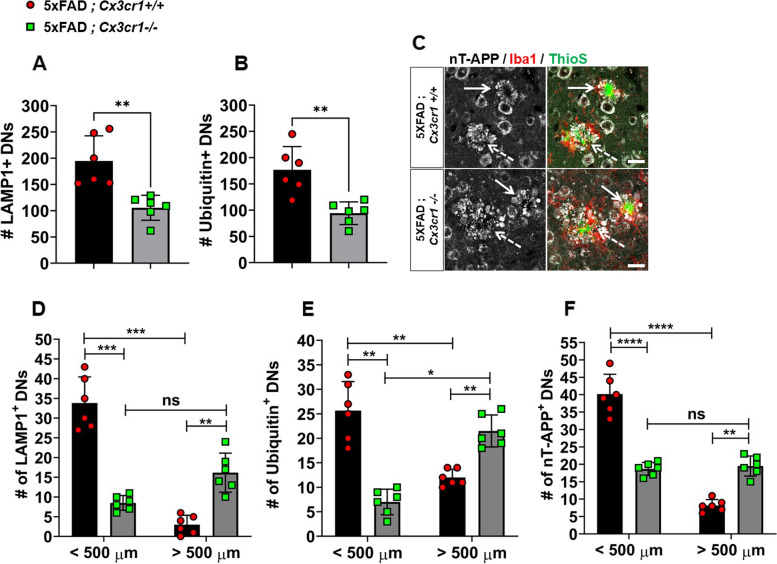
“Cx3cr1 deficiency leads to severe neuritic dystrophy Based on studies that have demonstrated the association of OC+ oAβ with dystrophic neurites [36], we next assessed whether increased oAβ in 5xFAD;Cx3cr1−/− mice (Fig.”
BackgroundLópez-López, A., Gelpí, E., Lopategui, D. M., & Vidal-Taboada, J. M. (2017) · Molecular Neurobiology · doi:10.1007/s12035-017-0489-3 · Cited by 35 · cited 1×
“TREM2, APOE, PLCγ2), diseasemodifying SNPs around microglial genes, particularly the loss-of-function CX3CR1-V249I variant, have been associated with increased neuronal loss and disease severity in macular degeneration, ALS and AD [7, 8, 38].”
BackgroundPuntambekar, S. S., Hinton, D. R., Yin, X., Savarin, C., Bergmann, C. C., Trapp, B. D., & Stohlman, S. A. (2015) · Glia · doi:10.1002/glia.22880 · Cited by 31 · cited 1×
“Brain-derived mononuclear cells were prepared as previously described [30].”
MethodSelf-citationCorrelative Light and Electron Microscopy to Study Microglial Interactions with β-Amyloid Plaques
Bisht, K., Hajj, H. E., Savage, J. C., Sanchez, M. G., & Tremblay, M. (2016) · Journal of Visualized Experiments · doi:10.3791/54060 · Cited by 28
Calvo, A., Moglia, C., Canosa, A., Cammarosano, S., Ilardi, A., Bertuzzo, D., Traynor, B. J., Brunetti, M., Barberis, M., Mora, G., et al. (2017) · Muscle & Nerve · doi:10.1002/mus.25653 · Cited by 26
Zhang, R., Wang, L., Wang, Y.-F., Wu, C., Lei, C.-L., Wang, M., & Ma, L. (2015) · Investigative Ophthalmology & Visual Science · doi:10.1167/iovs.15-16830 · Cited by 18 · cited 1×
“TREM2, APOE, PLCγ2), diseasemodifying SNPs around microglial genes, particularly the loss-of-function CX3CR1-V249I variant, have been associated with increased neuronal loss and disease severity in macular degeneration, ALS and AD [7, 8, 38].”
BackgroundIn Vivo Phagocytosis Analysis of Amyloid Beta
Tejera, D., & Heneka, M. T. (2019) · Methods in molecular biology · doi:10.1007/978-1-4939-9658-2_21 · Cited by 16 · cited 1×
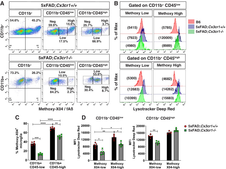
“and FACS‐based microglial purification fAβ internalized by resident microglia was analyzed using methoxy-X04, a brain-penetrant dye that specifically binds to fibrillar β-sheet deposits [29].”
Method
Cited by
Sorted by citations
Tau and neuroinflammation in Alzheimer’s disease: interplay mechanisms and clinical translation
Chen, Y., & Yu, Y. (2023) · Journal of Neuroinflammation · doi:10.1186/s12974-023-02853-3 · Cited by 516
Microglia and Astrocytes in Alzheimer’s Disease: Significance and Summary of Recent Advances
Deng, Q., Wu, C., Parker, E., Liu, T. C.-Y., Duan, R., & Yang, L. (2023) · Aging and disease · doi:10.14336/ad.2023.0907 · Cited by 102
“Cx3cr1 deficiency impairs the uptake of and degradation of fibrillar Aβ by microglia, resulting in increased accumulation of neurotoxic Aβ species [212].”
BackgroundOxygen metabolism abnormality and Alzheimer's disease: An update
Liu, G., Yang, C., Wang, X., Chen, X., Wang, Y., & Le, W. (2023) · Redox Biology · doi:10.1016/j.redox.2023.102955 · Cited by 87
Wu, X., Gao, Y.-Z., Shi, C., Tong, J.-H., Ma, D., Shen, J.-C., Yang, J., & Ji, M. (2023) · Neuropharmacology · doi:10.1016/j.neuropharm.2023.109646 · Cited by 77
Brain high-throughput multi-omics data reveal molecular heterogeneity in Alzheimer’s disease
Eteleeb, A. M., Novotny, B. C., Tarraga, C. S., Sohn, C., Dhungel, E., Brase, L., Nallapu, A., Buss, J., Farias, F., Bergmann, K., et al. (2024) · PLOS Biology · doi:10.1371/journal.pbio.3002607 · Cited by 64
Metadata
- Authors
- Puntambekar, S. S., Moutinho, M., Lin, P. B.-C., Jadhav, V., Tumbleson-Brink, D., Balaji, A., Benito, M. A., Xu, G., Oblak, A., Lasagna-Reeves, C. A., Landreth, G. E., & Lamb, B. T.
- Affiliations
- Indiana University Indianapolis (US)
- Indiana University School of Medicine (US)
- University of Washington (US)
- Indiana Clinical and Translational Sciences Institute (US)
- Corresponding author
- Puntambekar, Shweta S.
- Lamb, Bruce T.
- ORCID
- Venue
- Molecular Neurodegeneration
- Published
- 2022-06-28
- Volume
- 17
- Issue
- 1
- Pages
- 47-47
- ISSN
- 1750-1326 (ISSN-L)
- PMC
- PMC9241248
- OpenAlex
- W4283643624
- Cited by
- 113
- References
- 55
- Citations by year
- 2026: 26 · 2025: 33 · 2024: 23 · 2023: 26 · 2022: 5
- FWCI
- 13.45
- Type
- journal article
- Language
- en
- Open access
- gold
- doi.org
- Locations
- Molecular Neurodegeneration (publishedVersion, cc-by)
- PubMed Central (publishedVersion, cc-by)
- PubMed (publishedVersion)
- Purdue University Indianapolis (Indiana University) (submittedVersion, cc-by)
- DOAJ (DOAJ: Directory of Open Access Journals) (submittedVersion, cc-by-sa)
- IUScholarWorks (Indiana University) (submittedVersion, cc-by)
- Funders
- National Institutes of Health (NIH RF1AG051495, 1RF1AG069425-01)
- Topics
- Life Sciences › Neuroscience › Cellular and Molecular Neuroscience › Alzheimer's disease research and treatments
- Life Sciences › Neuroscience › Neurology › Neuroinflammation and Neurodegeneration Mechanisms
- Life Sciences › Neuroscience › Neurology › Neurological Disease Mechanisms and Treatments
- Keywords
- CX3CR1, Alzheimer's disease, amyloid-beta, microglial dysfunction, 5xFAD mice, tau pathology, neurodegeneration, cognitive decline, TGF-beta signaling, oxidative stress, microglial phagocytosis, lysosomal acidification, microglial polarization, synaptic loss, working memory, neuroinflammation, amyloid plaques, amyloid-beta oligomers
- MeSH terms
- CX3C Chemokine Receptor 1, Alzheimer Disease, Amyloidosis, Animals, Neurons, Transforming Growth Factor beta, Mice, Plaque, Amyloid, Amyloidogenic Proteins, Cognitive Dysfunction
About these checks
Registry, retraction, Cites and statistics results are computed, not judged, with one exception: a printed reference that no index lists is read once by a model before it counts as a citation error. Claims are extracted by a model and kept only when their quote appears verbatim in the text; each cited source is then checked, with a model’s help, for whether it says what the claim relies on. Those verdicts can be wrong — read the claim’s quote and the cited source before relying on one.
Statistics and arithmetic are recomputed, not judged.
Confidence bands are shown only for fields calibrated on our annotated test set. None is calibrated yet, so no band is shown.
What counts as a claim ›How we measure confidence ›How we check this ›
Are you an author, or do you see a problem with this page? Request a review
