

The DOI matches its Crossref record; 6 of the 6 references it cites were found in a registry or an index.
miR-212-5p attenuates ferroptotic neuronal death after traumatic brain injury by targeting Ptgs2
Abstract
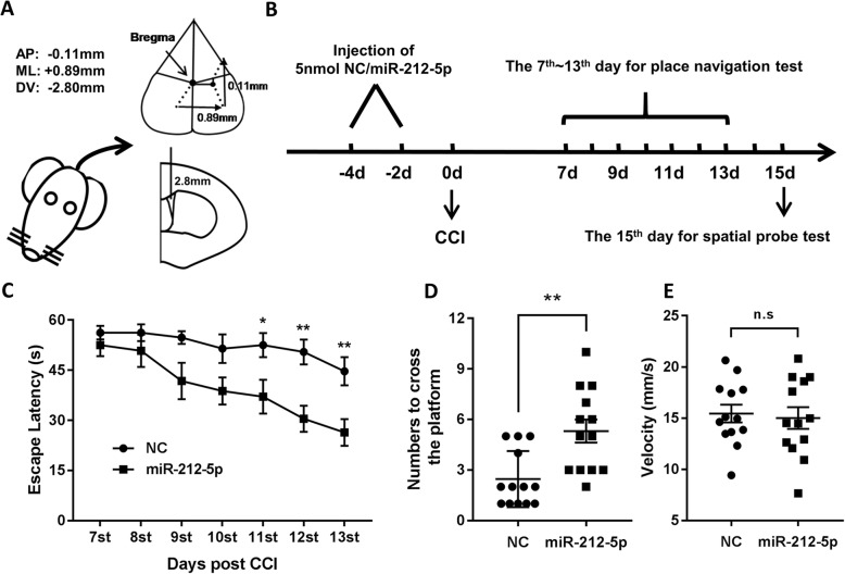
Ferroptosis, a newly discovered form of iron-dependent regulated cell death, has been implicated in traumatic brain injury (TBI). MiR-212-5p has previously been reported to be downregulated in extracellular vesicles following TBI. To investigate whether miR-212-5p is involved in the ferroptotic neuronal death in TBI mice, we first examined the accumulation of malondialdehyde (MDA) and ferrous ion, and the expression of ferroptosis-related molecules at 6 h, 12 h, 24 h, 48 h and 72 h following controlled cortical impact (CCI) in mice. There was a significant upregulation in the expression of Gpx4 and Acsl4 at 6 h, Slc7a11 from 12 h to 72 h, and Nox2 and Sat1 from 6 h to 72 h post injury. Similarly, an upregulation in the expression of Gpx4 at 6 h, Nox2 from 6 h to 72 h, xCT from 12 h to 72 h, and Sat1 at 72 h after CCI was observed at the protein level. Interestingly, MDA and ferrous ion were increased whereas miR-212-5p was decreased in the CCI group compared to the sham group. Furthermore, we found that overexpression of miR-212-5p attenuated ferroptosis while downregulation of miR-212-5p promoted ferroptotic cell death partially by targeting prostaglandin-endoperoxide synthase-2 (Ptgs2) in HT-22 and Neuro-2a cell lines. In addition, administration of miR-212-5p in CCI mice significantly improved learning and spatial memory. Collectively, these findings indicate that miR-212-5p may protect against ferroptotic neuronal death in CCI mice partially by targeting Ptgs2.
Overview
The claims and statistics of this paper haven’t been checked yet.
Run the check — free, no account needed. It reads the open-access text and checks the sources it cites.
Integrity signals
Registry
Cites
Statistics
Figures
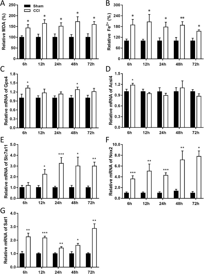
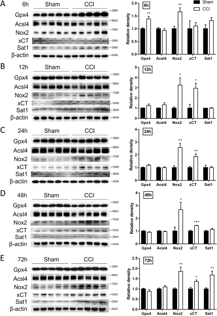
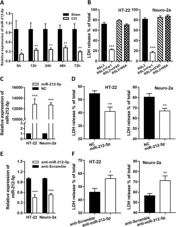
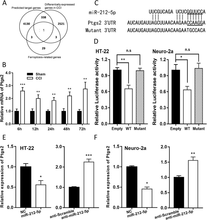
Verification
Claims the paper makes
The claims and statistics of this paper haven’t been checked yet.
Run the check — free, no account needed. It reads the open-access text and checks the sources it cites.
Claims other papers make about this one
Each is checked against this paper’s own text: does it say what the citing paper says it does?
The cited source says what the claim attributes to it.
In the citing paper
“Additional studies suggest that targeting miR-212-5p expression is neuroprotective in models of PD, AD, and traumatic brain injury [63–65].”
From A role for astrocytic miR-129-5p in frontotemporal dementia (2025) · cites this paper as [64]
Self-check
Percentages, sums and reported differences re-computed from the paper’s own numbers.
The self-check needs the paper’s full text.
Citations
Cites
Feigin, V. L., Nichols, E., Alam, T., Bannick, M. S., Beghi, E., Blake, N., Culpepper, W. J., Dorsey, E. R., Elbaz, A., Ellenbogen, R. G., et al. (2019) · The Lancet Neurology · doi:10.1016/s1474-4422(18)30499-x · Cited by 5,644
Self-citationThe impact of microRNAs on protein output
Baek, D., Villén, J., Shin, C., Camargo, F. D., Gygi, S. P., & Bartel, D. P. (2008) · Nature · doi:10.1038/nature07242 · Cited by 3,592
Traumatic brain injury: integrated approaches to improve prevention, clinical care, and research
Maas, A. I. R., Menon, D. K., Adelson, P. D., Andelic, N., Bell, M. J., Belli, A., Bragge, P., Brazinova, A., Büki, A., Chesnut, R. M., et al. (2017) · The Lancet Neurology · doi:10.1016/s1474-4422(17)30371-x · Cited by 2,609
The chronic and evolving neurological consequences of traumatic brain injury
Wilson, L., Stewart, W., Dams-O'Connor, K., Diaz-Arrastia, R., Horton, L., Menon, D. K., & Polinder, S. (2017) · The Lancet Neurology · doi:10.1016/s1474-4422(17)30279-x · Cited by 607
NCOA4-Mediated Ferritinophagy: A Potential Link to Neurodegeneration
Quiles del Rey, M., & Mancias, J. D. (2019) · Frontiers in Neuroscience · doi:10.3389/fnins.2019.00238 · Cited by 227
Harrison, E. B., Hochfelder, C. G., Lamberty, B. G., Meays, B. M., Morsey, B. M., Kelso, M. L., Fox, H. S., & Yelamanchili, S. V. (2016) · FEBS Open Bio · doi:10.1002/2211-5463.12092 · Cited by 151
Research works printed but not linked: 67
- Rubiano AM, Carney N, Chesnut R, Puyana JC. Global neurotrauma research challenges and opportunities. Nature. 2015;527(7578):S193–7. · DOI
- Iaccarino C, Carretta A, Nicolosi F, Morselli C. Epidemiology of severe traumatic brain injury. J Neurosurg Sci. 2018;62:535–41. · DOI
- Majdan M, Plancikova D, Maas A, Polinder S, Feigin V, Theadom A, et al. Years of life lost due to traumatic brain injury in Europe: a cross-sectional analysis of 16 countries. PLoS Med. 2017;14(7):e1002331. · DOI
- Cheng P, Yin P, Ning P, Wang L, Cheng X, Liu Y, et al. Trends in traumatic brain injury mortality in China, 2006–2013: a population-based longitudinal study. PLoS Med. 2017;14(7):e1002332. · DOI
- Ved R, Zaben M. Biomarkers for traumatic brain injury. J Neurol. 2018;265(5):1241–3. · DOI
- Stockwell BR, Friedmann Angeli JP, Bayir H, Bush AI, Conrad M, Dixon SJ, et al. Ferroptosis: a regulated cell death Nexus linking metabolism, redox biology, and disease. Cell. 2017;171:273–85. · DOI · Paper page
- Fricker M, Tolkovsky AM, Borutaite V, Coleman M, Brown GC. Neuronal cell death. Physiol Rev. 2018;98(2):813–80. https://doi.org/10.1152/physrev.00011.2017 . · DOI
- Gaschler MM, Hu F, Feng H, Linkermann A, Min W, Stockwell BR. Determination of the subcellular localization and mechanism of action of Ferrostatins in suppressing Ferroptosis. ACS Chem Biol. 2018;13(4):1013–20. · DOI
- Wenzel SE, Tyurina YY, Zhao J, St Croix CM, Dar HH, Mao G, et al. PEBP1 Wardens Ferroptosis by Enabling Lipoxygenase Generation of Lipid Death Signals. Cell. 2017;171(3):628–641.e26. · DOI
- Cao JY, Dixon SJ. Mechanisms of ferroptosis. Cell Mol Life Sci. 2016;73:2195–209. · DOI · Paper page
- Lane DJR, Ayton S, Bush AI. Iron and Alzheimer’s disease: an update on emerging mechanisms. J Alzheimers Dis. 2018;64(s1):S379–95. Available from: http://www.medra.org/servlet/aliasResolver?alias=iospress&doi=10.3233/JAD-179944 . · DOI
- Becker RE, Kapogiannis D, Greig NH. Does traumatic brain injury hold the key to the Alzheimer’s disease puzzle? Alzheimers Dement. 2018;14(4):431–43. https://doi.org/10.1016/j.jalz.2017.11.007 . · DOI
- Rosenfeld JV, McFarlane AC, Bragge P, Armonda RA, Grimes JB, Ling GS. Blast-related traumatic brain injury. Lancet Neurol. 2013;12(9):882–93. · DOI
- Pan YB, Sun ZL, Feng DF. The role of MicroRNA in traumatic brain injury. Neuroscience. 2017;367:189–99. · DOI
- Wanet A, Tacheny A, Arnould T, Renard P. MiR-212/132 expression and functions: within and beyond the neuronal compartment. Nucleic Acids Res. 2012;40(11):4742–53. · DOI
- Aten S, Hansen KF, Hoyt KR, Obrietan K. The miR-132/212 locus: a complex regulator of neuronal plasticity, gene expression and cognition. RNA Dis. 2016;3:e1375.
- Hansen KF, Sakamoto K, Aten S, Snider KH, Loeser J, Hesse AM, et al. Targeted deletion of miR-132/−212 impairs memory and alters the hippocampal transcriptome. Learn Mem. 2016;23(2):61–71. · DOI · Paper page
- Hernandez-Rapp J, Smith PY, Filali M, Goupil C, Planel E, Magill ST, et al. Memory formation and retention are affected in adult miR-132/212 knockout mice. Behav Brain Res. 2015;287:15–26. · DOI · Paper page
- Burek M, König A, Lang M, Fiedler J, Oerter S, Roewer N, et al. Hypoxia-induced MicroRNA-212/132 Alter blood-brain barrier integrity through inhibition of tight junction-associated proteins in human and mouse brain microvascular endothelial cells. Transl Stroke Res. 2019. https://doi.org/10.1007/s12975-018-0683-2 . · DOI
- Smith PY, Hernandez-Rapp J, Jolivette F, Lecours C, Bisht K, Goupil C, et al. MiR-132/212 deficiency impairs tau metabolism and promotes pathological aggregation in vivo. Hum Mol Genet. 2015;24(23):6721–35. · DOI · Paper page
- Hernandez-Rapp J, Rainone S, Goupil C, Dorval V, Smith PY, Saint-Pierre M, et al. MicroRNA-132/212 deficiency enhances Aβ production and senile plaque deposition in Alzheimer’s disease triple transgenic mice. Sci Rep. 2016;6:30953. · DOI
- Lighthall JW. Controlled cortical impact: a New experimental brain injury model. J Neurotrauma. 2009;5(1):1–15. · DOI
- Dean DD, Frank JA, Turtzo LC. Controlled cortical impact in the rat. Curr Protoc Neurosci. 2017;81:9.62.1–9.62.12. · DOI
- Romine J, Gao X, Chen J. Controlled cortical impact model for traumatic brain injury. J Vis Exp. 2014;(90):e51781. https://doi.org/10.3791/51781 . · DOI
- Lyeth BG, Ray M, Hamm RJ, Schnabel J, Saady JJ, Poklis A, et al. Postinjury scopolamine administration in experimental traumatic brain injury. Brain Res. 1992;569(2):281–6. · DOI
- Bermpohl D, You Z, Korsmeyer SJ, Moskowitz MA, Whalen MJ. Traumatic brain injury in mice deficient in bid: effects on histopathology and functional outcome. J Cereb Blood Flow Metab. 2006;26(5):625–33. · DOI
- He J, Yang X, Men B, Wang D. Interfacial mechanisms of heterogeneous Fenton reactions catalyzed by iron-based materials: a review. J Environ Sci (China). 2016;39:97–109. · DOI
- Sticht C, De La Torre C, Parveen A, Gretz N. Mirwalk: an online resource for prediction of microrna binding sites. PLoS One. 2018;13(10):e0206239. · DOI · Paper page
- Jiang YN, Yang SW, Zhang X, Luo LM, Zhang Z, Chen NH. Mechanism of ferroptosis and its role in neurological diseases. Chinese Pharmacol Bull. 2018;34(2):166–9.
- Xie BS, Wang YQ, Lin Y, Mao Q, Feng JF, Gao GY, et al. Inhibition of ferroptosis attenuates tissue damage and improves long-term outcomes after traumatic brain injury in mice. CNS Neurosci Ther. 2018;25:465–75. · DOI
- Kenny EM, Fidan E, Yang Q, Anthonymuthu TS, New LA, Meyer EA, et al. Ferroptosis contributes to neuronal death and functional outcome after traumatic brain injury. Crit Care Med. 2019;47(3):410–8. · DOI
- Devos D, Moreau C, Kyheng M, Garçon G, Rolland AS, Blasco H, et al. A ferroptosis–based panel of prognostic biomarkers for amyotrophic lateral sclerosis. Sci Rep. 2019;9(1):2198. · DOI
- Wu Y, Song J, Wang Y, Wang X, Culmsee C, Zhu C. The potential role of Ferroptosis in neonatal brain injury. Front Neurosci. 2019;13:115. · DOI
- Magtanong L, Dixon SJ. Ferroptosis and brain injury. Dev Neurosci. 2019;40:382–95. · DOI
- Hu C, Dong ZL. MicroRNA-212 promotes the recovery function and vascular regeneration of endothelial progenitor cells in mice with ischemic stroke through inactivation of the notch signaling pathway via downregulating MMP9 expression. J Cell Physiol. 2019;234(5):7090–103. · DOI
- Mendoza-Viveros L, Chiang CK, Ong JLK, Hegazi S, Cheng AH, Bouchard-Cannon P, et al. miR-132/212 modulates seasonal adaptation and dendritic morphology of the central circadian clock. Cell Rep. 2017;19(3):505–20. · DOI
- Wang CY, Deneen B, Tzeng SF. MicroRNA-212 inhibits oligodendrocytes during maturation by down-regulation of differentiation-associated gene expression. J Neurochem. 2017;143(1):112–25. · DOI
- Garcia-Concejo A, Jimenez-Gonzalez A, Rodriguez RE. Opioid and notch signaling pathways are reciprocally regulated through miR- 29a and miR-212 expression. Biochim Biophys Acta Gen Subj. 2018;1862(12):2605–12. · DOI
- Weigelt K, Bergink V, Burgerhout KM, Pescatori M, Wijkhuijs A, Drexhage HA. Down-regulation of inflammation-protective microRNAs 146a and 212 in monocytes of patients with postpartum psychosis. Brain Behav Immun. 2013;29:147–55. · DOI
- Keller RF, Kanlikilicer P, Dragomir A, Fan Y, Akay YM, Akay M. Investigating the effect of perinatal nicotine exposure on dopaminergic neurons in the VTA using miRNA expression profiles. IEEE Trans Nanobiosci. 2017;16(8):843–9. · DOI
- Wang Y, Veremeyko T, Wong AHK, El Fatimy R, Wei Z, Cai W, et al. Downregulation of miR-132/212 impairs S-nitrosylation balance and induces tau phosphorylation in Alzheimer’s disease. Neurobiol Aging. 2017;51:156–66. · DOI · Paper page
- Pichler S, Gu W, Hartl D, Gasparoni G, Leidinger P, Keller A, et al. The miRNome of Alzheimer’s disease: consistent downregulation of the miR-132/212 cluster. Neurobiol Aging. 2017;50:167.e1–167.e10. · DOI · Paper page
- Amadio P, Tarantino E, Sandrini L, Tremoli E, Barbieri SS. Prostaglandin-Endoperoxide Synthase-2 deletion affects the natural trafficking of annexin A2 in monocytes and favours venous thrombosis in mice. Thromb Haemost. 2017;117(8):1486–97. · DOI
- Hua X, Chi W, Su L, Li J, Zhang Z, Yuan X. ROS-induced oxidative injury involved in pathogenesis of fungal keratitis via p38 MAPK activation. Sci Rep. 2017;7(1):10421. · DOI
- Kong Z, Liu R, Cheng Y. Artesunate alleviates liver fibrosis by regulating ferroptosis signaling pathway. Biomed Pharmacother. 2019;109:2043–53. · DOI
- Zhang Z, Yao Z, Wang L, Ding H, Shao J, Chen A, et al. Activation of ferritinophagy is required for the RNA-binding protein ELAVL1/HuR to regulate ferroptosis in hepatic stellate cells. Autophagy. 2018;14(12):2083–103. · DOI
- Li Q, Han X, Lan X, Gao Y, Wan J, Durham F, et al. Inhibition of neuronal ferroptosis protects hemorrhagic brain. JCI Insight. 2017;2(7):e90777. · DOI
- Dixon SJ, Lemberg KM, Lamprecht MR, Skouta R, Zaitsev EM, Gleason CE, et al. Ferroptosis: an iron-dependent form of nonapoptotic cell death. Cell. 2012;149(5):1060–72. · DOI · Paper page
- Sui X, Zhang R, Liu S, Duan T, Zhai L, Zhang M, et al. RSL3 drives ferroptosis through GPX4 inactivation and ros production in colorectal cancer. Front Pharmacol. 2018;9:1371. · DOI · Paper page
- Hirschhorn T, Stockwell BR. The development of the concept of ferroptosis. Free Radic Biol Med. 2019;133:130–43. · DOI · Paper page
- Liu X, Moon SH, Jenkins CM, Sims HF, Gross RW. Cyclooxygenase-2 mediated oxidation of 2-Arachidonoyl-Lysophospholipids identifies unknown lipid signaling pathways. Cell Chem Biol. 2016;23(10):1217–27. · DOI
- Bretscher P, Egger J, Shamshiev A, Trotzmuller M, Kofeler H, Carreira EM, et al. Phospholipid oxidation generates potent anti-inflammatory lipid mediators that mimic structurally related pro-resolving eicosanoids by activating Nrf2. EMBO Mol Med. 2015;7(5):593–607. · DOI
- Hambright WS, Fonseca RS, Chen L, Na R, Ran Q. Ablation of ferroptosis regulator glutathione peroxidase 4 in forebrain neurons promotes cognitive impairment and neurodegeneration. Redox Biol. 2017;12:8–17. · DOI · Paper page
- Guiney SJ, Adlard PA, Bush AI, Finkelstein DI, Ayton S. Ferroptosis and cell death mechanisms in Parkinson’s disease. Neurochem Int. 2017;104:34–48. · DOI
- Ilmaniemi S, Taipale H, Tanskanen A, Tiihonen J, Hartikainen S, Tolppanen AM. Incidence of head injury and traumatic brain injury among people with Alzheimer’s disease. J Epidemiol Community Health. 2019;73:451–4. · DOI
- Mohamed AZ, Cumming P, Götz J, Nasrallah F. Tauopathy in veterans with long-term posttraumatic stress disorder and traumatic brain injury. Eur J Nucl Med Mol Imaging. 2019;46:1139–51. · DOI
- Lee Y-S, Lee D-H, Choudry HA, Bartlett DL, Lee YJ. Ferroptosis-induced endoplasmic reticulum stress: crosstalk between Ferroptosis and apoptosis. Mol Cancer Res. 2018;16:1073–6. https://doi.org/10.1158/1541-7786.MCR-18-0055 . · DOI
- Sun D, Chen X, Gu G, Wang J, Zhang J. Potential roles of mitochondria-associated ER membranes (MAMs) in traumatic brain injury. Cell Mol Neurobiol. 2017;37:1349–57. · DOI
- Gao M, Monian P, Pan Q, Zhang W, Xiang J, Jiang X. Ferroptosis is an autophagic cell death process. Cell Res. 2016;26(9):1021–32. · DOI · Paper page
- Kang R, Tang D. Autophagy and Ferroptosis - What’s the connection? Curr Pathobiol Rep. 2017;5(2):153–9 Available from: http://www.ncbi.nlm.nih.gov/pubmed/29038744 . · DOI
- Sun S, Han X, Li X, Song Q, Lu M, Jia M, et al. MicroRNA-212-5p prevents dopaminergic neuron death by inhibiting SIRT2 in MPTP-induced mouse model of Parkinson’s disease. Front Mol Neurosci. 2018;11:381. · DOI · Paper page
- Chen W, Zhou Y, Zhi X, Ma T, Liu H, Chen BW, et al. Delivery of miR-212 by chimeric peptide-condensed supramolecular nanoparticles enhances the sensitivity of pancreatic ductal adenocarcinoma to doxorubicin. Biomaterials. 2019;192:590–600. · DOI
- Zhang X, Lu L, Zhang X, Zhang B, Wu J. Synaptic acetylcholinesterase targeted by microRNA-212 functions as a tumor suppressor in non-small cell lung cancer. Int J Biochem Cell Biol. 2013;45:2530–40. · DOI
- Guo Y, Yu J, Wang C, Li K, Liu B, Du Y, et al. MiR-212-5p suppresses lipid accumulation by targeting FAS and SCD1. J Mol Endocrinol. 2017;59:205–17. · DOI
- Xiao J, Bei Y, Liu J, Dimitrova-Shumkovska J, Kuang D, Zhou Q, et al. miR-212 downregulation contributes to the protective effect of exercise against non-alcoholic fatty liver via targeting FGF-21. J Cell Mol Med. 2016;20:204–16. · DOI
- Fiedler SD, Carletti MZ, Hong X, Christenson LK. Hormonal regulation of MicroRNA expression in Periovulatory mouse mural granulosa Cells1. Biol Reprod. 2008;79:1030–7. · DOI
- Shang J, Li J, Keller MP, Hohmeier HE, Wang Y, Feng Y, et al. Induction of miR-132 and miR-212 expression by glucagon-like peptide 1 (GLP-1) in rodent and human pancreatic β-cells. Mol Endocrinol. 2015;29:1243–53. · DOI
Cited by
Sorted by citations
A role for astrocytic miR-129-5p in frontotemporal dementia
Kaurani, L., Pradhan, R., Schröder, S., Burkhardt, S., Schuetz, A.-L., Krüger, D. M., Pena, T., Heutink, P., Sananbenesi, F., & Fischer, A. (2025) · Translational Psychiatry · doi:10.1038/s41398-025-03338-y · Cited by 10
Metadata
- Authors
- Xiao, X., Jiang, Y., Liang, W., Wang, Y., Cao, S., Yan, H., Gao, L., & Zhang, L.
- Venue
- Molecular Brain
- Published
- 2019-09-18
- Volume
- 12
- Issue
- 1
- ISSN
- 1756-6606
- PMC
- PMC6749650
- Cited by
- 163
- References
- 6
- Language
- en
- Funders
- Fundamental Research Funds for the Central Universities (No. 2018SCUH0053)
- Science and Technology Major Projects of Sichuan Province of China (No.2017SZDZX0013)
- Science and Technology Support Program of Sichuan Province of China (No.2016SZ0013)
About these checks
Registry, retraction, Cites and statistics results are computed, not judged, with one exception: a printed reference that no index lists is read once by a model before it counts as a citation error. Claims are extracted by a model and kept only when their quote appears verbatim in the text; each cited source is then checked, with a model’s help, for whether it says what the claim relies on. Those verdicts can be wrong — read the claim’s quote and the cited source before relying on one.
Statistics and arithmetic are recomputed, not judged.
Confidence bands are shown only for fields calibrated on our annotated test set. None is calibrated yet, so no band is shown.
What counts as a claim ›How we measure confidence ›How we check this ›
Are you an author, or do you see a problem with this page? Request a review
