

The DOI matches its Crossref record; 64 of the 64 references it cites were found in a registry or an index.
Glycoprotein NMB: a novel Alzheimer’s disease associated marker expressed in a subset of activated microglia
Open accessAbstract
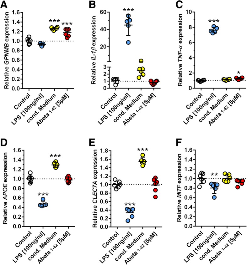
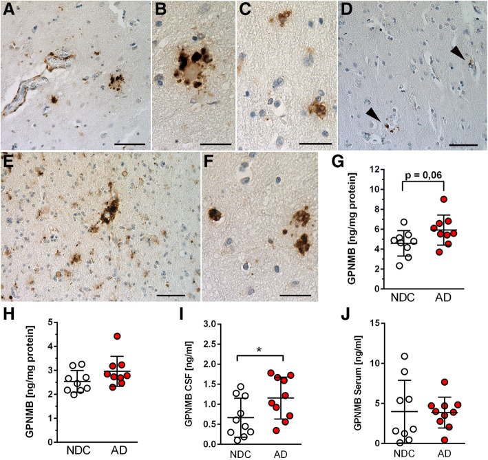
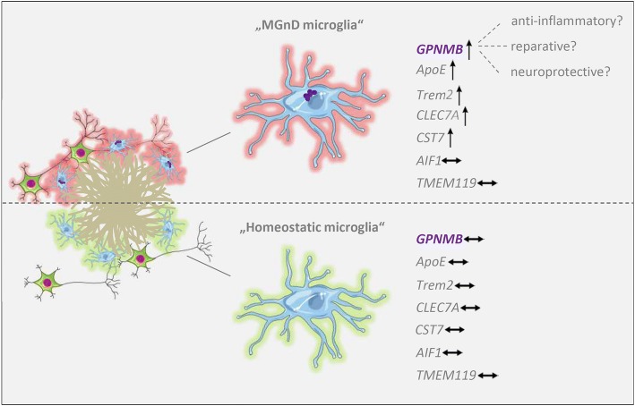
Alzheimer's disease (AD) is an irreversible, devastating neurodegenerative brain disorder characterized by the loss of neurons and subsequent cognitive decline. Despite considerable progress in the understanding of the pathophysiology of AD, the precise molecular mechanisms that cause the disease remain elusive. By now, there is ample evidence that activated microglia have a critical role in the initiation and progression of AD. The present study describes the identification of Glycoprotein nonmetastatic melanoma protein B (GPNMB) as a novel AD-related factor in both transgenic mice and sporadic AD patients by expression profiling, immunohistochemistry and ELISA measurements. We show that GPNMB levels increase in an age-dependent manner in transgenic AD models showing profound cerebral neuron loss and demonstrate that GPNMB co-localizes with a distinct population of IBA1-positive microglia cells that cluster around amyloid plaques. Our data further indicate that GPNMB is part of a microglia activation state that is only present under neurodegenerative conditions and that is characterized by the up-regulation of a subset of genes including TREM2, APOE and CST7. In agreement, we provide in vitro evidence that soluble Aβ has a direct effect on GPNMB expression in an immortalized microglia cell line. Importantly, we show for the first time that GPNMB is elevated in brain samples and cerebrospinal fluid (CSF) of sporadic AD patients when compared to non-demented controls.The current findings indicate that GPNMB represents a novel disease-associated marker that appears to play a role in the neuroinflammatory response of AD.
Overview
The claims and statistics of this paper haven’t been checked yet.
Run the check — free, no account needed. It reads the open-access text and checks the sources it cites.
Integrity signals
Registry
Cites
Statistics
Figures
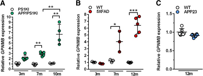
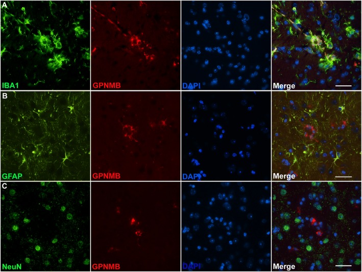
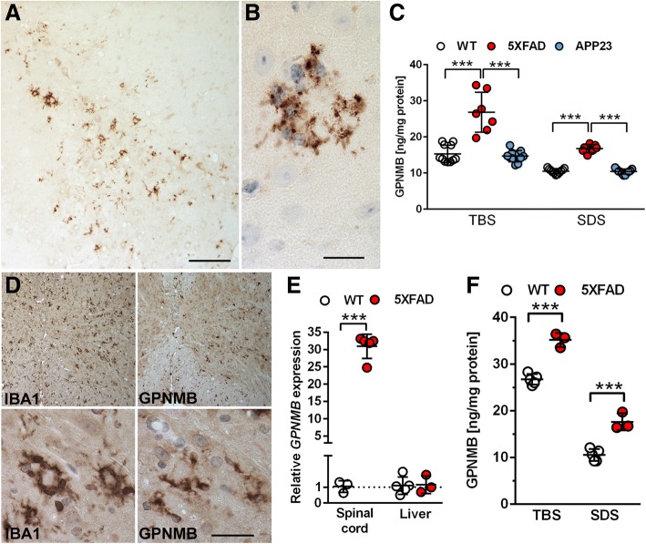
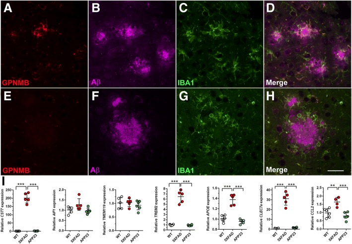
Verification
Claims the paper makes
The claims and statistics of this paper haven’t been checked yet.
Run the check — free, no account needed. It reads the open-access text and checks the sources it cites.
Claims other papers make about this one
Each is checked against this paper’s own text: does it say what the citing paper says it does?
The cited source says what the claim attributes to it.
In the citing paper
“In the list, 4 proteins (GPNMB, SMOC1, NPTX2 and VGF) have previously been reported in AD CSF (Dayon et al., 2018; Huttenrauch et al., 2018; Llano et al., 2019; Xiao et al., 2017)”
From Deep Multilayer Brain Proteomics Identifies Molecular Networks in Alzheimer’s Disease Progression (2020) · cites this paper as Huttenrauch et al., 2018
The cited source says what the claim attributes to it.
In the citing paper
“While, SMOC1 and GPNMB have been reported as putative CSF biomarkers in previous studies [18, 33, 62]”
From Integrated analysis of ultra-deep proteomes in cortex, cerebrospinal fluid and serum reveals a mitochondrial signature in Alzheimer’s disease (2020) · cites this paper as [62]
The cited source says what the claim attributes to it.
In the citing paper
“Consistently, SMOC1 and GPNMB were reported to be CSF biomarker candidates for AD in other recent biomarker studies [62, 69].”
From Integrated analysis of ultra-deep proteomes in cortex, cerebrospinal fluid and serum reveals a mitochondrial signature in Alzheimer’s disease (2020) · cites this paper as [62]
Self-check
Percentages, sums and reported differences re-computed from the paper’s own numbers.
The self-check needs the paper’s full text.
Citations
Cites
Analyzing real-time PCR data by the comparative CT method
Schmittgen, T. D., & Livak, K. J. (2008) · Nature Protocols · doi:10.1038/nprot.2008.73 · Cited by 26,190
The amyloid hypothesis of Alzheimer's disease at 25 years
Selkoe, D. J., & Hardy, J. (2016) · EMBO Molecular Medicine · doi:10.15252/emmm.201606210 · Cited by 6,164
A Unique Microglia Type Associated with Restricting Development of Alzheimer’s Disease
Keren-Shaul, H., Spinrad, A., Weiner, A., Matcovitch-Natan, O., Dvir-Szternfeld, R., Ulland, T. K., David, E., Baruch, K., Lara-Astaiso, D., Toth, B., et al. (2017) · Cell · doi:10.1016/j.cell.2017.05.018 · Cited by 5,493
Zhang, Y., Chen, K., Sloan, S. A., Bennett, M. L., Scholze, A. R., O'Keeffe, S., Phatnani, H. P., Guarnieri, P., Caneda, C., Ruderisch, N., et al. (2014) · The Journal of Neuroscience · doi:10.1523/jneurosci.1860-14.2014 · Cited by 4,875
Oakley, H., Cole, S. L., Logan, S., Maus, E., Shao, P., Craft, J., Guillozet-Bongaarts, A., Ohno, M., Disterhoft, J., Van Eldik, L., et al. (2006) · The Journal of Neuroscience · doi:10.1523/jneurosci.1202-06.2006 · Cited by 3,530 · cited 1×
“At 12 months of age, the model shows a massive Aβ plaque load in various brain regions, which is accompanied by extensive astro- and microgliosis [19, 33].”
BackgroundHarold, D., Abraham, R., Hollingworth, P., Sims, R., Gerrish, A., Hamshere, M. L., Pahwa, J. S., Moskvina, V., Dowzell, K., Williams, A., et al. (2009) · Nature Genetics · doi:10.1038/ng.440 · Cited by 3,326
Self-citationTREM2 Variants in Alzheimer's Disease
Guerreiro, R., Wojtas, A., Bras, J., Carrasquillo, M., Rogaeva, E., Majounie, E., Cruchaga, C., Sassi, C., Kauwe, J. S. K., Younkin, S., et al. (2013) · New England Journal of Medicine · doi:10.1056/nejmoa1211851 · Cited by 3,020
Krasemann, S., Madore, C., Cialic, R., Baufeld, C., Calcagno, N., El Fatimy, R., Beckers, L., O’Loughlin, E., Xu, Y., Fanek, Z., et al. (2017) · Immunity · doi:10.1016/j.immuni.2017.08.008 · Cited by 2,979
Genome-wide association study identifies variants at CLU and CR1 associated with Alzheimer's disease
the European Alzheimer's Disease Initiative Investigators, Lambert, J.-C., Heath, S., Even, G., Campion, D., Sleegers, K., Hiltunen, M., Combarros, O., Zelenika, D., Bullido, M. J., et al. (2009) · Nature Genetics · doi:10.1038/ng.439 · Cited by 2,234
Initiative, T. A. D. N., Hollingworth, P., CHARGE consortium, EADI1 consortium, Harold, D., Sims, R., Gerrish, A., Lambert, J.-C., Carrasquillo, M. M., Abraham, R., et al. (2011) · Nature Genetics · doi:10.1038/ng.803 · Cited by 1,997
Self-citationMicroglia in Alzheimer’s disease
Hansen, D. V., Hanson, J. E., & Sheng, M. (2018) · Journal of Cell Biology · doi:10.1083/jcb.201709069 · Cited by 1,935 · cited 1×
“disease, protective microglial activities supposedly occur in the early disease stages [10].”
BackgroundNaj, A. C., Jun, G., Beecham, G. W., Wang, L.-S., Vardarajan, B. N., Buros, J., Gallins, P. J., Buxbaum, J. D., Jarvik, G. P., Crane, P. K., et al. (2011) · Nature Genetics · doi:10.1038/ng.801 · Cited by 1,901
Two amyloid precursor protein transgenic mouse models with Alzheimer disease-like pathology
Sturchler-Pierrat, C., Abramowski, D., Duke, M., Wiederhold, K.-H., Mistl, C., Rothacher, S., Ledermann, B., Bürki, K., Frey, P., Paganetti, P. A., et al. (1997) · Proceedings of the National Academy of Sciences · doi:10.1073/pnas.94.24.13287 · Cited by 1,507
Alzheimer’s Disease Risk Genes and Mechanisms of Disease Pathogenesis
Karch, C. M., & Goate, A. M. (2015) · Biological Psychiatry · doi:10.1016/j.biopsych.2014.05.006 · Cited by 1,357
Jawhar, S., Trawicka, A., Jenneckens, C., Bayer, T. A., & Wirths, O. (2012) · Neurobiology of Aging · doi:10.1016/j.neurobiolaging.2010.05.027 · Cited by 623 · cited 4×
“While 5XFAD mice show cortical neuron loss at 12 months of age [7, 19], even higher cortical neuron numbers have been reported in 8-month-old APP23 compared to WT mice, which decrease in 27-month-old APP23 mice back to WT levels [2].”
BackgroundInfluentialSelf-citationHopperton, K. E., Mohammad, D., Trépanier, M. O., Giuliano, V, & Bazinet, R. P. (2018) · Molecular Psychiatry · doi:10.1038/mp.2017.246 · Cited by 567
Walker, D. G., & Lue, L.-F. (2015) · Alzheimer's Research & Therapy · doi:10.1186/s13195-015-0139-9 · Cited by 562
Suárez‐Calvet, M., Kleinberger, G., Araque Caballero, M. Á., Brendel, M., Rominger, A., Alcolea, D., Fortea, J., Lleó, A., Blesa, R., Gispert, J. D., et al. (2016) · EMBO Molecular Medicine · doi:10.15252/emmm.201506123 · Cited by 522
Eimer, W. A., & Vassar, R. (2013) · Molecular Neurodegeneration · doi:10.1186/1750-1326-8-2 · Cited by 425 · cited 2×
“While 5XFAD mice show cortical neuron loss at 12 months of age [7, 19], even higher cortical neuron numbers have been reported in 8-month-old APP23 compared to WT mice, which decrease in 27-month-old APP23 mice back to WT levels [2].”
BackgroundCasas, C., Sergeant, N., Itier, J.-M., Blanchard, V., Wirths, O., van der Kolk, N., Vingtdeux, V., van de Steeg, E., Ret, G., Canton, T., et al. (2004) · The American Journal of Pathology · doi:10.1016/s0002-9440(10)63388-3 · Cited by 400
Self-citationIsolation of glia from Alzheimer’s mice reveals inflammation and dysfunction
Orre, M., Kamphuis, W., Osborn, L. M., Jansen, A. H. P., Kooijman, L., Bossers, K., & Hol, E. M. (2014) · Neurobiology of Aging · doi:10.1016/j.neurobiolaging.2014.06.004 · Cited by 394
Association of Microglia with Amyloid Plaques in Brains of APP23 Transgenic Mice
Stalder, M., Phinney, A., Probst, A., Sommer, B., Staufenbiel, M., & Jucker, M. (1999) · The American Journal of Pathology · doi:10.1016/s0002-9440(10)65423-5 · Cited by 345
Cerebrospinal fluid soluble TREM2 is higher in Alzheimer disease and associated with mutation status
Piccio, L., Deming, Y., Del-Águila, J. L., Ghezzi, L., Holtzman, D. M., Fagan, A. M., Fenoglio, C., Galimberti, D., Borroni, B., & Cruchaga, C. (2016) · Acta Neuropathologica · doi:10.1007/s00401-016-1533-5 · Cited by 337
A Two-Stage Meta-Analysis Identifies Several New Loci for Parkinson's Disease
Wellcome Trust Case Control Consortium (2011) · PLoS Genetics · doi:10.1371/journal.pgen.1002142 · Cited by 314
Increased cerebrospinal fluid soluble TREM2 concentration in Alzheimer’s disease
Heslegrave, A., Heywood, W., Paterson, R., Magdalinou, N., Svensson, J., Johansson, P., Öhrfelt, A., Blennow, K., Hardy, J., Schott, J., et al. (2016) · Molecular Neurodegeneration · doi:10.1186/s13024-016-0071-x · Cited by 303
Munter, L. M., Voigt, P., Harmeier, A., Kaden, D., Gottschalk, K. E., Weise, C., Pipkorn, R., Schaefer, M. K. E., Langosch, D., & Multhaup, G. (2007) · The EMBO Journal · doi:10.1038/sj.emboj.7601616 · Cited by 296
nmb , a novel gene, is expressed in low‐metastatic human melanoma cell lines and xenografts
Weterman, M. A. J., Ajubi, N. E., van Dinter, I. M. R., Degen, W. G. J., van Muijen, G. N. P., Ruiter, D. J., & Bloemers, H. P. J. (1995) · International Journal of Cancer · doi:10.1002/ijc.2910600111 · Cited by 256
Ripoll, V. M., Irvine, K. M., Ravasi, T., Sweet, M. J., & Hume, D. A. (2007) · The Journal of Immunology · doi:10.4049/jimmunol.178.10.6557 · Cited by 238
Immune hyperreactivity of Aβ plaque-associated microglia in Alzheimer's disease
Yin, Z., Raj, D., Saiepour, N., Van Dam, D., Brouwer, N., Holtman, I. R., Eggen, B. J. L., Möller, T., Tamm, J. A., Abdourahman, A., et al. (2017) · Neurobiology of Aging · doi:10.1016/j.neurobiolaging.2017.03.021 · Cited by 238
Rose, A. A. N., Annis, M. G., Dong, Z., Pepin, F., Hallett, M. T., Park, M., & Siegel, P. M. (2010) · PLoS ONE · doi:10.1371/journal.pone.0012093 · Cited by 220
Bondolfi, L., Calhoun, M., Ermini, F., Kuhn, H. G., Wiederhold, K.-H., Walker, L., Staufenbiel, M., & Jucker, M. (2002) · The Journal of Neuroscience · doi:10.1523/jneurosci.22-02-00515.2002 · Cited by 213
Kamphuis, W., Kooijman, L., Schetters, S., Orre, M., & Hol, E. M. (2016) · Biochimica et Biophysica Acta (BBA) - Molecular Basis of Disease · doi:10.1016/j.bbadis.2016.07.007 · Cited by 209
The glycoprotein GPNMB attenuates astrocyte inflammatory responses through the CD44 receptor
Neal, M. P., Boyle, A. M., Budge, K. M., Safadi, F. F., & Richardson, J. R. (2018) · Journal of Neuroinflammation · doi:10.1186/s12974-018-1100-1 · Cited by 182
Landel, V., Baranger, K., Virard, I., Loriod, B., Khrestchatisky, M., Rivera, S., Benech, P., & Féron, F. (2014) · Molecular Neurodegeneration · doi:10.1186/1750-1326-9-33 · Cited by 176
Kuan, C.-T., Wakiya, K., Dowell, J. M., Herndon, J. E., Reardon, D. A., Graner, M. W., Riggins, G. J., Wikstrand, C. J., & Bigner, D. D. (2006) · Clinical Cancer Research · doi:10.1158/1078-0432.ccr-05-2797 · Cited by 176
Mitochondrial Dysfunction and Neurodegeneration in Lysosomal Storage Disorders
Plotegher, N., & Duchen, M. R. (2017) · Trends in Molecular Medicine · doi:10.1016/j.molmed.2016.12.003 · Cited by 161
Cerebrospinal fluid soluble TREM2 in aging and Alzheimer’s disease
Henjum, K., Almdahl, I. S., Årskog, V., Minthon, L., Hansson, O., Fladby, T., & Nilsson, L. N. G. (2016) · Alzheimer s Research & Therapy · doi:10.1186/s13195-016-0182-1 · Cited by 145
Osteoactivin, an anabolic factor that regulates osteoblast differentiation and function
ABDEL-MAGID, S. S., Barbe, M. F., Rico, M. C., Salihoglu, S., Arango‐Hisijara, I., Selim, A., Anderson, M. G., Owen, T. A., Popoff, S. N., & Safadi, F. F. (2008) · Experimental Cell Research · doi:10.1016/j.yexcr.2008.02.006 · Cited by 142
Zhou, L., Zhuo, H., Ouyang, H., Liu, Y., Fang, Y., Sun, L., Liu, F., & Liu, H. (2017) · Cellular Immunology · doi:10.1016/j.cellimm.2017.03.006 · Cited by 131
The potential of GPNMB as novel neuroprotective factor in amyotrophic lateral sclerosis
Tanaka, H., Shimazawa, M., Kimura, M., Takata, M., Tsuruma, K., Yamada, M., Takahashi, H., Hozumi, I., Niwa, J., Iguchi, Y., et al. (2012) · Scientific Reports · doi:10.1038/srep00573 · Cited by 129
Christensen, D. Z., Kraus, S. L., Flohr, A., Cotel, M.-C., Wirths, O., & Bayer, T. A. (2008) · Acta Neuropathologica · doi:10.1007/s00401-008-0451-6 · Cited by 126 · cited 1×
“hippocampal and cortical neuron loss [3, 6].”
Self-citationWirths, O., Breyhan, H., Schäfer, S., Roth, C., & Bayer, T. A. (2008) · Neurobiology of Aging · doi:10.1016/j.neurobiolaging.2006.12.004 · Cited by 97
Self-citationOgawa, T., Nikawa, T., Furochi, H., Kosyoji, M., Hirasaka, K., Suzue, N., Sairyo, K., Nakano, S., Yamaoka, T., Itakura, M., et al. (2005) · American Journal of Physiology-Cell Physiology · doi:10.1152/ajpcell.00565.2004 · Cited by 96
Intraneuronal Aβ accumulation and neurodegeneration: Lessons from transgenic models
Wirths, O., & Bayer, T. A. (2012) · Life Sciences · doi:10.1016/j.lfs.2012.02.001 · Cited by 93
Self-citationSaul, A., Sprenger, F., Bayer, T. A., & Wirths, O. (2013) · Neurobiology of Aging · doi:10.1016/j.neurobiolaging.2013.05.003 · Cited by 88
Self-citationAPP/PS1KI bigenic mice develop early synaptic deficits and hippocampus atrophy
Breyhan, H., Wirths, O., Duan, K., Marcello, A., Rettig, J., & Bayer, T. A. (2009) · Acta Neuropathologica · doi:10.1007/s00401-009-0539-7 · Cited by 87
Self-citationHüttenrauch, M., Baches, S., Gerth, J., Bayer, T. A., Weggen, S., & Wirths, O. (2015) · Journal of Alzheimer s Disease · doi:10.3233/jad-142463 · Cited by 86
Self-citationTseveleki, V., Rubio, R., Vamvakas, S. M., White, J., Taoufik, E., Petit, E., Quackenbush, J. F., & Probert, L. (2010) · Genomics · doi:10.1016/j.ygeno.2010.04.004 · Cited by 86
Identification of a Biomarker in Cerebrospinal Fluid for Neuronopathic Forms of Gaucher Disease
Zigdon, H., Savidor, A., Levin, Y., Meshcheriakova, A., Schiffmann, R., & Futerman, A. H. (2015) · PLoS ONE · doi:10.1371/journal.pone.0120194 · Cited by 72
Kramer, G., Wegdam, W., Donker-Koopman, W., Ottenhoff, R., Gaspar, P., Verhoek, M., Nelson, J., Gabriel, T., Kallemeijn, W., Boot, R., et al. (2016) · FEBS Open Bio · doi:10.1002/2211-5463.12078 · Cited by 72
Huang, J., Ma, W., & Yokoyama, S. (2012) · Brain and Behavior · doi:10.1002/brb3.39 · Cited by 66
Schieb, H., Kratzin, H., Jahn, O., Möbius, W., Rabe, S., Staufenbiel, M., Wiltfang, J., & Klafki, H. W. (2011) · Journal of Biological Chemistry · doi:10.1074/jbc.m111.246561 · Cited by 65
Self-citationThe transcription factor MITF is a critical regulator of GPNMB expression in dendritic cells
Gutknecht, M. F., Geiger, J., Joas, S., Dörfel, D., Salih, H. R., Müller, M., Grünebach, F., & Rittig, S. M. (2015) · Cell Communication and Signaling · doi:10.1186/s12964-015-0099-5 · Cited by 63
Gpnmb Is a Potential Marker for the Visceral Pathology in Niemann-Pick Type C Disease
Marques, A., Gabriel, T., Aten, J., van Roomen, C., Ottenhoff, R., Claessen, N., Alfonso, P., Irún, P., Giraldo, P., Aerts, J., et al. (2016) · PLoS ONE · doi:10.1371/journal.pone.0147208 · Cited by 54
GPNMB expression in uveal melanoma: a potential for targeted therapy
Williams, M. D., Esmaeli, B., Soheili, A., Simantov, R., Gombos, D. S., Bedikian, A. Y., & Hwu, P. (2010) · Melanoma Research · doi:10.1097/cmr.0b013e3283364a08 · Cited by 47
Glycoprotein NMB: an Emerging Role in Neurodegenerative Disease
Budge, K. M., Neal, M. P., Richardson, J. R., & Safadi, F. F. (2017) · Molecular Neurobiology · doi:10.1007/s12035-017-0707-z · Cited by 47
Reinert, J., Richard, B. C., Klafki, H., Friedrich, B., Bayer, T. A., Wiltfang, J., Kovács, G. G., Ingelsson, M., Lannfelt, L., Paetau, A. E., et al. (2016) · Acta Neuropathologica Communications · doi:10.1186/s40478-016-0294-7 · Cited by 44
Self-citationYu, B., Alboslemy, T. A., Safadi, F. F., & Kim, M. (2017) · Journal of Investigative Dermatology · doi:10.1016/j.jid.2017.08.034 · Cited by 42
Lipopolysaccharide suppresses RANK gene expression in macrophages by down‐regulating PU.1 and MITF
Ishii, J., Kitazawa, R., Mori, K., McHugh, K. P., Morii, E., Kondo, T., & Kitazawa, S. (2008) · Journal of Cellular Biochemistry · doi:10.1002/jcb.21886 · Cited by 40
GPNMB ameliorates mutant TDP‐43‐induced motor neuron cell death
Nagahara, Y., Shimazawa, M., Ohuchi, K., Itô, J., Takahashi, H., Tsuruma, K., Kakita, A., & Hara, H. (2016) · Journal of Neuroscience Research · doi:10.1002/jnr.23999 · Cited by 39
Lipid microdomain modification sustains neuronal viability in models of Alzheimer’s disease
Herzer, S., Meldner, S., Rehder, K. F., Gröne, H., & Nordström, V. (2016) · Acta Neuropathologica Communications · doi:10.1186/s40478-016-0354-z · Cited by 33
Gene Expression Profiling in the APP/PS1KI Mouse Model of Familial Alzheimer’s Disease
Weißmann, R., Hüttenrauch, M., Kacprowski, T., Bouter, Y., Pradier, L., Bayer, T. A., Kuss, A. W., & Wirths, O. (2016) · Journal of Alzheimer s Disease · doi:10.3233/jad-150745 · Cited by 21
Self-citationWu, H., Chen, C., Chen, Y., Fung, H., Chang, K., & Wu, Y. (2017) · Neurobiology of Aging · doi:10.1016/j.neurobiolaging.2017.11.016 · Cited by 19
Xu, Y., Chen, Y., Ou, R., Wei, Q., Cao, B., Chen, K., & Shang, H. (2016) · Neuroscience Letters · doi:10.1016/j.neulet.2016.04.060 · Cited by 13
Cited by
Sorted by citations
Microglia Biology: One Century of Evolving Concepts
Prinz, M., Jung, S., & Priller, J. (2019) · Cell · doi:10.1016/j.cell.2019.08.053 · Cited by 1,525
Deep Multilayer Brain Proteomics Identifies Molecular Networks in Alzheimer’s Disease Progression
Bai, B., Wang, X., Li, Y., Chen, P.-C., Yu, K., Dey, K. K., Yarbro, J. M., Han, X., Lutz, B. M., Rao, S., et al. (2020) · Neuron · doi:10.1016/j.neuron.2019.12.015 · Cited by 565
“(F) Consistent protein changes of 8 proteins in AD brain tissue and CSF.”
BackgroundInfluentialSmajić, S., Prada-Medina, C. A., Landoulsi, Z., Ghelfi, J., Delcambre, S., Dietrich, C., Jarazo, J., Henck, J., Balachandran, S., Pachchek, S., et al. (2022) · Brain · doi:10.1093/brain/awab446 · Cited by 590
McQuade, A., Kang, Y. J., Hasselmann, J., Jairaman, A., Sotelo, A., Coburn, M., Shabestari, S. K., Chadarevian, J. P., Fote, G., Tu, C. H., et al. (2020) · Nature Communications · doi:10.1038/s41467-020-19227-5 · Cited by 361
Microglia in Brain Development, Homeostasis, and Neurodegeneration
Bohlen, C. J., Friedman, B. A., Dejanovic, B., & Sheng, M. (2019) · Annual Review of Genetics · doi:10.1146/annurev-genet-112618-043515 · Cited by 212
Metadata
- Authors
- Hüttenrauch, M., Ogorek, I., Klafki, H., Otto, M., Stadelmann, C., Weggen, S., Wiltfang, J., & Wirths, O.
- Affiliations
- Universitätsmedizin Göttingen (DE)
- Heinrich Heine University Düsseldorf (DE)
- Universität Ulm (DE)
- German Center for Neurodegenerative Diseases (DE)
- Corresponding author
- Wirths, Oliver
- Venue
- Acta Neuropathologica Communications
- Published
- 2018-10-19
- Volume
- 6
- Issue
- 1
- Pages
- 108-108
- ISSN
- 2051-5960 (ISSN-L)
- PMC
- PMC6194687
- OpenAlex
- W2898010909
- Cited by
- 166
- References
- 64
- Citations by year
- 2026: 22 · 2025: 23 · 2024: 27 · 2023: 22 · 2022: 15 · 2021: 34 · 2020: 12 · 2019: 8 · 2018: 1
- FWCI
- 6.18
- Type
- journal article
- Language
- en
- Open access
- gold
- doi.org
- Licence
- Locations
- Acta Neuropathologica Communications (publishedVersion, cc-by)
- Europe PMC (PubMed Central) (publishedVersion, other-oa)
- PubMed Central (publishedVersion, cc-by)
- PubMed (publishedVersion)
- DOAJ (DOAJ: Directory of Open Access Journals) (submittedVersion, cc-by-sa)
- GoeScholar The Publication Server of the Georg-August-Universität Göttingen (Georg-August-Universität Göttingen) (submittedVersion, cc-by)
- DZNE Pub (submittedVersion, cc-by)
- Funders
- Alzheimer Forschung Initiative (16013)
- Topics
- Life Sciences › Neuroscience › Neurology › Neuroinflammation and Neurodegeneration Mechanisms
- Life Sciences › Neuroscience › Cellular and Molecular Neuroscience › Alzheimer's disease research and treatments
- Health Sciences › Medicine › Psychiatry and Mental health › Dementia and Cognitive Impairment Research
- Keywords
- GPNMB, Alzheimer's disease, microglial activation, disease biomarkers, amyloid plaques, TREM2, apolipoprotein E, cystatin F, amyloid-beta, neuroinflammation, cerebrospinal fluid, transgenic mouse model, sporadic Alzheimer's disease
- MeSH terms
- Age Factors, Aged, Aged, 80 and over, Alzheimer Disease, Animals, Brain, Cell Line, Transformed, Disease Models, Animal, Female, Glial Fibrillary Acidic Protein, Humans, Lipopolysaccharides, Male, Membrane Glycoproteins, Mice, Transgenic, Mutation, Peptide Fragments, Phosphopyruvate Hydratase, Up-Regulation, Amyloid beta-Peptides, Amyloid beta-Protein Precursor, Microglia, Mice, Presenilin-1
About these checks
Registry, retraction, Cites and statistics results are computed, not judged, with one exception: a printed reference that no index lists is read once by a model before it counts as a citation error. Claims are extracted by a model and kept only when their quote appears verbatim in the text; each cited source is then checked, with a model’s help, for whether it says what the claim relies on. Those verdicts can be wrong — read the claim’s quote and the cited source before relying on one.
Statistics and arithmetic are recomputed, not judged.
Confidence bands are shown only for fields calibrated on our annotated test set. None is calibrated yet, so no band is shown.
What counts as a claim ›How we measure confidence ›How we check this ›
Are you an author, or do you see a problem with this page? Request a review
