

The DOI matches its Crossref record; 10 of the 10 references it cites were found in a registry or an index.
miR-139-5p is a regulator of metastatic pathways in breast cancer
Abstract
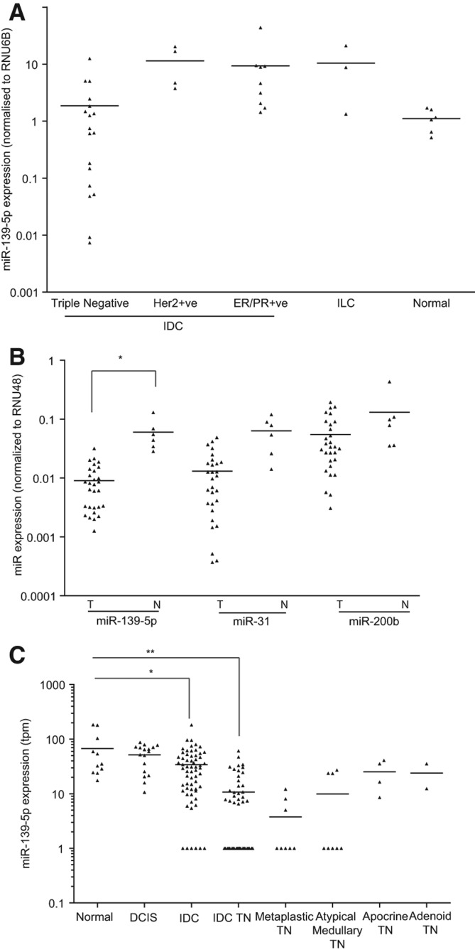
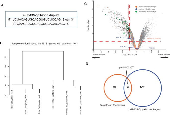
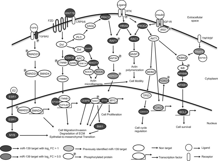
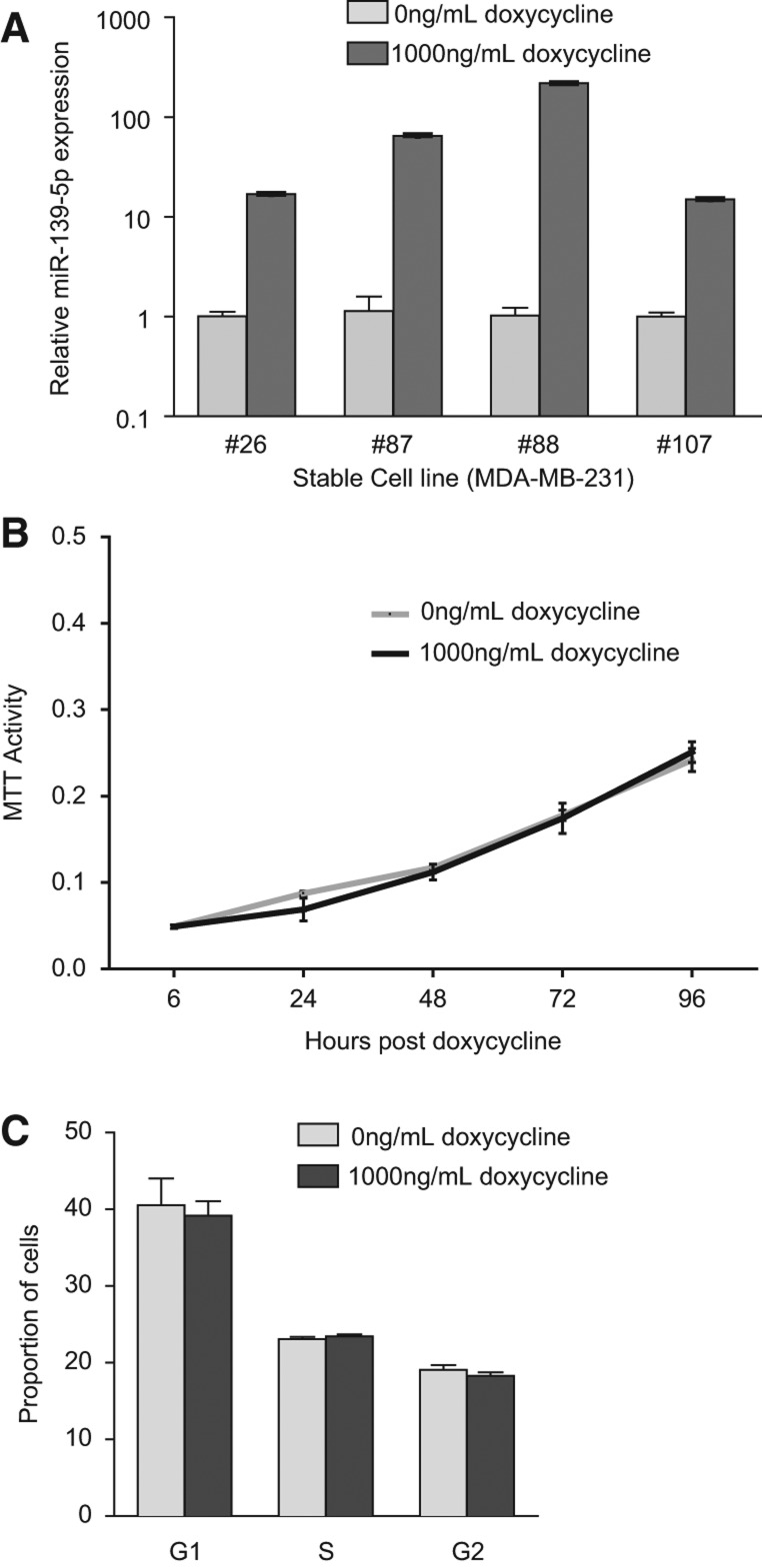
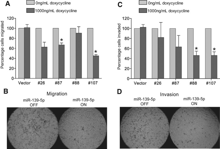
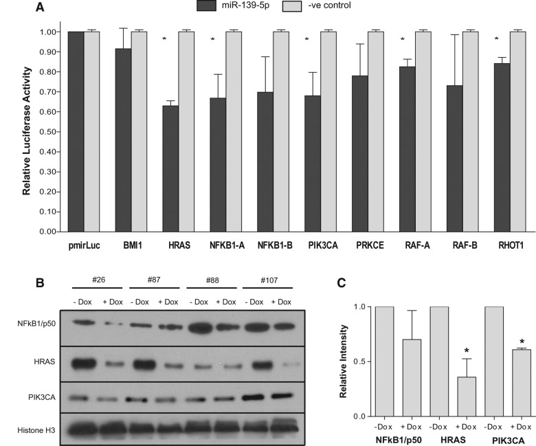
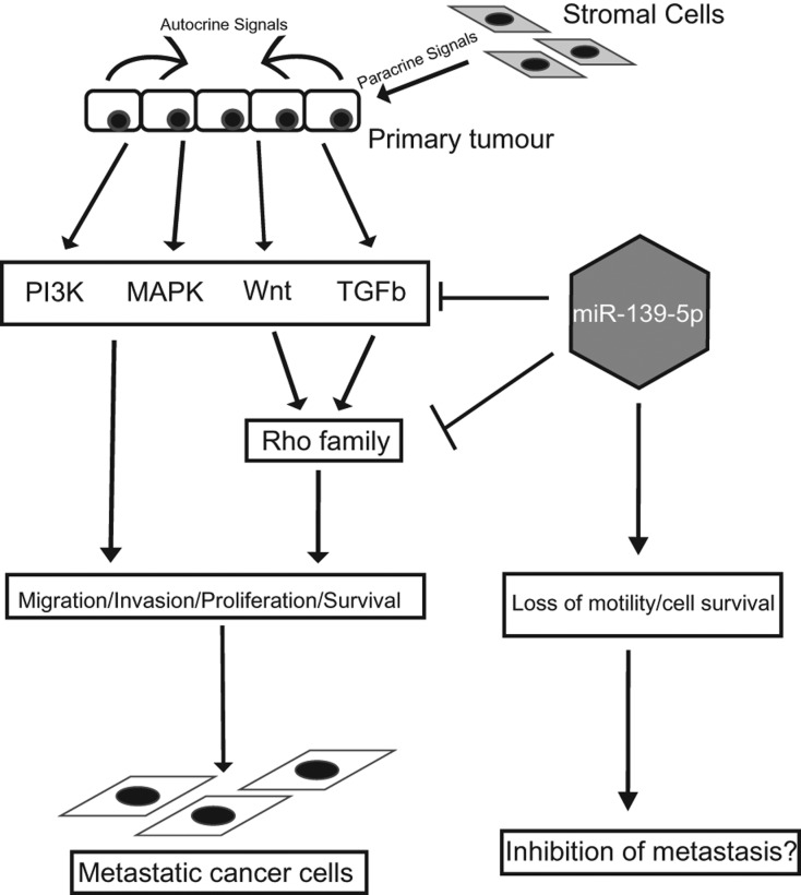
Metastasis is a complex, multistep process involved in the progression of cancer from a localized primary tissue to distant sites, often characteristic of the more aggressive forms of this disease. Despite being studied in great detail in recent years, the mechanisms that govern this process remain poorly understood. In this study, we identify a novel role for miR-139-5p in the inhibition of breast cancer progression. We highlight its clinical relevance by reviewing miR-139-5p expression across a wide variety of breast cancer subtypes using in-house generated and online data sets to show that it is most frequently lost in invasive tumors. A biotin pull-down approach was then used to identify the mRNA targets of miR-139-5p in the breast cancer cell line MCF7. Functional enrichment analysis of the pulled-down targets showed significant enrichment of genes in pathways previously implicated in breast cancer metastasis (P< 0.05). Further bioinformatic analysis revealed a predicted disruption to the TGFβ, Wnt, Rho, and MAPK/PI3K signaling cascades, implying a potential role for miR-139-5p in regulating the ability of cells to invade and migrate. To corroborate this finding, using the MDA-MB-231 breast cancer cell line, we show that overexpression of miR-139-5p results in suppression of these cellular phenotypes. Furthermore, we validate the interaction between miR-139-5p and predicted targets involved in these pathways. Collectively, these results suggest a significant functional role for miR-139-5p in breast cancer cell motility and invasion and its potential to be used as a prognostic marker for the aggressive forms of breast cancer.
Overview
The claims and statistics of this paper haven’t been checked yet.
Run the check — free, no account needed. It reads the open-access text and checks the sources it cites.
Integrity signals
Registry
Cites
Statistics
Figures
Verification
Claims the paper makes
The claims and statistics of this paper haven’t been checked yet.
Run the check — free, no account needed. It reads the open-access text and checks the sources it cites.
Claims other papers make about this one
Each is checked against this paper’s own text: does it say what the citing paper says it does?
In the citing paper
“miR-139-5p has been known to be a tumor suppressor miRNA by inhibiting the metastasis pathway [46]”
From Bioinformatic Interrogation of 5p-arm and 3p-arm Specific miRNA Expression Using TCGA Datasets (2015) · cites this paper as [46]
Self-check
Percentages, sums and reported differences re-computed from the paper’s own numbers.
The self-check needs the paper’s full text.
Citations
Cites
Subramanian, A., Tamayo, P., Mootha, V. K., Mukherjee, S., Ebert, B. L., Gillette, M. A., Paulovich, A., Pomeroy, S. L., Golub, T. R., Lander, E. S., et al. (2005) · Proceedings of the National Academy of Sciences · doi:10.1073/pnas.0506580102 · Cited by 53,625
Smyth, G. K. (2004) · Statistical Applications in Genetics and Molecular Biology · doi:10.2202/1544-6115.1027 · Cited by 11,829
Wnt/β-catenin signaling and disease
Clevers, H., & Nusse, R. (2012) · Cell · doi:10.1016/j.cell.2012.05.012 · Cited by 5,554
The pathogenesis of cancer metastasis: the ‘seed and soil’ hypothesis revisited
Fidler, I. J. (2003) · Nature Reviews Cancer · doi:10.1038/nrc1098 · Cited by 4,686
Regulation of mRNA Translation and Stability by microRNAs
Fabian, M. R., Sonenberg, N., & Filipowicz, W. (2010) · Annual Review of Biochemistry · doi:10.1146/annurev-biochem-060308-103103 · Cited by 3,214
Tumour invasion and metastasis initiated by microRNA-10b in breast cancer
Ma, L., Teruya-Feldstein, J., & Weinberg, R. A. (2007) · Nature · doi:10.1038/nature06174 · Cited by 2,605
Gene silencing by microRNAs: contributions of translational repression and mRNA decay
Huntzinger, E., & Izaurralde, E. (2011) · Nature Reviews Genetics · doi:10.1038/nrg2936 · Cited by 2,339
Asangani, I. A., Rasheed, S. A. K., Nikolova, D. A., Leupold, J. H., Colburn, N. H., Post, S., & Allgayer, H. (2008) · Oncogene · doi:10.1038/sj.onc.1210856 · Cited by 1,882
MicroRNAs and their isomiRs function cooperatively to target common biological pathways
Cloonan, N., Wani, S., Xu, Q., Gu, J., Lea, K., Heater, S., Barbacioru, C., Steptoe, A. L., Martin, H. C., Nourbakhsh, E., et al. (2011) · Genome Biology · doi:10.1186/gb-2011-12-12-r126 · Cited by 345
Self-citationThe miR-17-5p microRNA is a key regulator of the G1/S phase cell cycle transition
Cloonan, N., Brown, M. K., Steptoe, A. L., Wani, S., Chan, W. L., Forrest, A. R., Kolle, G., Gabrielli, B., & Grimmond, S. M. (2008) · Genome Biology · doi:10.1186/gb-2008-9-8-r127 · Cited by 304
Self-citation
Research works printed but not linked: 76
- 10.1158/0008-5472.CAN-07-1183 · DOI
- 2005, Altered expression of the hormone- and xenobiotic-metabolizing sulfotransferase enzymes 1A2 and 1C1 in malignant breast tissue, Int J Oncol, 26, 1079
- 10.1053/j.gastro.2011.08.050 · DOI
- 2006, PI3K and RAC signalling in leukocyte and cancer cell migration, Bull Cancer, 93, E44
- 1995, Controlling the false discovery rate: A practical and powerful approach to multiple testing, J R Stat Soc Series B (Methodological), 57, 289, 10.1111/j.2517-6161.1995.tb02031.x · DOI · Paper page
- 10.1016/j.febslet.2005.09.040 · DOI
- 10.1038/nature03096 · DOI
- 10.1186/bcr2831 · DOI
- 10.1158/1078-0432.CCR-05-0885 · DOI
- 10.1196/annals.1386.024 · DOI
- 10.1038/sj.onc.1206216 · DOI
- 2009, Locally recurrent or metastatic breast cancer: ESMO clinical recommendations for diagnosis, treatment and follow-up, Ann Oncol, 20, 15, 10.1093/annonc/mdp115 · DOI
- 10.1016/S0960-9822(00)00475-9 · DOI
- 10.1158/1078-0432.CCR-07-1658 · DOI
- 2011, Defining MAP3 kinases required for MDA-MB-231 cell tumor growth and metastasis, Oncogene, 31, 3889
- 10.1093/bioinformatics/btn224 · DOI
- 10.1371/journal.pone.0015940 · DOI
- 2012, Derepression of c-Fos caused by microRNA-139 down-regulation contributes to the metastasis of human hepatocellular carcinoma, Cell Biochem Funct, 31, 319
- 10.1158/0008-5472.CAN-11-0608 · DOI
- 10.1101/gr.130435.111 · DOI
- 10.1038/ncb1722 · DOI
- 10.1016/j.biocel.2012.05.015 · DOI
- 10.1016/j.bbrc.2009.10.135 · DOI
- 10.1371/journal.pone.0051056 · DOI
- 10.1158/0008-5472.CAN-10-2914 · DOI
- 10.1242/jcs.01481 · DOI
- 10.1371/journal.pone.0033421 · DOI
- 10.1091/mbc.E07-02-0182 · DOI
- 10.1016/j.bbrc.2011.10.154 · DOI
- 10.1074/jbc.C800074200 · DOI
- 10.1038/nm.2401 · DOI
- 10.1261/rna.034926.112 · DOI
- 10.1093/glycob/cwn071 · DOI · Paper page
- 10.3390/ijms140611560 · DOI
- 10.1111/cns.12089 · DOI
- 10.1093/nar/gkm1075 · DOI
- 10.1002/path.4000 · DOI
- 10.1038/ncomms2276 · DOI
- 1996, Association of tumor cell proliferation with lymph node metastasis in early gastric cancer, Oncology, 53, 1, 10.1159/000227525 · DOI
- 10.1186/1476-4598-10-118 · DOI
- 10.1136/jcp.2009.073908 · DOI
- 10.1093/carcin/bgl129 · DOI
- 10.1183/09031936.00084108 · DOI
- 10.1074/jbc.M105316200 · DOI
- 10.1200/JCO.2011.38.2010 · DOI
- 2008, The glucocorticoid receptor signalling in breast cancer, J Cell Mol Med, 12, 145, 10.1111/j.1582-4934.2007.00177.x · DOI
- 10.1007/s12013-011-9245-x · DOI
- 10.1016/j.cell.2008.01.046 · DOI
- 10.1177/106689690000800208 · DOI
- 10.3349/ymj.2010.51.6.864 · DOI
- 10.1016/j.ejca.2010.02.036 · DOI
- 10.1038/sj.bjc.6606062 · DOI
- 10.1002/1097-0142(197710)40:4<1365::AID-CNCR2820400402>3.0.CO;2-N · DOI
- 10.1038/43474 · DOI · Paper page
- 10.1371/journal.pone.0031904 · DOI
- 10.1016/j.cell.2011.04.029 · DOI
- 10.1038/onc.2011.453 · DOI
- 10.1186/bcr1769 · DOI
- 10.1101/gad.1760809 · DOI
- 10.1242/jcs.122895 · DOI
- 10.1016/S0960-9822(99)80310-8 · DOI
- 10.1016/j.bcp.2012.04.017 · DOI
- 10.1371/journal.pone.0017429 · DOI
- 10.1038/onc.2011.275 · DOI
- 10.1073/pnas.0511045103 · DOI · Paper page
- 10.1016/j.bbrc.2010.04.127 · DOI
- 10.1074/jbc.270.30.17656 · DOI
- 10.1016/j.cell.2009.03.047 · DOI
- 10.1016/j.febslet.2008.04.039 · DOI
- 10.1002/iub.1051 · DOI
- 10.1053/j.gastro.2010.10.006 · DOI
- 10.1186/gb-2002-3-9-research0048 · DOI
- 10.1158/2159-8290.CD-11-0270 · DOI
- 10.1093/annonc/mdp245 · DOI
- 10.1038/sj.onc.1208927 · DOI
- 10.1038/cr.2008.24 · DOI · Paper page
Cited by
Sorted by citations
Bioinformatic Interrogation of 5p-arm and 3p-arm Specific miRNA Expression Using TCGA Datasets
Kuo, W.-T., Su, M.-W., Lee, Y., Chen, C.-H., Wu, C.-W., Fang, W.-L., Huang, K.-H., & Lin, W.-C. (2015) · Journal of Clinical Medicine · doi:10.3390/jcm4091798 · Cited by 30
Metadata
- Authors
- Krishnan, K., Steptoe, A. L., Martin, H. C., Pattabiraman, D. R., Nones, K., Waddell, N., Mariasegaram, M., Simpson, P. T., Lakhani, S. R., Vlassov, A., Grimmond, S. M., & Cloonan, N.
- Venue
- RNA
- Publisher
- Cold Spring Harbor Laboratory
- Published
- 2013-10-24
- Volume
- 19
- Issue
- 12
- Pages
- 1767-1780
- ISSN
- 1355-8382
- 1469-9001
- PMC
- PMC3884652
- Cited by
- 134
- References
- 10
- Language
- en
About these checks
Registry, retraction, Cites and statistics results are computed, not judged, with one exception: a printed reference that no index lists is read once by a model before it counts as a citation error. Claims are extracted by a model and kept only when their quote appears verbatim in the text; each cited source is then checked, with a model’s help, for whether it says what the claim relies on. Those verdicts can be wrong — read the claim’s quote and the cited source before relying on one.
Statistics and arithmetic are recomputed, not judged.
Confidence bands are shown only for fields calibrated on our annotated test set. None is calibrated yet, so no band is shown.
What counts as a claim ›How we measure confidence ›How we check this ›
Are you an author, or do you see a problem with this page? Request a review
