

The DOI matches its Crossref record; 65 of the 65 references it cites were found in a registry or an index.
A Becn1 mutation mediates hyperactive autophagic sequestration of amyloid oligomers and improved cognition in Alzheimer's disease
Open accessAbstract
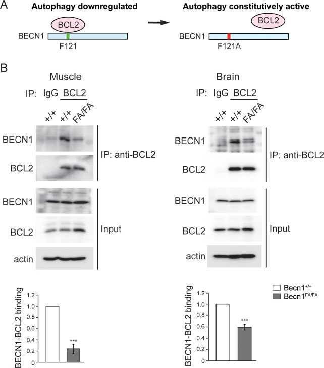
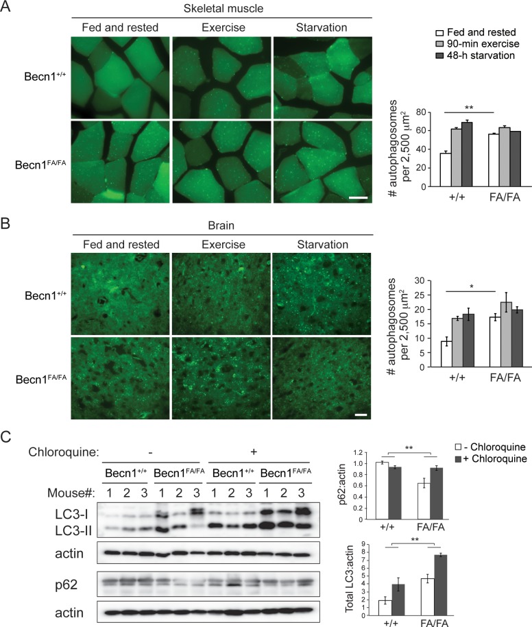
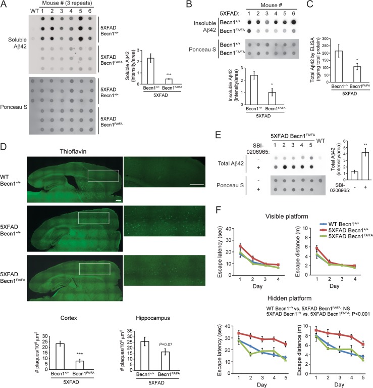
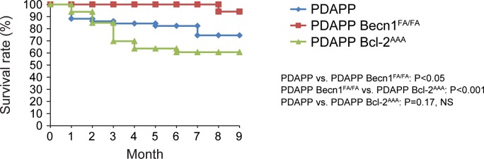
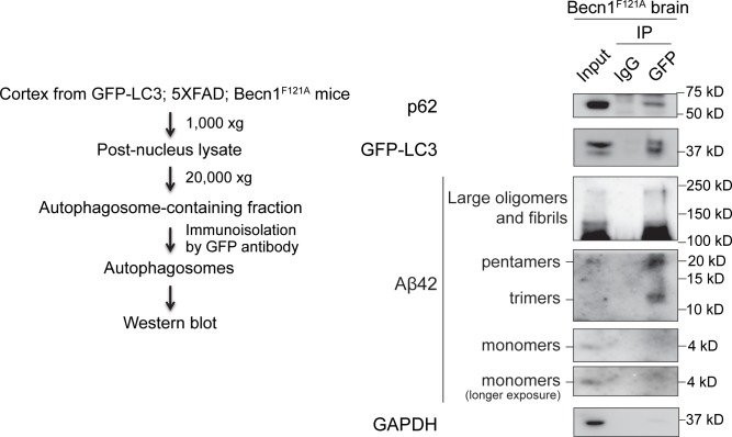
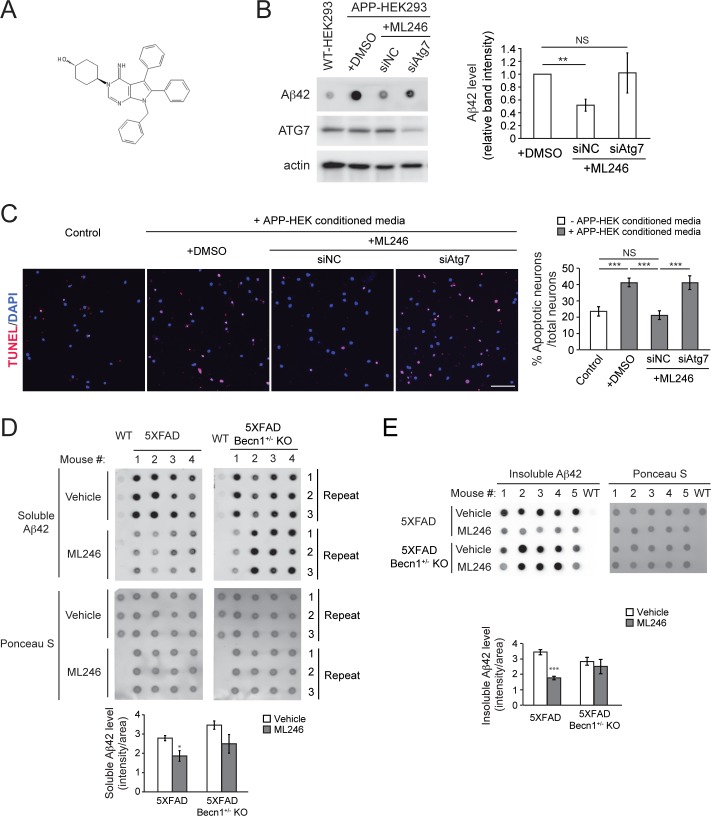
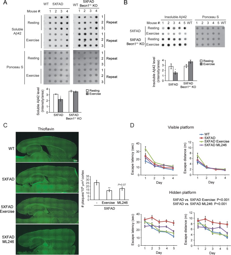
Impairment of the autophagy pathway has been observed during the pathogenesis of Alzheimer's disease (AD), a neurodegenerative disorder characterized by abnormal deposition of extracellular and intracellular amyloid β (Aβ) peptides. Yet the role of autophagy in Aβ production and AD progression is complex. To study whether increased basal autophagy plays a beneficial role in Aβ clearance and cognitive improvement, we developed a novel genetic model to hyperactivate autophagy in vivo. We found that knock-in of a point mutation F121A in the essential autophagy gene Beclin 1/Becn1 in mice significantly reduces the interaction of BECN1 with its inhibitor BCL2, and thus leads to constitutively active autophagy even under non-autophagy-inducing conditions in multiple tissues, including brain. Becn1F121A-mediated autophagy hyperactivation significantly decreases amyloid accumulation, prevents cognitive decline, and restores survival in AD mouse models. Using an immunoisolation method, we found biochemically that Aβ oligomers are autophagic substrates and sequestered inside autophagosomes in the brain of autophagy-hyperactive AD mice. In addition to genetic activation of autophagy by Becn1 gain-of-function, we also found that ML246, a small-molecule autophagy inducer, as well as voluntary exercise, a physiological autophagy inducer, exert similar Becn1-dependent protective effects on Aβ removal and memory in AD mice. Taken together, these results demonstrate that genetically disrupting BECN1-BCL2 binding hyperactivates autophagy in vivo, which sequestrates amyloid oligomers and prevents AD progression. The study establishes new approaches to activate autophagy in the brain, and reveals the important function of Becn1-mediated autophagy hyperactivation in the prevention of AD.
Overview
The claims and statistics of this paper haven’t been checked yet.
Run the check — free, no account needed. It reads the open-access text and checks the sources it cites.
Integrity signals
Registry
Cites
Statistics
Figures
Verification
Claims the paper makes
The claims and statistics of this paper haven’t been checked yet.
Run the check — free, no account needed. It reads the open-access text and checks the sources it cites.
Claims other papers make about this one
Each is checked against this paper’s own text: does it say what the citing paper says it does?
In the citing paper
“Evidence indicates that Aβ peptides can accumulate intracellularly while their extracellular release decreases when autophagy is impaired [85, 93].”
From Senescence-related impairment of autophagy induces toxic intraneuronal amyloid-β accumulation in a mouse model of amyloid pathology (2023) · cites this paper as [93]
Self-check
Percentages, sums and reported differences re-computed from the paper’s own numbers.
The self-check needs the paper’s full text.
Citations
Cites
Repertoires of Autophagy in the Pathogenesis of Ocular Diseases
Li, Y., Jiang, Q. L., Cao, G.-F., Yao, J., & Yan, B. (2015) · Cellular Physiology and Biochemistry · doi:10.1159/000373980 · Cited by 16,351
Genome engineering using the CRISPR-Cas9 system
Ran, F. A., Hsu, P. D., Wright, J., Agarwala, V., Scott, D. A., & Zhang, F. (2013) · Nature Protocols · doi:10.1038/nprot.2013.143 · Cited by 11,133
Sperling, R. A., Aisen, P. S., Beckett, L. A., Bennett, D. A., Craft, S., Fagan, A. M., Iwatsubo, T., Jack, C. R., Kaye, J., Montine, T. J., et al. (2011) · Alzheimer's & Dementia · doi:10.1016/j.jalz.2011.03.003 · Cited by 7,155
Autophagy: Renovation of Cells and Tissues
Mizushima, N., & Komatsu, M. (2011) · Cell · doi:10.1016/j.cell.2011.10.026 · Cited by 6,615
Cui, C., Cui, J., Jin, F., Cui, Y., Li, R., Jiang, X., Tian, Y., Wang, R. C., Jiang, P., & Gao, J. (2017) · Cellular Physiology and Biochemistry · doi:10.1159/000479571 · Cited by 5,214
Exercise training increases size of hippocampus and improves memory
Erickson, K. I., Voss, M. W., Prakash, R. S., Basak, C., Szabo, A., Chaddock, L., Kim, J. S., Heo, S., Alves, H., White, S. M., et al. (2011) · Proceedings of the National Academy of Sciences · doi:10.1073/pnas.1015950108 · Cited by 4,855
Suppression of basal autophagy in neural cells causes neurodegenerative disease in mice
Hara, T., Nakamura, K., Matsui, M., Yamamoto, A., Nakahara, Y., Suzuki-Migishima, R., Yokoyama, M., Mishima, K., Saito, I., Okano, H., et al. (2006) · Nature · doi:10.1038/nature04724 · Cited by 3,882
Levine, B., & Klionsky, D. J. (2004) · Developmental Cell · doi:10.1016/s1534-5807(04)00099-1 · Cited by 3,741
Self-citationBcl-2 Antiapoptotic Proteins Inhibit Beclin 1-Dependent Autophagy
Pattingre, S., Tassa, A., Qu, X., Garuti, R., Liang, X. H., Mizushima, N., Packer, M., Schneider, M. D., & Levine, B. (2005) · Cell · doi:10.1016/j.cell.2005.07.002 · Cited by 3,536
Self-citationOakley, H., Cole, S. L., Logan, S., Maus, E., Shao, P., Craft, J., Guillozet-Bongaarts, A., Ohno, M., Disterhoft, J., Van Eldik, L., et al. (2006) · The Journal of Neuroscience · doi:10.1523/jneurosci.1202-06.2006 · Cited by 3,530
Self-citation
Cited by
Sorted by citations
Biological Functions of Autophagy Genes: A Disease Perspective
Levine, B., & Kroemer, G. (2019) · Cell · doi:10.1016/j.cell.2018.09.048 · Cited by 2,832
Self-citationAutophagy in healthy aging and disease
Aman, Y., Schmauck-Medina, T., Hansen, M., Morimoto, R. I., Simon, A. K., Bjedov, I., Palikaras, K., Simonsen, A., Johansen, T., Tavernarakis, N., et al. (2021) · Nature Aging · doi:10.1038/s43587-021-00098-4 · Cited by 1,313
The Amyloid-β Oligomer Hypothesis: Beginning of the Third Decade
Cline, E. N., Bicca, M. A., Viola, K. L., & Klein, W. L. (2018) · Journal of Alzheimer's Disease · doi:10.3233/jad-179941 · Cited by 870
Disruption of the beclin 1–BCL2 autophagy regulatory complex promotes longevity in mice
Fernández, Á. F., Sebti, S., Wei, Y., Zou, Z., Shi, M., McMillan, K. L., He, C., Ting, T., Liu, Y., Chiang, W.-C., et al. (2018) · Nature · doi:10.1038/s41586-018-0162-7 · Cited by 630
Self-citationPromoting the clearance of neurotoxic proteins in neurodegenerative disorders of ageing
Boland, B., Yu, W. H., Corti, O., Mollereau, B., Henriques, A., Bézard, E., Pastores, G., Rubinsztein, D. C., Nixon, R. A., Duchen, M. R., et al. (2018) · Nature Reviews Drug Discovery · doi:10.1038/nrd.2018.109 · Cited by 566
Self-citation
Metadata
- Authors
- Rocchi, A., Yamamoto, S., Ting, T., Fan, Y., Sadleir, K., Wang, Y., Zhang, W., Huang, S., Levine, B., Vassar, R., & He, C.
- Affiliations
- Northwestern University (US)
- Howard Hughes Medical Institute (US)
- Corresponding author
- He, Congcong
- ORCID
- Venue
- PLOS Genetics
- Publisher
- Public Library of Science (PLoS)
- Published
- 2017-08-14
- Volume
- 13
- Issue
- 8
- Pages
- e1006962
- ISSN
- 1553-7390 (ISSN-L)
- 1553-7404
- PMC
- PMC5570506
- OpenAlex
- W2749882665
- Cited by
- 166
- References
- 65
- Citations by year
- 2026: 11 · 2025: 24 · 2024: 12 · 2023: 21 · 2022: 24 · 2021: 29 · 2020: 19 · 2019: 10 · 2018: 16
- FWCI
- 9.10
- Type
- journal article
- Language
- en
- Update notices
- new_version (2017-08-24)
- Open access
- gold
- doi.org
- Licence
- Locations
- PLoS Genetics (publishedVersion, cc-by)
- Europe PMC (PubMed Central) (publishedVersion, cc-by)
- PubMed Central (publishedVersion, cc-by)
- PubMed (publishedVersion)
- DOAJ (DOAJ: Directory of Open Access Journals) (submittedVersion, cc-by-sa)
- Figshare (submittedVersion, cc-by)
- Funders
- Cancer Prevention and Research Institute of Texas (RP120718)
- Northwestern University (AG13854)
- Northeast Normal University
- National Institutes of Health (U19AI199725, 5r01gm078555-08, AG13854, RO1 CA109618)
- University of North Carolina at Chapel Hill
- Fundamental Research Funds for the Central Universities (2412017FZ018)
- National Institute on Aging (AG13854, R01AG030142, R01AG022560, P30AG013854)
- National Cancer Institute (R01CA109618)
- National Institute of General Medical Sciences (R01 GM-078555)
- National Institute of Allergy and Infectious Diseases (U19AI109725, T32AI007163)
- Topics
- Life Sciences › Neuroscience › Cellular and Molecular Neuroscience › Alzheimer's disease research and treatments
- Life Sciences › Biochemistry, Genetics and Molecular Biology › Cell Biology › Autophagy in Disease and Therapy
- Life Sciences › Neuroscience › Neurology › Neuroinflammation and Neurodegeneration Mechanisms
- Keywords
- Alzheimer's disease, autophagy, Beclin-1, Bcl-2, amyloid-beta oligomers, autophagosomes, cognitive decline, voluntary exercise, autophagy inducers, genetic mouse models
- MeSH terms
- Beclin-1, Alzheimer Disease, Animals, Autophagy, Cognition, Disease Models, Animal, HeLa Cells, Humans, Male, Memory, Mice, Inbred C57BL, Neurons, Amyloid beta-Peptides, Point Mutation, Sequence Analysis, DNA, Proto-Oncogene Proteins c-bcl-2, In Situ Nick-End Labeling, Mice, Gene Knock-In Techniques, HEK293 Cells
About these checks
Registry, retraction, Cites and statistics results are computed, not judged, with one exception: a printed reference that no index lists is read once by a model before it counts as a citation error. Claims are extracted by a model and kept only when their quote appears verbatim in the text; each cited source is then checked, with a model’s help, for whether it says what the claim relies on. Those verdicts can be wrong — read the claim’s quote and the cited source before relying on one.
Statistics and arithmetic are recomputed, not judged.
Confidence bands are shown only for fields calibrated on our annotated test set. None is calibrated yet, so no band is shown.
What counts as a claim ›How we measure confidence ›How we check this ›
Are you an author, or do you see a problem with this page? Request a review
