

The DOI matches its Crossref record; 80 of the 81 references it cites were found in a registry or an index; none of the 59 sources its claims cite is checked for support yet.
What Are You Feeling? Using Functional Magnetic Resonance Imaging to Assess the Modulation of Sensory and Affective Responses during Empathy for Pain
Open accessAbstract
BACKGROUND: Recent neuroscientific evidence suggests that empathy for pain activates similar neural representations as the first-hand experience of pain. However, empathy is not an all-or-none phenomenon but it is strongly malleable by interpersonal, intrapersonal and situational factors. This study investigated how two different top-down mechanisms - attention and cognitive appraisal - affect the perception of pain in others and its neural underpinnings. METHODOLOGY/PRINCIPAL FINDINGS: We performed one behavioral (N = 23) and two functional magnetic resonance imaging (fMRI) experiments (N = 18). In the first fMRI experiment, participants watched photographs displaying painful needle injections, and were asked to evaluate either the sensory or the affective consequences of these injections. The role of cognitive appraisal was examined in a second fMRI experiment in which participants watched injections that only appeared to be painful as they were performed on an anesthetized hand. Perceiving pain in others activated the affective-motivational and sensory-discriminative aspects of the pain matrix. Activity in the somatosensory areas was specifically enhanced when participants evaluated the sensory consequences of pain. Perceiving non-painful injections into the anesthetized hand also led to signal increase in large parts of the pain matrix, suggesting an automatic affective response to the putatively harmful stimulus. This automatic response was modulated by areas involved in self/other distinction and valence attribution - including the temporo-parietal junction and medial orbitofrontal cortex. CONCLUSIONS/SIGNIFICANCE: Our findings elucidate how top-down control mechanisms and automatic bottom-up processes interact to generate and modulate other-oriented responses. They stress the role of cognitive processing in empathy, and shed light on how emotional and bodily awareness enable us to evaluate the sensory and affective states of others.
Overview
AI snapshot
Written from the paper’s full text.
TL;DR
This paper used one behavioral and two fMRI experiments to test how attention (sensory vs. affective evaluation) and cognitive appraisal (numbed vs. non-numbed hand injections) modulate empathy for pain. It reports that perceiving pain in others activates large parts of the pain matrix, including somatosensory areas, and that top-down appraisal modulates regions involved in self/other distinction, valence attribution, and affective processing.
Key findings
- In the behavioral experiment (N = 23), painful needle-injection photographs produced higher pain intensity and unpleasantness ratings than non-painful control photographs: intensity/painful 64.84 ± 19.065 vs. intensity/non-painful 1.444 ± 2.221; unpleasantness/painful 69.033 ± 14.225 vs. unpleasantness/non-painful 9.164 ± 14.304; main effect stimulus F(1,22) = 510.641, P < 0.001, η² = 0.959.
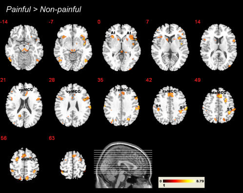
- In fMRI experiment I, painful > non-painful stimuli activated affective-motivational and sensory-discriminative pain-matrix regions, including dorsal/ventral aMCC, bilateral anterior insula, and somatosensory areas. The interaction Intensity > Unpleasantness showed strongest modulation in right postcentral gyrus, contralateral to the stimulated hand, plus inferior parietal/supramarginal and ventral premotor clusters; the reverse interaction yielded only a right lingual gyrus cluster at the main threshold, with subcallosal cingulate at a lower threshold.
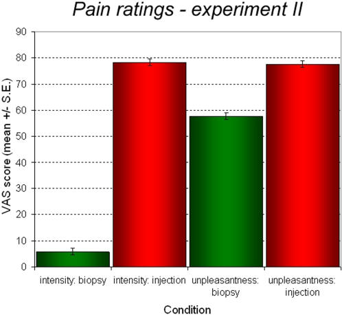
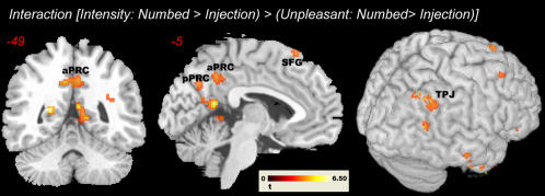
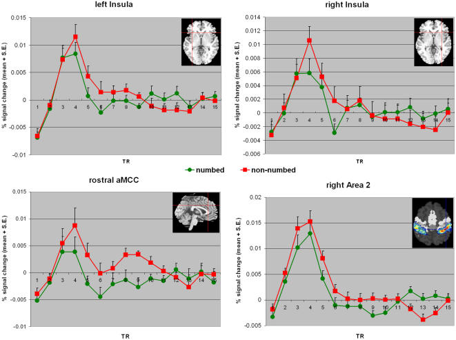
- In fMRI experiment II, numbed-hand injections were rated non-painful but unpleasant. The interaction Intensity (Numbed > Injection) > Unpleasantness yielded precuneus and bilateral temporo-parietal junction clusters; the reverse interaction yielded left anterior insula, cerebellum, OFC, and basal ganglia. ROI analyses showed a significant raMCC interaction (F(1,13) = 5.069, P = 0.042), a right anterior insula trend (F(1,16) = 3.45, P = 0.082), a significant right insula effect for intensity-only trials (F(1,16) = 6.34, P = 0.023), and a late raMCC response at TRs 9–11 (F(1,13) = 6.96, P = 0.02).
- Dispositional measures showed emotional contagion correlated with activation in bilateral anterior insula and aMCC, whereas IRI empathic concern correlated with premotor, somatosensory, precuneus, and fusiform regions but not insular or cingulate cortices.
Methods
- Design: one behavioral experiment for stimulus validation/design optimization (N = 23; 19 females; mean age 27.69 years) and two subsequent fMRI experiments (N = 18; 9 females; age 19–35, mean 23.67 years). fMRI experiment I manipulated evaluative focus (pain intensity vs. unpleasantness) and stimulus type (painful vs. non-painful); fMRI experiment II manipulated cognitive appraisal (numbed vs. non-numbed hand injections) and rating type (intensity vs. unpleasantness).
- Data/measures: participants viewed photographs of needle injections and rated pain intensity or unpleasantness on a VAS; they also completed the Interpersonal Reactivity Index, Emotional Contagion Scale, and Sensitivity to Pain Questionnaire.
- Analysis: MRI at 3T with EPI; preprocessing and GLM analysis in SPM2; second-level random-effects analyses. Physical stimulus contrasts used voxel threshold P = 0.01, k = 10, FDR-corrected; subtler psychological contrasts used P = 0.001 uncorrected, k = 5, with lower thresholds for a priori regions. ROI analyses used MarsBaR with a priori functional ROIs.
Limitations
- Stated: the low temporal resolution of hemodynamic responses may not precisely reveal when and how top-down modulation affects pain-matrix activity; the authors are replicating the paradigm with event-related potentials.
- Stated: the second raMCC peak could not be explained by subsequent trials because interstimulus intervals and stimulus order were randomized and counterbalanced, but future studies with longer interstimulus intervals are needed to exclude this confound; one participant in experiment II was excluded due to excessive movement.
- Evident from the text: the fMRI samples were small (N = 18, with one excluded from experiment II), and several subtle psychological contrasts relied on liberal uncorrected thresholds; the authors also note that future studies might use a separate localizer task for first-hand somatosensory representations.
Model-written summary. Check it against the paper before relying on it.
Integrity signals
Registry
Cites
Statistics
Figures
Verification
Claims the paper makes
85 Own result · 39 Not checked yet · 28 Own interpretation · 16 Own method
A result the paper reports from its own data — it cites no other source for it, so there is none to check. The quote shows it is in the paper.
In the paper
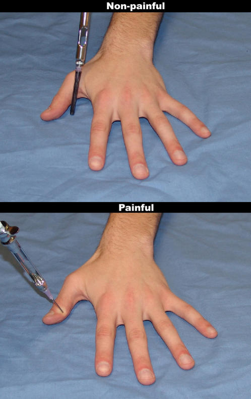
“Photographs depicting needle injections led to higher pain intensity and pain unpleasantness ratings than the photographs in which the needle was covered by the black protector cap (main effect stimulus (painful vs. non-painful), F(1,22) = 510.641, P<0.001, η2 = 0.959).”
§ What Are You Feeling? Using Functional Magnetic Resonance Imaging to Assess the Modulation of Sensory and Affective Responses during Empathy for Pain > Results > Behavioral experiment
plaindirect- Asserted: passes — The authors put it forward as true.
- Checkable: passes — It can be checked against evidence.
- Load-bearing: passes — The paper’s conclusions depend on it.
- Based: passes — The paper gives a reason why it holds.
A result the paper reports from its own data — it cites no other source for it, so there is none to check. The quote shows it is in the paper.
In the paper
“In addition, P(A) showed a significant correlation with the response bias value B of the SPQ (r = 0.648, P = 0.004).”
§ What Are You Feeling? Using Functional Magnetic Resonance Imaging to Assess the Modulation of Sensory and Affective Responses during Empathy for Pain > Results > Functional MRI experiments
plaincorrelational- Asserted: passes — The authors put it forward as true.
- Checkable: passes — It can be checked against evidence.
- Load-bearing: unclear — It is unclear whether the paper’s conclusions depend on it.
- Based: passes — The paper gives a reason why it holds.
A result the paper reports from its own data — it cites no other source for it, so there is none to check. The quote shows it is in the paper.
In the paper
“Activity in the somatosensory areas was specifically enhanced when participants evaluated the sensory consequences of pain.”
§ What Are You Feeling? Using Functional Magnetic Resonance Imaging to Assess the Modulation of Sensory and Affective Responses during Empathy for Pain > Abstract > Methodology/Principal Findings
plaindirectn = 18- Asserted: passes — The authors put it forward as true.
- Checkable: passes — It can be checked against evidence.
- Load-bearing: passes — The paper’s conclusions depend on it.
- Based: passes — The paper gives a reason why it holds.
A result the paper reports from its own data — it cites no other source for it, so there is none to check. The quote shows it is in the paper.
In the paper
“Activation clusters were detected in areas coding the affective, the sensory and the motor aspects associated with nociception (Figure 4).”
§ What Are You Feeling? Using Functional Magnetic Resonance Imaging to Assess the Modulation of Sensory and Affective Responses during Empathy for Pain > Results > fMRI experiment I – effects of evaluative focus
plaincorrelational- Asserted: passes — The authors put it forward as true.
- Checkable: passes — It can be checked against evidence.
- Load-bearing: passes — The paper’s conclusions depend on it.
- Based: passes — The paper gives a reason why it holds.
A result the paper reports from its own data — it cites no other source for it, so there is none to check. The quote shows it is in the paper.
In the paper
“This was the case for both intensity and unpleasantness ratings (mean±S.D. for intensity/painful stimulus: 69.789±14.654; intensity/non-painful stimulus: 3.548±9.68; unpleasantness/painful: 71.237±13.65; unpleasantness/non-painful: 2.054±4.005).”
§ What Are You Feeling? Using Functional Magnetic Resonance Imaging to Assess the Modulation of Sensory and Affective Responses during Empathy for Pain > Results > Functional MRI experiments
plaincorrelational- Asserted: passes — The authors put it forward as true.
- Checkable: passes — It can be checked against evidence.
- Load-bearing: unclear — It is unclear whether the paper’s conclusions depend on it.
- Based: passes — The paper gives a reason why it holds.
A result the paper reports from its own data — it cites no other source for it, so there is none to check. The quote shows it is in the paper.
In the paper
“Similar to the behavioral experiment, photographs depicting injections led to significantly higher rating scores than images of the needle with the protector cap (main effect stimulus, F(1,17) = 348.815, P<0.001, η2 = 0.954).”
§ What Are You Feeling? Using Functional Magnetic Resonance Imaging to Assess the Modulation of Sensory and Affective Responses during Empathy for Pain > Results > Functional MRI experiments
plaindirect- Asserted: passes — The authors put it forward as true.
- Checkable: passes — It can be checked against evidence.
- Load-bearing: passes — The paper’s conclusions depend on it.
- Based: passes — The paper gives a reason why it holds.
A result the paper reports from its own data — it cites no other source for it, so there is none to check. The quote shows it is in the paper.
In the paper
“When the non-painful stimuli were excluded from this calculation, the correlation remained basically unchanged (r = 0.797) - indicating that the two stimulus dimensions have similar correlation for both painful and non-painful stimuli.”
§ What Are You Feeling? Using Functional Magnetic Resonance Imaging to Assess the Modulation of Sensory and Affective Responses during Empathy for Pain > Results > Behavioral experiment
hedgedcorrelational- Asserted: passes — The authors put it forward as true.
- Checkable: passes — It can be checked against evidence.
- Load-bearing: unclear — It is unclear whether the paper’s conclusions depend on it.
- Based: passes — The paper gives a reason why it holds.
A result the paper reports from its own data — it cites no other source for it, so there is none to check. The quote shows it is in the paper.
In the paper
“Perceiving pain in others activated the affective-motivational and sensory-discriminative aspects of the pain matrix.”
§ What Are You Feeling? Using Functional Magnetic Resonance Imaging to Assess the Modulation of Sensory and Affective Responses during Empathy for Pain > Abstract > Methodology/Principal Findings
plaindirectn = 18- Asserted: passes — The authors put it forward as true.
- Checkable: passes — It can be checked against evidence.
- Load-bearing: passes — The paper’s conclusions depend on it.
- Based: passes — The paper gives a reason why it holds.
None of the sources cited for this claim have been checked yet.
In the paper
“This contrast indicated the involvement of large portions of the pain matrix [29], [30].”
§ What Are You Feeling? Using Functional Magnetic Resonance Imaging to Assess the Modulation of Sensory and Affective Responses during Empathy for Pain > Results > fMRI experiment I – effects of evaluative focus
What the sources say
hedgedcorrelational- Asserted: passes — The authors put it forward as true.
- Checkable: passes — It can be checked against evidence.
- Load-bearing: passes — The paper’s conclusions depend on it.
- Based: passes — The paper gives a reason why it holds.
A result the paper reports from its own data — it cites no other source for it, so there is none to check. The quote shows it is in the paper.
In the paper
“This automatic response was modulated by areas involved in self/other distinction and valence attribution – including the temporo-parietal junction and medial orbitofrontal cortex.”
§ What Are You Feeling? Using Functional Magnetic Resonance Imaging to Assess the Modulation of Sensory and Affective Responses during Empathy for Pain > Abstract > Methodology/Principal Findings
plaindirectn = 18- Asserted: passes — The authors put it forward as true.
- Checkable: passes — It can be checked against evidence.
- Load-bearing: passes — The paper’s conclusions depend on it.
- Based: passes — The paper gives a reason why it holds.
- Probably not a substantial claim. 39
- It may state two separate claims as one. 29
None of the sources cited for this claim have been checked yet.
In the paper
“Data for the IRI are well within published norms (as reported in detail in [5]), while the sample mean for the ECS was slightly below the norm average. SPQ sample means are comparable to a study collecting data from 96 normal controls [28].”
§ What Are You Feeling? Using Functional Magnetic Resonance Imaging to Assess the Modulation of Sensory and Affective Responses during Empathy for Pain > Results > Functional MRI experiments
What the sources say
- Not checked yetcites [5] C Lamm, 2007, The neural substrate of human empathy: effects of perspective-taking and cognitive appraisal., J Cogn Neurosci source
- Not checked yetcites [28] N Danziger, 2006, Is pain the price of empathy? The perception of others' pain in patients with congenital insensitivity to pain., Brain source
plain- Asserted: passes — The authors put it forward as true.
- Checkable: passes — It can be checked against evidence.
- Load-bearing: fails — The paper’s conclusions may not depend on it.
- Based: passes — The paper gives a reason why it holds.
Probably not a substantial claim. The paper’s conclusions may not depend on it. It may state two separate claims as one.
A result the paper reports from its own data — it cites no other source for it, so there is none to check. The quote shows it is in the paper.
In the paper
“In addition, the mean intensity and unpleasantness ratings were significantly different (main effect for rating, F(1,22) = 13.389, P = 0.001, η2 = 0.378), while no significant interaction term was found (P = 0.413).”
§ What Are You Feeling? Using Functional Magnetic Resonance Imaging to Assess the Modulation of Sensory and Affective Responses during Empathy for Pain > Results > Behavioral experiment
plain- Asserted: passes — The authors put it forward as true.
- Checkable: passes — It can be checked against evidence.
- Load-bearing: unclear — It is unclear whether the paper’s conclusions depend on it.
- Based: passes — The paper gives a reason why it holds.
It may state two separate claims as one.
A result the paper reports from its own data — it cites no other source for it, so there is none to check. The quote shows it is in the paper.
In the paper
“The Pearson correlation between intensity and unpleasantness ratings was r = 0.769 (P<0.001), showing that the two types of rating share about 50% of their variance.”
§ What Are You Feeling? Using Functional Magnetic Resonance Imaging to Assess the Modulation of Sensory and Affective Responses during Empathy for Pain > Results > Behavioral experiment
plaincorrelational- Asserted: passes — The authors put it forward as true.
- Checkable: passes — It can be checked against evidence.
- Load-bearing: unclear — It is unclear whether the paper’s conclusions depend on it.
- Based: passes — The paper gives a reason why it holds.
It may state two separate claims as one.
A result the paper reports from its own data — it cites no other source for it, so there is none to check. The quote shows it is in the paper.
In the paper
“No significant change in scores across the two imaging runs was observed, indicating the absence of strong habituation.”
§ What Are You Feeling? Using Functional Magnetic Resonance Imaging to Assess the Modulation of Sensory and Affective Responses during Empathy for Pain > Results > Functional MRI experiments
hedged- Asserted: passes — The authors put it forward as true.
- Checkable: passes — It can be checked against evidence.
- Load-bearing: unclear — It is unclear whether the paper’s conclusions depend on it.
- Based: passes — The paper gives a reason why it holds.
It may state two separate claims as one.
A result the paper reports from its own data — it cites no other source for it, so there is none to check. The quote shows it is in the paper.
In the paper
“Correlation coefficients (Pearson) reveal that the ECS correlates significantly with the IRI Fantasy scale (r = 0.513, P = 0.029), the IRI Empathic Concern scale (r = 0.469, P = 0.049), and the IRI Personal Distress scale (r = 0.545, P = 0.019).”
§ What Are You Feeling? Using Functional Magnetic Resonance Imaging to Assess the Modulation of Sensory and Affective Responses during Empathy for Pain > Results > Functional MRI experiments
plaincorrelational- Asserted: passes — The authors put it forward as true.
- Checkable: passes — It can be checked against evidence.
- Load-bearing: unclear — It is unclear whether the paper’s conclusions depend on it.
- Based: passes — The paper gives a reason why it holds.
It may state two separate claims as one.
A result the paper reports from its own data — it cites no other source for it, so there is none to check. The quote shows it is in the paper.
In the paper
“The discrimination score (P(A)) of the SPQ was inversely related to the Personal Distress scale (r = −0.504, P = 0.033), and positively correlated with IRI Perspective Taking (r = 0.519, P = 0.027).”
§ What Are You Feeling? Using Functional Magnetic Resonance Imaging to Assess the Modulation of Sensory and Affective Responses during Empathy for Pain > Results > Functional MRI experiments
plaincorrelational- Asserted: passes — The authors put it forward as true.
- Checkable: passes — It can be checked against evidence.
- Load-bearing: unclear — It is unclear whether the paper’s conclusions depend on it.
- Based: passes — The paper gives a reason why it holds.
It may state two separate claims as one.
A result the paper reports from its own data — it cites no other source for it, so there is none to check. The quote shows it is in the paper.
In the paper
“B also significantly correlated with IRI's Personal Distress subscale (r = −0.605, P = 0.008), and a trend towards significance was observed for ECS (r = −0.454, P = 0.059).”
§ What Are You Feeling? Using Functional Magnetic Resonance Imaging to Assess the Modulation of Sensory and Affective Responses during Empathy for Pain > Results > Functional MRI experiments
plaincorrelational- Asserted: passes — The authors put it forward as true.
- Checkable: passes — It can be checked against evidence.
- Load-bearing: unclear — It is unclear whether the paper’s conclusions depend on it.
- Based: passes — The paper gives a reason why it holds.
It may state two separate claims as one.
A result the paper reports from its own data — it cites no other source for it, so there is none to check. The quote shows it is in the paper.
In the paper
“Again, scores did not significantly change over the course of the experiment.”
§ What Are You Feeling? Using Functional Magnetic Resonance Imaging to Assess the Modulation of Sensory and Affective Responses during Empathy for Pain > Results > Functional MRI experiments
plain- Asserted: passes — The authors put it forward as true.
- Checkable: passes — It can be checked against evidence.
- Load-bearing: fails — The paper’s conclusions may not depend on it.
- Based: passes — The paper gives a reason why it holds.
Probably not a substantial claim. The paper’s conclusions may not depend on it.
In the paper
“Pain intensity ratings were additionally associated with activation in contralateral precentral gyrus, and in dorsal posterior cingulate gyrus in a region involved in visuo-spatial attention [32].”
§ What Are You Feeling? Using Functional Magnetic Resonance Imaging to Assess the Modulation of Sensory and Affective Responses during Empathy for Pain > Results > fMRI experiment I – effects of evaluative focus
What the sources say
- Not checked yetcites [32] BA Vogt, 2005, Pain and emotion interactions in subregions of the cingulate gyrus., Nat Rev Neurosci source
plaincorrelational- Asserted: passes — The authors put it forward as true.
- Checkable: passes — It can be checked against evidence.
- Load-bearing: unclear — It is unclear whether the paper’s conclusions depend on it.
- Based: passes — The paper gives a reason why it holds.
It may state two separate claims as one.
A result the paper reports from its own data — it cites no other source for it, so there is none to check. The quote shows it is in the paper.
In the paper
“Activations were also found in the thalamus, in right medial frontal gyrus, and in the superior part of the periaqueductal grey.”
§ What Are You Feeling? Using Functional Magnetic Resonance Imaging to Assess the Modulation of Sensory and Affective Responses during Empathy for Pain > Results > fMRI experiment I – effects of evaluative focus
plain- Asserted: passes — The authors put it forward as true.
- Checkable: passes — It can be checked against evidence.
- Load-bearing: passes — The paper’s conclusions depend on it.
- Based: passes — The paper gives a reason why it holds.
It may state two separate claims as one.
A result the paper reports from its own data — it cites no other source for it, so there is none to check. The quote shows it is in the paper.
In the paper
“This comparison indicated excellent reproducibility of results, with experiment II yielding basically the same findings as experiment I for the painful injections.”
§ What Are You Feeling? Using Functional Magnetic Resonance Imaging to Assess the Modulation of Sensory and Affective Responses during Empathy for Pain > Results > fMRI experiment II – effects of cognitive appraisal
hedged- Asserted: passes — The authors put it forward as true.
- Checkable: passes — It can be checked against evidence.
- Load-bearing: unclear — It is unclear whether the paper’s conclusions depend on it.
- Based: passes — The paper gives a reason why it holds.
It may state two separate claims as one.
A result the paper reports from its own data — it cites no other source for it, so there is none to check. The quote shows it is in the paper.
In the paper
“Results differed, however, with respect to areas involved in self-awareness and mentalizing such as the posterior precuneus, temporo-parietal junction (TPJ) or medial prefrontal/paracingulate cortex, which – contrary to our expectations - did not correlate with the perspective taking scores (Table S4).”
§ What Are You Feeling? Using Functional Magnetic Resonance Imaging to Assess the Modulation of Sensory and Affective Responses during Empathy for Pain > Results > fMRI experiment II – effects of cognitive appraisal
plain- Asserted: passes — The authors put it forward as true.
- Checkable: passes — It can be checked against evidence.
- Load-bearing: unclear — It is unclear whether the paper’s conclusions depend on it.
- Based: passes — The paper gives a reason why it holds.
It may state two separate claims as one.
A result the paper reports from its own data — it cites no other source for it, so there is none to check. The quote shows it is in the paper.
In the paper
“While insular activation overlapped almost perfectly with the clusters detected by the contrast of painful with non-painful stimuli, activation in aMCC was considerably more rostral.”
§ What Are You Feeling? Using Functional Magnetic Resonance Imaging to Assess the Modulation of Sensory and Affective Responses during Empathy for Pain > Results > fMRI experiment I – effects of evaluative focus
plain- Asserted: passes — The authors put it forward as true.
- Checkable: passes — It can be checked against evidence.
- Load-bearing: unclear — It is unclear whether the paper’s conclusions depend on it.
- Based: passes — The paper gives a reason why it holds.
It may state two separate claims as one.
A result the paper reports from its own data — it cites no other source for it, so there is none to check. The quote shows it is in the paper.
In the paper
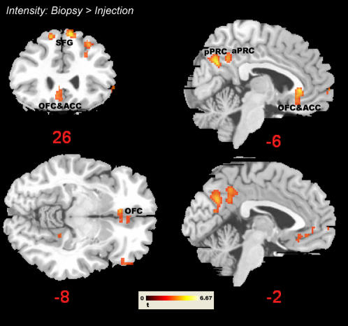
“In the frontal lobe, activation differed in medial and in superior frontal gyrus as well as in lateral OFC.”
§ What Are You Feeling? Using Functional Magnetic Resonance Imaging to Assess the Modulation of Sensory and Affective Responses during Empathy for Pain > Results > fMRI experiment II – effects of cognitive appraisal
plaincorrelational- Asserted: passes — The authors put it forward as true.
- Checkable: passes — It can be checked against evidence.
- Load-bearing: unclear — It is unclear whether the paper’s conclusions depend on it.
- Based: passes — The paper gives a reason why it holds.
It may state two separate claims as one.
A result the paper reports from its own data — it cites no other source for it, so there is none to check. The quote shows it is in the paper.
In the paper
“Lowering the threshold to P = 0.005 (uncorrected) yielded additional clusters in medial OFC and a small cluster encompassing right pre- and postcentral gyrus (Areas 3 and 4; see Figure 6 and Figure S2).”
§ What Are You Feeling? Using Functional Magnetic Resonance Imaging to Assess the Modulation of Sensory and Affective Responses during Empathy for Pain > Results > fMRI experiment II – effects of cognitive appraisal
plaindirect- Asserted: passes — The authors put it forward as true.
- Checkable: passes — It can be checked against evidence.
- Load-bearing: fails — The paper’s conclusions may not depend on it.
- Based: passes — The paper gives a reason why it holds.
Probably not a substantial claim. The paper’s conclusions may not depend on it.
A result the paper reports from its own data — it cites no other source for it, so there is none to check. The quote shows it is in the paper.
In the paper
“First, we observed consistent activation in bilateral somatosensory areas, with activation being more pronounced in the right hemisphere – i.e., contralateral to the stimulated hand.”
§ What Are You Feeling? Using Functional Magnetic Resonance Imaging to Assess the Modulation of Sensory and Affective Responses during Empathy for Pain > Discussion > The role of sensory and affective components in empathy for pain
plain- Asserted: passes — The authors put it forward as true.
- Checkable: passes — It can be checked against evidence.
- Load-bearing: passes — The paper’s conclusions depend on it.
- Based: passes — The paper gives a reason why it holds.
It may state two separate claims as one.
A result the paper reports from its own data — it cites no other source for it, so there is none to check. The quote shows it is in the paper.
In the paper
“Lowering the threshold to P = 0.005, k = 5, revealed additional clusters in the right cerebellum, and in subcallosal cingulate cortex (see Table 1).”
§ What Are You Feeling? Using Functional Magnetic Resonance Imaging to Assess the Modulation of Sensory and Affective Responses during Empathy for Pain > Results > fMRI experiment I – effects of evaluative focus
plaindirect- Asserted: passes — The authors put it forward as true.
- Checkable: passes — It can be checked against evidence.
- Load-bearing: fails — The paper’s conclusions may not depend on it.
- Based: passes — The paper gives a reason why it holds.
Probably not a substantial claim. The paper’s conclusions may not depend on it.
A result the paper reports from its own data — it cites no other source for it, so there is none to check. The quote shows it is in the paper.
In the paper
“Lowering the threshold to P = 0.005 yielded additional clusters in right anterior insular cortex, and in the inferior parietal cortex/supramarginal gyrus.”
§ What Are You Feeling? Using Functional Magnetic Resonance Imaging to Assess the Modulation of Sensory and Affective Responses during Empathy for Pain > Results > fMRI experiment II – effects of cognitive appraisal
plaindirect- Asserted: passes — The authors put it forward as true.
- Checkable: passes — It can be checked against evidence.
- Load-bearing: fails — The paper’s conclusions may not depend on it.
- Based: passes — The paper gives a reason why it holds.
Probably not a substantial claim. The paper’s conclusions may not depend on it.
A result the paper reports from its own data — it cites no other source for it, so there is none to check. The quote shows it is in the paper.
In the paper
“Interestingly, these effects resulted from a relative difference in deactivation between conditions – with the target contrast (numbed hand>baseline during intensity trials) being the only condition that showed activation and all the other conditions showing deactivation.”
§ What Are You Feeling? Using Functional Magnetic Resonance Imaging to Assess the Modulation of Sensory and Affective Responses during Empathy for Pain > Results > fMRI experiment II – effects of cognitive appraisal
plaindirect- Asserted: passes — The authors put it forward as true.
- Checkable: passes — It can be checked against evidence.
- Load-bearing: unclear — It is unclear whether the paper’s conclusions depend on it.
- Based: passes — The paper gives a reason why it holds.
It may state two separate claims as one.
A result the paper reports from its own data — it cites no other source for it, so there is none to check. The quote shows it is in the paper.
In the paper
“There were no significant clusters in occipital primary or secondary visual areas, not even when lowering the threshold to P = 0.05 (uncorrected).”
§ What Are You Feeling? Using Functional Magnetic Resonance Imaging to Assess the Modulation of Sensory and Affective Responses during Empathy for Pain > Results > fMRI experiment II – effects of cognitive appraisal
plain- Asserted: passes — The authors put it forward as true.
- Checkable: passes — It can be checked against evidence.
- Load-bearing: fails — The paper’s conclusions may not depend on it.
- Based: passes — The paper gives a reason why it holds.
Probably not a substantial claim. The paper’s conclusions may not depend on it.
A result the paper reports from its own data — it cites no other source for it, so there is none to check. The quote shows it is in the paper.
In the paper
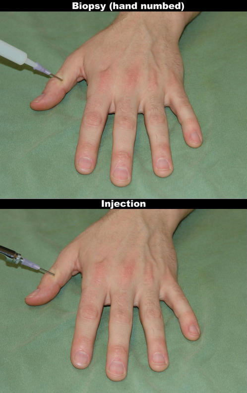
“While perceiving numbed vs. non-numbed hands resulted in clearly different pain intensity ratings, this effect was significantly reduced for unpleasantness ratings because – in line with the cover story - the biopsies on the numbed hand were evaluated as unpleasant for the target.”
§ What Are You Feeling? Using Functional Magnetic Resonance Imaging to Assess the Modulation of Sensory and Affective Responses during Empathy for Pain > Discussion > The role of appraisal in empathy for pain
boosteddirect- Asserted: passes — The authors put it forward as true.
- Checkable: passes — It can be checked against evidence.
- Load-bearing: passes — The paper’s conclusions depend on it.
- Based: passes — The paper gives a reason why it holds.
It may state two separate claims as one.
- Rests on an assumption: Participants believed the cover story that biopsies on the numbed hand were unpleasant for the target.
A result the paper reports from its own data — it cites no other source for it, so there is none to check. The quote shows it is in the paper.
In the paper
“Focusing on the sensory consequences also resulted in stronger activations in the action anticipation network outlined above (inferior parietal cortex and ventral premotor cortex), as well as in two distinct clusters in the anterior cingulate.”
§ What Are You Feeling? Using Functional Magnetic Resonance Imaging to Assess the Modulation of Sensory and Affective Responses during Empathy for Pain > Discussion > Effects of focusing on sensory vs. affective consequences of pain
plaindirect- Asserted: passes — The authors put it forward as true.
- Checkable: passes — It can be checked against evidence.
- Load-bearing: passes — The paper’s conclusions depend on it.
- Based: passes — The paper gives a reason why it holds.
It may state two separate claims as one.
A result the paper reports from its own data — it cites no other source for it, so there is none to check. The quote shows it is in the paper.
In the paper
“In the case of the biopsies on the numbed hand, however, no affect has to be shared and yet insular and cingulate cortices are clearly activated.”
§ What Are You Feeling? Using Functional Magnetic Resonance Imaging to Assess the Modulation of Sensory and Affective Responses during Empathy for Pain > Discussion > Conclusion
boosted- Asserted: passes — The authors put it forward as true.
- Checkable: passes — It can be checked against evidence.
- Load-bearing: passes — The paper’s conclusions depend on it.
- Based: passes — The paper gives a reason why it holds.
It may state two separate claims as one.
None of the sources cited for this claim have been checked yet.
In the paper
“In contrast, transcranial magnetic stimulation (TMS) [15], [16], electroencephalography [17], [18] and magnetoencephalographic measurements [2] suggest a role of sensorimotor representations during the perception of pain in others.”
§ What Are You Feeling? Using Functional Magnetic Resonance Imaging to Assess the Modulation of Sensory and Affective Responses during Empathy for Pain > Introduction
What the sources say
- Not checked yetcites [15] A Avenanti, 2005, Transcranial magnetic stimulation highlights the sensorimotor side of empathy for pain., Nat Neurosci source
- Not checked yetcites [16] A Avenanti, 2006, Stimulus-driven modulation of motor-evoked potentials during observation of others' pain., Neuroimage source
- Not checked yetcites [17] I Bufalari, 2007, Empathy for pain and touch in the human somatosensory cortex. source
hedgedcorrelational- Asserted: passes — The authors put it forward as true.
- Checkable: passes — It can be checked against evidence.
- Load-bearing: passes — The paper’s conclusions depend on it.
- Based: passes — The paper gives a reason why it holds.
Probably not a substantial claim.
None of the sources cited for this claim have been checked yet.
In the paper
“Research in developmental psychology shows that one's ability to engage in emotion regulation positively relates to feelings of concern for the other person [22], [23].”
§ What Are You Feeling? Using Functional Magnetic Resonance Imaging to Assess the Modulation of Sensory and Affective Responses during Empathy for Pain > Introduction
What the sources say
- Not checked yetcites [22] D Derryberry, 1998, Arousal, affect, and attention as components of temperament., J Pers Soc Psychol source
- Not checked yetcites [23] N Eisenberg, 2005, Empathy-related responding in children.
plaincorrelational- Asserted: passes — The authors put it forward as true.
- Checkable: passes — It can be checked against evidence.
- Load-bearing: fails — The paper’s conclusions may not depend on it.
- Based: passes — The paper gives a reason why it holds.
Probably not a substantial claim. The paper’s conclusions may not depend on it.
In the paper
“Another study recently demonstrated that the appraisal of others' pain is mediated by brain structures involved in stimulus evaluation and emotion regulation (such as the medial orbitofrontal cortex OFC and the right lateral prefrontal cortex [24]).”
§ What Are You Feeling? Using Functional Magnetic Resonance Imaging to Assess the Modulation of Sensory and Affective Responses during Empathy for Pain > Introduction
What the sources say
- Not checked yetcites [24] C Lamm, 2007, The neural substrate of human empathy: effects of perspective-taking and cognitive appraisal., J Cogn Neurosci source
boosteddirect- Asserted: passes — The authors put it forward as true.
- Checkable: passes — It can be checked against evidence.
- Load-bearing: passes — The paper’s conclusions depend on it.
- Based: passes — The paper gives a reason why it holds.
Probably not a substantial claim.
What the paper did — it cites no source for this, so there is none to check. The quote shows it is in the paper.
In the paper
“Note that due to excessive movement during experiment II, one participant had to be excluded from all analyses.”
§ What Are You Feeling? Using Functional Magnetic Resonance Imaging to Assess the Modulation of Sensory and Affective Responses during Empathy for Pain > Results > Functional MRI experiments
plaindirectSingle case- Asserted: passes — The authors put it forward as true.
- Checkable: passes — It can be checked against evidence.
- Load-bearing: fails — The paper’s conclusions may not depend on it.
- Based: passes — The paper gives a reason why it holds.
Probably not a substantial claim. The paper’s conclusions may not depend on it.
None of the sources cited for this claim have been checked yet.
In the paper
“These models emphasize that empathy is not an all-or-none phenomenon. Its experience is malleable by a number of factors including personality traits and the type of situation in which social interaction occurs.”
§ What Are You Feeling? Using Functional Magnetic Resonance Imaging to Assess the Modulation of Sensory and Affective Responses during Empathy for Pain > Introduction
What the sources say
plaindirect- Asserted: passes — The authors put it forward as true.
- Checkable: passes — It can be checked against evidence.
- Load-bearing: passes — The paper’s conclusions depend on it.
- Based: passes — The paper gives a reason why it holds.
Probably not a substantial claim. It may state two separate claims as one.
None of the sources cited for this claim have been checked yet.
In the paper
“Current neurobehavioral models of empathy (e.g., [12], [13], [45]) emphasize the contribution of both automatic and controlled processes to the conscious experience of empathy.”
§ What Are You Feeling? Using Functional Magnetic Resonance Imaging to Assess the Modulation of Sensory and Affective Responses during Empathy for Pain > Discussion > Correlations between brain activation and dispositional measures
What the sources say
- Not checked yetcites [12] J Decety, 2006, Human empathy through the lens of social neuroscience., ScientificWorldJournal source
- Not checked yetcites [13] L Goubert, 2005, Facing others in pain: the effects of empathy., Pain source
- Not checked yetcites [45] J Decety, 2004, The functional architecture of human empathy., Behav Cogn Neurosci Rev source
plaincorrelational- Asserted: passes — The authors put it forward as true.
- Checkable: passes — It can be checked against evidence.
- Load-bearing: fails — The paper’s conclusions may not depend on it.
- Based: passes — The paper gives a reason why it holds.
Probably not a substantial claim. The paper’s conclusions may not depend on it.
None of the sources cited for this claim have been checked yet.
In the paper
“In addition, part of the clusters in inferior parietal cortex might be related to the coding of nocifensive movements, and the visuospatial encoding of noxious threats [40], [41].”
§ What Are You Feeling? Using Functional Magnetic Resonance Imaging to Assess the Modulation of Sensory and Affective Responses during Empathy for Pain > Discussion > The role of sensory and affective components in empathy for pain
What the sources say
hedgedcorrelational- Asserted: passes — The authors put it forward as true.
- Checkable: passes — It can be checked against evidence.
- Load-bearing: unclear — It is unclear whether the paper’s conclusions depend on it.
- Based: passes — The paper gives a reason why it holds.
It may state two separate claims as one.
What the paper did — it cites no source for this, so there is none to check. The quote shows it is in the paper.
In the paper
“The consistency of the group analysis was confirmed by analyses on the single-subject level – as the five functional regions of interest (ROIs) described in the Material & Methods section were clearly activated in the majority of participants.”
§ What Are You Feeling? Using Functional Magnetic Resonance Imaging to Assess the Modulation of Sensory and Affective Responses during Empathy for Pain > Results > fMRI experiment I – effects of evaluative focus
boostedcorrelational- Asserted: passes — The authors put it forward as true.
- Checkable: passes — It can be checked against evidence.
- Load-bearing: unclear — It is unclear whether the paper’s conclusions depend on it.
- Based: passes — The paper gives a reason why it holds.
It may state two separate claims as one.
A conclusion the paper draws from its own results — it cites no other source for it, so there is none to check. The quote shows it is in the paper.
In the paper
“Moreover, both the sensory-discriminative and the affective-motivational component is modulated by the context in which pain has occurred, and by the consequences the observer is focusing on.”
§ What Are You Feeling? Using Functional Magnetic Resonance Imaging to Assess the Modulation of Sensory and Affective Responses during Empathy for Pain > Discussion > Conclusion
plaindirect- Asserted: passes — The authors put it forward as true.
- Checkable: passes — It can be checked against evidence.
- Load-bearing: passes — The paper’s conclusions depend on it.
- Based: passes — The paper gives a reason why it holds.
It may state two separate claims as one.
What the paper did — it cites no source for this, so there is none to check. The quote shows it is in the paper.
In the paper
“It should be acknowledged that the low temporal resolution of hemodynamic responses might not yield precise enough information about when and how this top-down modulation affects neural activities in the pain matrix.”
§ What Are You Feeling? Using Functional Magnetic Resonance Imaging to Assess the Modulation of Sensory and Affective Responses during Empathy for Pain > Discussion > Conclusion
hedgedconditional- Asserted: passes — The authors put it forward as true.
- Checkable: passes — It can be checked against evidence.
- Load-bearing: passes — The paper’s conclusions depend on it.
- Based: passes — The paper gives a reason why it holds.
Probably not a substantial claim.
A conclusion the paper draws from its own results — it cites no other source for it, so there is none to check. The quote shows it is in the paper.
In the paper
“This initial response might be down-regulated by cognitive mechanism of top-down control.”
§ What Are You Feeling? Using Functional Magnetic Resonance Imaging to Assess the Modulation of Sensory and Affective Responses during Empathy for Pain > Discussion > Conclusion
hedgedconditional- Asserted: passes — The authors put it forward as true.
- Checkable: passes — It can be checked against evidence.
- Load-bearing: fails — The paper’s conclusions may not depend on it.
- Based: passes — The paper gives a reason why it holds.
Probably not a substantial claim. The paper’s conclusions may not depend on it.
None of the sources cited for this claim have been checked yet.
In the paper
“The emotion contagion questionnaire assesses an individual's susceptibility to automatically mimic another's behavior – a mechanism that is also found in phylogenetically older species (e.g., [46], [47]).”
§ What Are You Feeling? Using Functional Magnetic Resonance Imaging to Assess the Modulation of Sensory and Affective Responses during Empathy for Pain > Discussion > Correlations between brain activation and dispositional measures
What the sources say
plain- Asserted: passes — The authors put it forward as true.
- Checkable: passes — It can be checked against evidence.
- Load-bearing: passes — The paper’s conclusions depend on it.
- Based: passes — The paper gives a reason why it holds.
Probably not a substantial claim.
In the paper
“In addition, electrical stimulation of the posterior part of the insula evokes painful sensations while stimulation of more anterior parts does not [50].”
§ What Are You Feeling? Using Functional Magnetic Resonance Imaging to Assess the Modulation of Sensory and Affective Responses during Empathy for Pain > Discussion > Effects of focusing on sensory vs. affective consequences of pain
What the sources say
- Not checked yetcites [50] K Ostrowsky, 2002, Representation of pain and somatic sensation in the human insula: a study of responses to direct electrical cortical stimulation., Cereb Cortex source
plaindirect- Asserted: passes — The authors put it forward as true.
- Checkable: passes — It can be checked against evidence.
- Load-bearing: unclear — It is unclear whether the paper’s conclusions depend on it.
- Based: passes — The paper gives a reason why it holds.
It may state two separate claims as one.
In the paper
“The contralateral middle insular cortex has intrinsic connections to the basal ganglia, and a meta-analysis of neuroimaging studies shows that it is most consistently activated during the first-hand experience of pain [48] – suggesting a specific role in coding the sensory-motor aspects associated with pain.”
§ What Are You Feeling? Using Functional Magnetic Resonance Imaging to Assess the Modulation of Sensory and Affective Responses during Empathy for Pain > Discussion > Effects of focusing on sensory vs. affective consequences of pain
What the sources say
- Not checked yetcites [48] TD Wager, 2004, From affect to control: functional specialization of the insula in motovation and regulation.
plaincorrelational- Asserted: passes — The authors put it forward as true.
- Checkable: passes — It can be checked against evidence.
- Load-bearing: unclear — It is unclear whether the paper’s conclusions depend on it.
- Based: passes — The paper gives a reason why it holds.
It may state two separate claims as one.
A conclusion the paper draws from its own results — it cites no other source for it, so there is none to check. The quote shows it is in the paper.
In the paper
“Finally, the correlation analyses corroborate and refine the findings of the contrast analyses.”
§ What Are You Feeling? Using Functional Magnetic Resonance Imaging to Assess the Modulation of Sensory and Affective Responses during Empathy for Pain > Discussion > The role of appraisal in empathy for pain
plaincorrelational- Asserted: passes — The authors put it forward as true.
- Checkable: passes — It can be checked against evidence.
- Load-bearing: passes — The paper’s conclusions depend on it.
- Based: passes — The paper gives a reason why it holds.
It may state two separate claims as one.
A conclusion the paper draws from its own results — it cites no other source for it, so there is none to check. The quote shows it is in the paper.
In the paper
“The correlation analyses also supports the interpretation that right anterior insula is more sensitive to affective variations, and that precuneus and TPJ play a specific role in distinguishing between the sensory painful and non-painful events.”
§ What Are You Feeling? Using Functional Magnetic Resonance Imaging to Assess the Modulation of Sensory and Affective Responses during Empathy for Pain > Discussion > The role of appraisal in empathy for pain
hedgedcorrelational- Asserted: passes — The authors put it forward as true.
- Checkable: passes — It can be checked against evidence.
- Load-bearing: passes — The paper’s conclusions depend on it.
- Based: passes — The paper gives a reason why it holds.
It may state two separate claims as one.
A conclusion the paper draws from its own results — it cites no other source for it, so there is none to check. The quote shows it is in the paper.
In the paper
“These time-courses suggest that an automatic response was triggered by the presentation of an aversive and putatively noxious stimulus, resulting in the mobilization of withdrawal-related neural response.”
§ What Are You Feeling? Using Functional Magnetic Resonance Imaging to Assess the Modulation of Sensory and Affective Responses during Empathy for Pain > Discussion > The role of appraisal in empathy for pain
hedgeddirect- Asserted: passes — The authors put it forward as true.
- Checkable: passes — It can be checked against evidence.
- Load-bearing: passes — The paper’s conclusions depend on it.
- Based: passes — The paper gives a reason why it holds.
It may state two separate claims as one.
In the paper
“The inter-stimuli interval was jittered (mean = 3.5 s, minimum/maximum = 2/5.8 s) to reduce stimulus predictability and to allow efficient event-related signal estimation [71].”
§ What Are You Feeling? Using Functional Magnetic Resonance Imaging to Assess the Modulation of Sensory and Affective Responses during Empathy for Pain > Materials and Methods > Experimental design and procedures
What the sources say
- Not checked yetcites [71] DI Donaldson, 2001, Effective paradigm design.
plaindirect- Asserted: passes — The authors put it forward as true.
- Checkable: passes — It can be checked against evidence.
- Load-bearing: fails — The paper’s conclusions may not depend on it.
- Based: passes — The paper gives a reason why it holds.
Probably not a substantial claim. The paper’s conclusions may not depend on it. It may state two separate claims as one.
In the paper
“the response bias indicates the degree to which the situations are considered as painful.”
§ What Are You Feeling? Using Functional Magnetic Resonance Imaging to Assess the Modulation of Sensory and Affective Responses during Empathy for Pain > Materials and Methods > Functional MRI experiments
What the sources say
- Not checked yetcites [27] WC Clark, 1983, Pain measurement and assessment. Applications of sensory detection theory to problems in laboratory and clinical pain.
plain- Asserted: passes — The authors put it forward as true.
- Checkable: passes — It can be checked against evidence.
- Load-bearing: passes — The paper’s conclusions depend on it.
- Based: passes — The paper gives a reason why it holds.
Probably not a substantial claim.
In the paper
“The Emotional Contagion Scale (ECS) assesses the susceptibility to other's emotions from afferent feedback generated by mimicry”
§ What Are You Feeling? Using Functional Magnetic Resonance Imaging to Assess the Modulation of Sensory and Affective Responses during Empathy for Pain > Materials and Methods > Functional MRI experiments
What the sources say
- Not checked yetcites [26] RW Doherty, 1997, The emotional contagion scale: a measure of individual differences., J Nonverb Behav source
plaindirect- Asserted: passes — The authors put it forward as true.
- Checkable: passes — It can be checked against evidence.
- Load-bearing: fails — The paper’s conclusions may not depend on it.
- Based: passes — The paper gives a reason why it holds.
Probably not a substantial claim. The paper’s conclusions may not depend on it.
In the paper
“In all cases, violations of the sphericity-assumption for these comparisons were accounted for by using specific error-variances [76].”
§ What Are You Feeling? Using Functional Magnetic Resonance Imaging to Assess the Modulation of Sensory and Affective Responses during Empathy for Pain > Materials and Methods > Function MRI data acquisition and analysis
What the sources say
- Not checked yetcites [76] RJ Boik, 1981, A priori tests in repeated measures designs: effects of nonsphericity., Psychometrika source
plain- Asserted: passes — The authors put it forward as true.
- Checkable: passes — It can be checked against evidence.
- Load-bearing: fails — The paper’s conclusions may not depend on it.
- Based: passes — The paper gives a reason why it holds.
Probably not a substantial claim. The paper’s conclusions may not depend on it.
What the paper did — it cites no source for this, so there is none to check. The quote shows it is in the paper.
In the paper
“The difference between the sensory and the affective consequences of the painful stimulations were explained using standardized written instructions and using a number of practice trials.”
- Also stated in: § Experimental design and procedures · plain
“The same standardized written instruction as in the behavioral experiment was used to explain the difference between these two aspects of the pain response.”
§ What Are You Feeling? Using Functional Magnetic Resonance Imaging to Assess the Modulation of Sensory and Affective Responses during Empathy for Pain > Materials and Methods > Behavioral experiment
plain- Asserted: passes — The authors put it forward as true.
- Checkable: passes — It can be checked against evidence.
- Load-bearing: passes — The paper’s conclusions depend on it.
- Based: passes — The paper gives a reason why it holds.
Probably not a substantial claim.
- Also stated in: § Experimental design and procedures · plain
None of the sources cited for this claim have been checked yet.
In the paper
“The choice of these thresholds was based upon exploratory data analyses and upon effect size considerations derived from similar experiments of our own and other groups [3], [4], [8], [9], [24].”
§ What Are You Feeling? Using Functional Magnetic Resonance Imaging to Assess the Modulation of Sensory and Affective Responses during Empathy for Pain > Materials and Methods > Function MRI data acquisition and analysis
What the sources say
- Not checked yetcites [3] P Jackson, 2006, Empathy examined through the neural mechanisms involved in imagining how I feel versus how you feel pain., Neuropsychologia source
- Not checked yetcites [4] P Jackson, 2005, How do we perceive the pain of others? A window into the neural processes involved in empathy., Neuroimage source
- Not checked yetcites [8] T Singer, 2004, Empathy for pain involves the affective but not the sensory components of pain., Science source
plain- Asserted: passes — The authors put it forward as true.
- Checkable: passes — It can be checked against evidence.
- Load-bearing: fails — The paper’s conclusions may not depend on it.
- Based: passes — The paper gives a reason why it holds.
Probably not a substantial claim. The paper’s conclusions may not depend on it.
What the paper did — it cites no source for this, so there is none to check. The quote shows it is in the paper.
In the paper
“The resulting first-level contrast images were entered into second-level random effects analyses to assess differences between conditions with population inference.”
§ What Are You Feeling? Using Functional Magnetic Resonance Imaging to Assess the Modulation of Sensory and Affective Responses during Empathy for Pain > Materials and Methods > Function MRI data acquisition and analysis
plain- Asserted: passes — The authors put it forward as true.
- Checkable: passes — It can be checked against evidence.
- Load-bearing: passes — The paper’s conclusions depend on it.
- Based: passes — The paper gives a reason why it holds.
Probably not a substantial claim.
In the paper
“The influence of in-plane susceptibility gradients in orbitofrontal regions was reduced by orienting image slices according to recommendations by [72].”
§ What Are You Feeling? Using Functional Magnetic Resonance Imaging to Assess the Modulation of Sensory and Affective Responses during Empathy for Pain > Materials and Methods > Function MRI data acquisition and analysis
What the sources say
- Not checked yetcites [72] R Deichmann, 2003, Optimized EPI for fMRI studies of the orbitofrontal cortex., Neuroimage source
plaindirect- Asserted: passes — The authors put it forward as true.
- Checkable: passes — It can be checked against evidence.
- Load-bearing: fails — The paper’s conclusions may not depend on it.
- Based: passes — The paper gives a reason why it holds.
Probably not a substantial claim. The paper’s conclusions may not depend on it.
What the paper did — it cites no source for this, so there is none to check. The quote shows it is in the paper.
In the paper
“Several practice trials were performed before entering the scanner to ensure appropriate understanding of the instructions and experimental procedures.”
§ What Are You Feeling? Using Functional Magnetic Resonance Imaging to Assess the Modulation of Sensory and Affective Responses during Empathy for Pain > Materials and Methods > Experimental design and procedures
plain- Asserted: passes — The authors put it forward as true.
- Checkable: passes — It can be checked against evidence.
- Load-bearing: fails — The paper’s conclusions may not depend on it.
- Based: passes — The paper gives a reason why it holds.
Probably not a substantial claim. The paper’s conclusions may not depend on it.
What the paper did — it cites no source for this, so there is none to check. The quote shows it is in the paper.
In the paper
“The more subtle differences in signal strength between conditions that differed psychologically (e.g., intensity vs. unpleasantness ratings, painful injection vs. numbed injection) were thresholded using a more liberal threshold of P = 0.001 (uncorrected for multiple comparisons) and an extent criterion of k = 5.”
§ What Are You Feeling? Using Functional Magnetic Resonance Imaging to Assess the Modulation of Sensory and Affective Responses during Empathy for Pain > Materials and Methods > Function MRI data acquisition and analysis
plain- Asserted: passes — The authors put it forward as true.
- Checkable: passes — It can be checked against evidence.
- Load-bearing: passes — The paper’s conclusions depend on it.
- Based: passes — The paper gives a reason why it holds.
Probably not a substantial claim.
In the paper
“The Interpersonal Reactivity Index (IRI) is probably the most widely used self-report measure of dispositional empathy.”
§ What Are You Feeling? Using Functional Magnetic Resonance Imaging to Assess the Modulation of Sensory and Affective Responses during Empathy for Pain > Materials and Methods > Functional MRI experiments
What the sources say
- Not checked yetcites [25] MH Davis, 1994, Empathy: a social psychological approach
hedged- Asserted: passes — The authors put it forward as true.
- Checkable: passes — It can be checked against evidence.
- Load-bearing: fails — The paper’s conclusions may not depend on it.
- Based: passes — The paper gives a reason why it holds.
Probably not a substantial claim. The paper’s conclusions may not depend on it.
In the paper
“P(A) indicates the extent to which participants are able to differentiate between painful and non-painful situations”
§ What Are You Feeling? Using Functional Magnetic Resonance Imaging to Assess the Modulation of Sensory and Affective Responses during Empathy for Pain > Materials and Methods > Functional MRI experiments
What the sources say
- Not checked yetcites [27] WC Clark, 1983, Pain measurement and assessment. Applications of sensory detection theory to problems in laboratory and clinical pain.
hedged- Asserted: passes — The authors put it forward as true.
- Checkable: passes — It can be checked against evidence.
- Load-bearing: passes — The paper’s conclusions depend on it.
- Based: passes — The paper gives a reason why it holds.
Probably not a substantial claim.
In the paper
“Based on signal detection theory, a discrimination score (P(A)) and a response bias score (B) is calculated.”
§ What Are You Feeling? Using Functional Magnetic Resonance Imaging to Assess the Modulation of Sensory and Affective Responses during Empathy for Pain > Materials and Methods > Functional MRI experiments
What the sources say
- Not checked yetcites [27] WC Clark, 1983, Pain measurement and assessment. Applications of sensory detection theory to problems in laboratory and clinical pain.
plain- Asserted: passes — The authors put it forward as true.
- Checkable: passes — It can be checked against evidence.
- Load-bearing: passes — The paper’s conclusions depend on it.
- Based: passes — The paper gives a reason why it holds.
Probably not a substantial claim. It may state two separate claims as one.
In the paper
“The Sensitivity to Pain Questionnaire (SPQ) assesses the participants' sensitivity to pain by asking them to assess the amount of stimulus-induced pain they would experience in fifteen painful and fifteen non-painful situations.”
§ What Are You Feeling? Using Functional Magnetic Resonance Imaging to Assess the Modulation of Sensory and Affective Responses during Empathy for Pain > Materials and Methods > Functional MRI experiments
What the sources say
- Not checked yetcites [27] WC Clark, 1983, Pain measurement and assessment. Applications of sensory detection theory to problems in laboratory and clinical pain.
plain- Asserted: passes — The authors put it forward as true.
- Checkable: passes — It can be checked against evidence.
- Load-bearing: passes — The paper’s conclusions depend on it.
- Based: passes — The paper gives a reason why it holds.
Probably not a substantial claim.
What the paper did — it cites no source for this, so there is none to check. The quote shows it is in the paper.
In the paper
“In accordance with other studies assessing brain-behavior relationships, a rather liberal significance threshold of P = 0.001 (uncorrected) and k = 5 was selected for these analyses.”
§ What Are You Feeling? Using Functional Magnetic Resonance Imaging to Assess the Modulation of Sensory and Affective Responses during Empathy for Pain > Materials and Methods > Function MRI data acquisition and analysis
plain- Asserted: passes — The authors put it forward as true.
- Checkable: passes — It can be checked against evidence.
- Load-bearing: fails — The paper’s conclusions may not depend on it.
- Based: passes — The paper gives a reason why it holds.
Probably not a substantial claim. The paper’s conclusions may not depend on it.
In the paper
“For fMRI experiment II individual ROIs of activations coding the affective-motivational consequences of painful stimulation were defined guided by a meta-analysis of insular and cingulate cortex activation during the perception of pain in self and others [34].”
§ What Are You Feeling? Using Functional Magnetic Resonance Imaging to Assess the Modulation of Sensory and Affective Responses during Empathy for Pain > Materials and Methods > Function MRI data acquisition and analysis
What the sources say
- Not checked yetcites [34] P Jackson, 2006, To what extent do we share the pain of others? Insight from the neural bases of pain empathy., Pain source
plain- Asserted: passes — The authors put it forward as true.
- Checkable: passes — It can be checked against evidence.
- Load-bearing: passes — The paper’s conclusions depend on it.
- Based: passes — The paper gives a reason why it holds.
Probably not a substantial claim.
What the paper did — it cites no source for this, so there is none to check. The quote shows it is in the paper.
In the paper
“In addition, the threshold was lowered to P = 0.005 (uncorrected), k = 5, for a priori defined regions involved in the perception of pain and in emotion regulation in order to assess whether they showed activation below threshold.”
§ What Are You Feeling? Using Functional Magnetic Resonance Imaging to Assess the Modulation of Sensory and Affective Responses during Empathy for Pain > Materials and Methods > Function MRI data acquisition and analysis
plain- Asserted: passes — The authors put it forward as true.
- Checkable: passes — It can be checked against evidence.
- Load-bearing: fails — The paper’s conclusions may not depend on it.
- Based: passes — The paper gives a reason why it holds.
Probably not a substantial claim. The paper’s conclusions may not depend on it.
In the paper
“Its four subscales (Empathic Concern, Perspective Taking, Fantasy Scale and Personal Distress) assess different aspects of interpersonal affective responses.”
§ What Are You Feeling? Using Functional Magnetic Resonance Imaging to Assess the Modulation of Sensory and Affective Responses during Empathy for Pain > Materials and Methods > Functional MRI experiments
What the sources say
- Not checked yetcites [25] MH Davis, 1994, Empathy: a social psychological approach
plain- Asserted: passes — The authors put it forward as true.
- Checkable: passes — It can be checked against evidence.
- Load-bearing: fails — The paper’s conclusions may not depend on it.
- Based: passes — The paper gives a reason why it holds.
Probably not a substantial claim. The paper’s conclusions may not depend on it.
In the paper
“Activity differences between the presentation of physically distinct stimuli (painful vs. non-painful photographs, and photographs vs. fixation) were interpreted using a voxel-level threshold of P = 0.01 and a spatial extent threshold of k = 10, corrected for multiple comparisons across the whole volume using the false discovery rate (FDR) approach [73].”
§ What Are You Feeling? Using Functional Magnetic Resonance Imaging to Assess the Modulation of Sensory and Affective Responses during Empathy for Pain > Materials and Methods > Function MRI data acquisition and analysis
What the sources say
- Not checked yetcites [73] CR Genovese, 2002, Thresholding of statistical maps in functional neuroimaging using the false discovery rate., Neuroimage source
plain- Asserted: passes — The authors put it forward as true.
- Checkable: passes — It can be checked against evidence.
- Load-bearing: passes — The paper’s conclusions depend on it.
- Based: passes — The paper gives a reason why it holds.
Probably not a substantial claim.
What the paper did — it cites no source for this, so there is none to check. The quote shows it is in the paper.
In the paper
“Stimuli were presented in eight blocks containing 41 randomly interspersed painful or non-painful stimuli each.”
§ What Are You Feeling? Using Functional Magnetic Resonance Imaging to Assess the Modulation of Sensory and Affective Responses during Empathy for Pain > Materials and Methods > Behavioral experiment
plain- Asserted: passes — The authors put it forward as true.
- Checkable: passes — It can be checked against evidence.
- Load-bearing: fails — The paper’s conclusions may not depend on it.
- Based: passes — The paper gives a reason why it holds.
Probably not a substantial claim. The paper’s conclusions may not depend on it.
What the paper did — it cites no source for this, so there is none to check. The quote shows it is in the paper.
In the paper
“Each run was preceded by several dummy scans ensuring steady state magnetization conditions.”
§ What Are You Feeling? Using Functional Magnetic Resonance Imaging to Assess the Modulation of Sensory and Affective Responses during Empathy for Pain > Materials and Methods > Function MRI data acquisition and analysis
plaindirect- Asserted: passes — The authors put it forward as true.
- Checkable: passes — It can be checked against evidence.
- Load-bearing: fails — The paper’s conclusions may not depend on it.
- Based: passes — The paper gives a reason why it holds.
Probably not a substantial claim. The paper’s conclusions may not depend on it.
What the paper did — it cites no source for this, so there is none to check. The quote shows it is in the paper.
In the paper
“The order of blocks was counterbalanced across participants.”
- Also stated in: § Experimental design and procedures · plain
“The order of blocks was counterbalanced across participants.”
§ What Are You Feeling? Using Functional Magnetic Resonance Imaging to Assess the Modulation of Sensory and Affective Responses during Empathy for Pain > Materials and Methods > Experimental design and procedures
plain- Asserted: passes — The authors put it forward as true.
- Checkable: passes — It can be checked against evidence.
- Load-bearing: fails — The paper’s conclusions may not depend on it.
- Based: passes — The paper gives a reason why it holds.
Probably not a substantial claim. The paper’s conclusions may not depend on it.
- Also stated in: § Experimental design and procedures · plain
What the paper did — it cites no source for this, so there is none to check. The quote shows it is in the paper.
In the paper
“Experiment II was always performed after experiment I. The reason for this was to avoid potential confusion and carry-over effects from the numbed hand stimuli to the non-painful stimuli from experiment I.”
§ What Are You Feeling? Using Functional Magnetic Resonance Imaging to Assess the Modulation of Sensory and Affective Responses during Empathy for Pain > Materials and Methods > Function MRI data acquisition and analysis
plaindirect- Asserted: passes — The authors put it forward as true.
- Checkable: passes — It can be checked against evidence.
- Load-bearing: passes — The paper’s conclusions depend on it.
- Based: passes — The paper gives a reason why it holds.
Probably not a substantial claim.
What the paper did — it cites no source for this, so there is none to check. The quote shows it is in the paper.
In the paper
“Individuals who participated in the behavioral study were not involved in the fMRI experiments to avoid learning and habituation effects.”
§ What Are You Feeling? Using Functional Magnetic Resonance Imaging to Assess the Modulation of Sensory and Affective Responses during Empathy for Pain > Materials and Methods > General design
plain- Asserted: passes — The authors put it forward as true.
- Checkable: passes — It can be checked against evidence.
- Load-bearing: passes — The paper’s conclusions depend on it.
- Based: passes — The paper gives a reason why it holds.
Probably not a substantial claim.
The paper relies on this without support.
Little is known about the neural mechanisms underlying the modulation of empathy.
“However, little is known about the neural mechanisms underlying the modulation of empathy.”
The paper relies on this without support.
It remains unclear whether the finding that evaluating sensory or affective consequences of first-hand pain recruits distinct neural pathways also holds true for the perception of pain in others.
“It remains unclear whether this also hold true for the perception of pain in others.”
The paper relies on this without support.
We have only cursory knowledge about how cognitive processes such as deliberate appraisal of the other's situation modulate the empathic reaction to the pain of others.
“We also have only cursory knowledge about how cognitive processes such as deliberate appraisal of the other's situation modulate the empathic reaction to the pain of others.”
The paper relies on this without support.
Neuroscientific evidence concerning the modulation of the empathic response by cognitive appraisal and emotion regulation is rather sparse.
“Neuroscientific evidence concerning the modulation of the empathic response by cognitive appraisal and emotion regulation is, however, rather sparse.”
The authors state this as an assumption.
The interaction analyses depend on the assumption of no or negligible differences for the unpleasantness evaluations of injections and numbed hands.
“whose results also depend upon the assumption of no or negligible differences for the unpleasantness evaluations of injections and numbed hands.”
The paper relies on this without support.
The empathic concern scale measures the more sophisticated aspect of empathy under cognitive control.
“Conversely, the empathic concern scale measures the more sophisticated aspect of empathy under cognitive control.”
The paper relies on this without support.
Participants believed the cover story that biopsies on the numbed hand were unpleasant for the target.
“While perceiving numbed vs. non-numbed hands resulted in clearly different pain intensity ratings, this effect was significantly reduced for unpleasantness ratings because – in line with the cover story - the biopsies on the numbed hand were evaluated as unpleasant for the target.”
The paper relies on this without support.
The IRI perspective taking subscale validly measures perspective taking.
“A similar result was expected when correlating the scores of the IRI perspective taking subscale with the activation differences between numbed and non-numbed stimuli (again, for intensity trials only).”
The paper relies on this without support.
Numbing of the hand resulted in complete elimination of the somatosensation of pain for the target, while the targets experienced unpleasantness and discomfort triggered by the surgical procedure.
“While the numbing of the hand resulted in complete elimination of the somatosensation of pain for the target, the targets experienced unpleasantness and discomfort triggered by the surgical procedure (in the same way as dental work on anesthetized teeth might not be painful, but still unpleasant).”
Claims other papers make about this one
Each is checked against this paper’s own text: does it say what the citing paper says it does?
In the citing paper
“Moreover, activation in AI and aMCC has repeatedly been observed to covary with negative affect ratings, both during self-experienced pain and when observing others suffering (Jackson et al., 2005;Cheng et al., 2007;Lamm et al., 2007;Saarela et al., 2007;Singer et al., 2008;Akitsuki and Decety, 2009).”
From Differential pattern of functional brain plasticity after compassion and empathy training (2014) · cites this paper as Lamm et al., 2007
Statistics recomputed
Every reported test result, recomputed from its test statistic and degrees of freedom. A result shows as not adding up only once an independent check has confirmed it.
- ConsistentF(1, 22) = 510.641reported p < .001 · recomputed p < .001
“F(1,22) = 510.641, p < .001”
- ConsistentF(1, 22) = 13.389reported p = .001 · recomputed p = .001
“F(1,22) = 13.389, p = .001”
- ConsistentF(1, 17) = 348.815reported p < .001 · recomputed p < .001
“F(1,17) = 348.815, p < .001”
- ConsistentF(1, 16) = 404.426reported p < .001 · recomputed p < .001
“F(1,16) = 404.426, p < .001”
- ConsistentF(1, 16) = 90.444reported p < .001 · recomputed p < .001
“F(1,16) = 90.444, p < .001”
- ConsistentF(1, 16) = 145.33reported p < .001 · recomputed p < .001
“F(1,16) = 145.33, p < .001”
- ConsistentF(1, 13) = 5.069reported p = .042 · recomputed p = .042
“F(1,13) = 5.069, p = .042”
- ConsistentF(1, 16) = 3.45reported p = .082 · recomputed p = .082
“F(1,16) = 3.45, p = .082”
- ConsistentF(1, 16) = 6.34reported p = .023 · recomputed p = .023
“F(1,16) = 6.34, p = .023”
- ConsistentF(1, 16) = 3.755reported p = .07 · recomputed p = .071
“F(1,16) = 3.755, p = .07”
- ConsistentF(1, 13) = 6.96reported p = .02 · recomputed p = .020
“F(1,13) = 6.96, p = .02”
Self-check
Percentages, sums and reported differences re-computed from the paper’s own numbers.
Each contradiction between two passages listed here was confirmed by an independent check.
Rated major: it would touch a result — a value a result rests on, reported and recomputed p-values on opposite sides of the significance level, two passages that conflict with no sign which is right, or a cited work retracted before this paper was published.
In the paper
§ What Are You Feeling? Using Functional Magnetic Resonance Imaging to Assess the Modulation of Sensory and Affective Responses during Empathy for Pain > Results > fMRI experiment I – effects of evaluative focus
“The reverse interaction [Unpleasantness (Painful>Non-Painful)>Intensity (Painful>Non-Painful)] only yielded a significant cluster in visual cortex (right lingual gyrus, MNI 23/−82/8).”
§ What Are You Feeling? Using Functional Magnetic Resonance Imaging to Assess the Modulation of Sensory and Affective Responses during Empathy for Pain > Results > fMRI experiment I – effects of evaluative focus
“| Insula | L | 7 | 4.77 | −30 | 24 | 8 |”
Rated minor: it would not touch a result — a value no result rests on, a p-value that stays on the same side of the significance level, or a conflict that is clerical, resolved elsewhere in the paper, or outside its own findings.
In the paper
§ What Are You Feeling? Using Functional Magnetic Resonance Imaging to Assess the Modulation of Sensory and Affective Responses during Empathy for Pain > Results > fMRI experiment II – effects of cognitive appraisal
“a second hemodynamic response about 9 image volumes (TRs) after stimulus onset for the painful injections (in both rating conditions, see Figure 7).”
§ What Are You Feeling? Using Functional Magnetic Resonance Imaging to Assess the Modulation of Sensory and Affective Responses during Empathy for Pain > Discussion > The role of appraisal in empathy for pain
“the occurrence of a second peak during intensity ratings only cannot be explained by subsequent trials”
Citations
Cites
Tzourio-Mazoyer, N., Landeau, B., Papathanassiou, D., Crivello, F., Etard, O., Delcroix, N., Mazoyer, B., & Joliot, M. (2002) · NeuroImage · doi:10.1006/nimg.2001.0978 · Cited by 16,319
A default mode of brain function
Raichle, M. E., MacLeod, A. M., Snyder, A. Z., Powers, W. J., Gusnard, D. A., & Shulman, G. L. (2001) · Proceedings of the National Academy of Sciences · doi:10.1073/pnas.98.2.676 · Cited by 12,190
How do you feel? Interoception: The sense of the physiological condition of the body
Craig, A. D. (2002) · Nature Reviews Neuroscience · doi:10.1038/nrn894 · Cited by 6,006
The precuneus: a review of its functional anatomy and behavioural correlates
Cavanna, A. E., & Trimble, M. R. (2006) · Brain · doi:10.1093/brain/awl004 · Cited by 5,128
Thresholding of statistical maps in functional neuroimaging using the false discovery rate
Genovese, C. R., Lazar, N. A., & Nichols, T. (2002) · NeuroImage · doi:10.1006/nimg.2001.1037 · Cited by 4,923
The cognitive control of emotion
OCHSNER, K., & GROSS, J. (2005) · Trends in Cognitive Sciences · doi:10.1016/j.tics.2005.03.010 · Cited by 4,495
Empathy: its ultimate and proximate bases
Preston, S. D., & de Waal, F. B. M. (2002) · Behavioral and Brain Sciences · doi:10.1017/s0140525x02000018 · Cited by 4,126
A new SPM toolbox for combining probabilistic cytoarchitectonic maps and functional imaging data
Eickhoff, S. B., Stephan, K. E., Mohlberg, H., Grefkes, C., Fink, G. R., Amunts, K., & Zilles, K. (2005) · NeuroImage · doi:10.1016/j.neuroimage.2004.12.034 · Cited by 4,033
Empathy for Pain Involves the Affective but not Sensory Components of Pain
Singer, T., Seymour, B., O'Doherty, J., Kaube, H., Dolan, R. J., & Frith, C. D. (2004) · Science · doi:10.1126/science.1093535 · Cited by 3,929
The functional architecture of human empathy
Decety, J., & Jackson, P. L. (2004) · Behavioral and Cognitive Neuroscience Reviews · doi:10.1177/1534582304267187 · Cited by 3,407 · cited 1×
“, [12,13,45]) emphasize the contribution of both automatic and controlled processes to the conscious experience of empathy.”
Background
Research works printed but not linked: 1
- C Lamm, 2007, The neural substrate of human empathy: effects of perspective-taking and cognitive appraisal., J Cogn Neurosci, 19, 42, 10.1162/jocn.2007.19.1.42 · DOI · Paper page
Cited by
Sorted by citations
Items 401–405 of 405
Diminished neural responses for greater numbers of lives at risk and lives lost in foreign countries
Campbell-Meiklejohn, D. K., & Cutler, J. (2026) · Social Cognitive and Affective Neuroscience · doi:10.1093/scan/nsag049
A Knowledge Argument Without “Knowledge”
Watson, J. J. (2026) · Analytic Philosophy · doi:10.1111/phib.70018
Pfaff, D., & Sherman, S. (2026) · Hormones and Behavior · doi:10.1016/j.yhbeh.2026.105981
He, Z., Song, X., Shi, Y., Zhou, J., Wang, Y., Wei, Q., Wang, D., Zhu, L., Lou, Q., Li, M., et al. (2026) · Communications Biology · doi:10.1038/s42003-026-11086-y
Pain generated by observation of others in pain
Osborn, J. (2011) · University of Birmingham Institutional Research Archive (University of Birmingham)
Metadata
- Authors
- Lamm, C., Nusbaum, H. C., Meltzoff, A. N., & Decety, J.
- Affiliations
- University of Chicago (US)
- University of Washington (US)
- Venue
- PLoS ONE
- Publisher
- Public Library of Science (PLoS)
- Published
- 2007-12-12
- Volume
- 2
- Issue
- 12
- Pages
- e1292
- ISSN
- 1932-6203 (ISSN-L)
- PMC
- PMC2144768
- OpenAlex
- W2125566732
- Semantic Scholar
- f55504298bf6efb8cab3d78aeda9d48af2331aaa
- Cited by
- 411
- References
- 81
- Citations by year
- 2026: 8 · 2025: 6 · 2024: 13 · 2023: 15 · 2022: 7 · 2021: 16 · 2020: 66 · 2019: 15 · 2018: 25 · 2017: 24 · 2016: 24 · 2015: 29 · 2014: 20 · 2013: 37 · 2012: 19
- FWCI
- 29.75
- Type
- journal article
- Language
- en
- Open access
- gold
- doi.org
- Locations
- PLoS ONE (publishedVersion, cc-by)
- PubMed Central (publishedVersion, cc-by)
- PubMed (publishedVersion)
- Knowledge@UChicago (University of Chicago) (submittedVersion, cc-by-nc-nd)
- DOAJ (DOAJ: Directory of Open Access Journals) (submittedVersion, cc-by-sa)
- Figshare (submittedVersion, cc-by)
- University of Chicago (cc-by)
- Funders
- National Science Foundation (0354453, 0718480)
- Topics
- Social Sciences › Psychology › Social Psychology › Action Observation and Synchronization
- Health Sciences › Medicine › Physiology (medical) › Pain Mechanisms and Treatments
- Health Sciences › Medicine › Psychiatry and Mental health › Empathy and Medical Education
- Keywords
- empathy for pain, cognitive appraisal, attention, pain matrix, somatosensory cortex, temporoparietal junction, medial orbitofrontal cortex, self-other distinction, top-down control, needle injection
- MeSH terms
- Brain, Humans, Magnetic Resonance Imaging, Pain
About these checks
Registry, retraction, Cites and statistics results are computed, not judged, with one exception: a printed reference that no index lists is read once by a model before it counts as a citation error. Claims are extracted by a model and kept only when their quote appears verbatim in the text; each cited source is then checked, with a model’s help, for whether it says what the claim relies on. Those verdicts can be wrong — read the claim’s quote and the cited source before relying on one.
A language model drew the claims from the paper on 2026-10-10, under version 3 of the claim definition (model deepseek-v4.1-flash, prompt extract-v3).
A second model labelled each claim on 2026-10-10 (model jev-1.13-20260917, definition revision gold-guideline-v0.2).
Statistics and arithmetic are recomputed, not judged.
Confidence bands are shown only for fields calibrated on our annotated test set. None is calibrated yet, so no band is shown.
What counts as a claim ›How we measure confidence ›How we check this ›
Are you an author, or do you see a problem with this page? Request a review
