

A p53-Dependent Response Limits Epidermal Stem Cell Functionality and Organismal Size in Mice with Short Telomeres
Open accessThe DOI matches its Crossref record; 46 of the 46 references it cites were found in a registry or an index.
Abstract
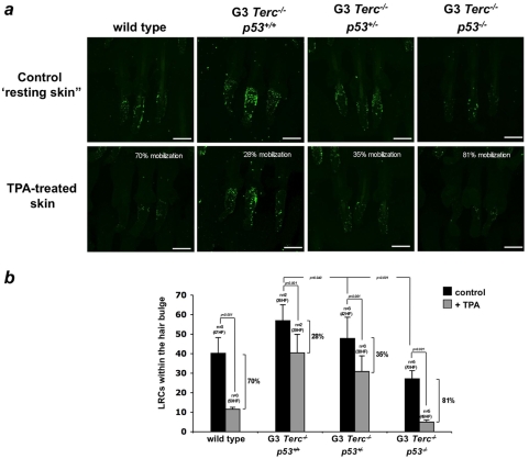
Telomere maintenance is essential to ensure proper size and function of organs with a high turnover. In particular, a dwarf phenotype as well as phenotypes associated to premature loss of tissue regeneration, including the skin (hair loss, hair graying, decreased wound healing), are found in mice deficient for telomerase, the enzyme responsible for maintaining telomere length. Coincidental with the appearance of these phenotypes, p53 is found activated in several tissues from these mice, where is thought to trigger cellular senescence and/or apoptotic responses. Here, we show that p53 abrogation rescues both the small size phenotype and restitutes the functionality of epidermal stem cells (ESC) of telomerase-deficient mice with dysfunctional telomeres. In particular, p53 ablation restores hair growth, skin renewal and wound healing responses upon mitogenic induction, as well as rescues ESCmobilization defects in vivo and defective ESC clonogenic activity in vitro. This recovery of ESC functions is accompanied by a downregulation of senescence markers and an increased proliferation in the skin and kidney of telomerase-deficient mice with critically short telomeres without changes in apoptosis rates. Together, these findings indicate the existence of a p53-dependent senescence response acting on stem/progenitor cells with dysfunctional telomeres that is actively limiting their contribution to tissue regeneration, thereby impinging on tissue fitness.
Overview
The claims and statistics of this paper haven’t been checked yet.
Free: it reads the open-access text and checks the first 5 claims that cite a source we can check. Create a free account to check every claim.
Integrity signals
Registry
Cites
Statistics
Figures




Verification
Claims the paper makes
The claims and statistics of this paper haven’t been checked yet.
Free: it reads the open-access text and checks the first 5 claims that cite a source we can check. Create a free account to check every claim.
Claims other papers make about this one
Each is checked against this paper’s own text: does it say what the citing paper says it does?
The source backs part of the claim, not all of it — or backs it for a different population, condition or comparison.
In the citing paper
“Skin aging associated with ROS accumulation, thus, was found to be rescued in the skin of late-generation p53 knockout mice [120].”
From Ultraviolet Radiation‐Induced Skin Aging: The Role of DNA Damage and Oxidative Stress in Epidermal Stem Cell Damage Mediated Skin Aging (2016) · cites this paper as [120]
Experimental — accuracy not yet measuredThe source backs part of the claim, not all of it — or backs it for a different population, condition or comparison.
In the citing paper
“In these mice, there were improved wound healing, hair growth, and skin renewal to levels comparable to those of wild-type mice, as well as increased ESC numbers and improved mobilization capacity [120].”
From Ultraviolet Radiation‐Induced Skin Aging: The Role of DNA Damage and Oxidative Stress in Epidermal Stem Cell Damage Mediated Skin Aging (2016) · cites this paper as [120]
Experimental — accuracy not yet measured
Self-check
Percentages, sums and reported differences re-computed from the paper’s own numbers.
The self-check needs the paper’s full text.
Citations
Cites
Shelterin: the protein complex that shapes and safeguards human telomeres
de Lange, T. (2005) · Genes & Development · doi:10.1101/gad.1346005 · Cited by 3,056
A DNA damage checkpoint response in telomere-initiated senescence
Fagagna, F. D. D., Reaper, P. M., Clay-Farrace, L., Fiegler, H., Carr, P., von Zglinicki, T., Saretzki, G., Carter, N. P., & Jackson, S. P. (2003) · Nature · doi:10.1038/nature02118 · Cited by 2,917
Cotsarelis, G., Sun, T.-T., & Lavker, R. M. (1990) · Cell · doi:10.1016/0092-8674(90)90696-c · Cited by 2,276
Defining the Epithelial Stem Cell Niche in Skin
Tumbar, T., Guasch, G., Greco, V., Blanpain, C., Lowry, W. E., Rendl, M., & Fuchs, E. (2004) · Science · doi:10.1126/science.1092436 · Cited by 2,031 · cited 1×
“Confocal microscopy of skin whole-mounts revealed that LRCs accumulated at the bulge area of the hair follicle in all the examined genotypes, in agreement with the known location of the hair follicle stem cell niche [9,33–36] (Fig.”
ResultCellular senescence in cancer and aging.
Collado, M., Blasco, M. A., & Serrano, M. (2007) · Cell · doi:10.1016/j.cell.2007.07.003 · Cited by 1,708 · cited 2×
“Senescence is associated with a decline in proliferation that culminates in a permanent arrest of the cell cycle [41].”
BackgroundSelf-citationBlanpain, C., Lowry, W. E., Geoghegan, A., Polak, L., & Fuchs, E. (2004) · Cell · doi:10.1016/j.cell.2004.08.012 · Cited by 1,428
DNA Damage Foci at Dysfunctional Telomeres
Takai, H., Smogorzewska, A., & de Lange, T. (2003) · Current Biology · doi:10.1016/s0960-9822(03)00542-6 · Cited by 1,409
Longevity, Stress Response, and Cancer in Aging Telomerase-Deficient Mice
Rudolph, K. L., Chang, S., Lee, H.-W., Blasco, M., Gottlieb, G. J., Greider, C., & DePinho, R. A. (1999) · Cell · doi:10.1016/s0092-8674(00)80580-2 · Cited by 1,349 · cited 5×
“, all mice were dead with in the first 40 weeks of life; not shown) prevented the analysis of the role of p53 in additional skin defects associated to critically short telomeres, such as hair graying and alopecia [27,28].”
BackgroundMethodInfluentialSelf-citationTelomeric Repeat–Containing RNA and RNA Surveillance Factors at Mammalian Chromosome Ends
Azzalin, C. M., Reichenbach, P., Khoriauli, L., Giulotto, E., & Lingner, J. (2007) · Science · doi:10.1126/science.1147182 · Cited by 1,329
Three clonal types of keratinocyte with different capacities for multiplication.
Barrandon, Y., & Green, H. (1987) · Proceedings of the National Academy of Sciences · doi:10.1073/pnas.84.8.2302 · Cited by 1,294
Capturing and profiling adult hair follicle stem cells
Morris, R. J., Liu, Y., Marles, L., Yang, Z., Trempus, C., Li, S., Lin, J. S., Sawicki, J. A., & Cotsarelis, G. (2004) · Nature Biotechnology · doi:10.1038/nbt950 · Cited by 1,283 · cited 1×
“Confocal microscopy of skin whole-mounts revealed that LRCs accumulated at the bulge area of the hair follicle in all the examined genotypes, in agreement with the known location of the hair follicle stem cell niche [9,33–36] (Fig.”
ResultYAP1 increases organ size and expands undifferentiated progenitor cells
Camargo, F. D., Gokhale, S., Johnnidis, J. B., Fu, D., Bell, G. W., Jaenisch, R., & Brummelkamp, T. R. (2007) · Current Biology · doi:10.1016/j.cub.2007.10.039 · Cited by 1,252 · cited 1×
“described that when activated result both in increased organ size and expanded stem/progenitor cell pools, such as components of the IGF-1 and the Hippo/YAP pathways [3–5].”
BackgroundCellular Senescence in Aging Primates
Herbig, U., Ferreira, M., Condel, L., Carey, D., & Sedivy, J. M. (2006) · Science · doi:10.1126/science.1122446 · Cited by 1,101 · cited 1×
“. Interestingly, an age-dependent accumulation cells with damaged telomeres (ie, cells showing co-localization of gamma-H2AX foci and telomeres) has been reported in primates, suggesting that telomere dysfunction may act as a chronological clock”
BackgroundTelomere dysfunction promotes non-reciprocal translocations and epithelial cancers in mice
Artandi, S. E., Chang, S., Lee, S.-L., Alson, S., Gottlieb, G. J., Chin, L., & DePinho, R. A. (2000) · Nature · doi:10.1038/35020592 · Cited by 1,085
Morphogenesis and Renewal of Hair Follicles from Adult Multipotent Stem Cells
Oshima, H., Rochat, A., Kedzia, C., Kobayashi, K., & Barrandon, Y. (2001) · Cell · doi:10.1016/s0092-8674(01)00208-2 · Cited by 1,046
Chin, L., Artandi, S. E., Shen, Q., Tam, A., Lee, S.-L., Gottlieb, G. J., Greider, C. W., & DePinho, R. A. (1999) · Cell · doi:10.1016/s0092-8674(00)80762-x · Cited by 979
Scratching the surface of skin development
Fuchs, E. (2007) · Nature · doi:10.1038/nature05659 · Cited by 930 · cited 1×
“2a–c), known locations for stem/progenitor cells in mouse epidermis [29,30] , suggesting that p53 is activated in stem/progenitor cells in the presence of short telomeres.”
BackgroundStems Cells and the Pathways to Aging and Cancer
Rossi, D. J., Jamieson, C. H. M., & Weissman, I. L. (2008) · Cell · doi:10.1016/j.cell.2008.01.036 · Cited by 895
Developmentally regulated transcription of mammalian telomeres by DNA-dependent RNA polymerase II
Schoeftner, S., & Blasco, M. A. (2008) · Nature Cell Biology · doi:10.1038/ncb1685 · Cited by 791
Self-citationAugmented Wnt signaling in a mammalian model of accelerated aging
Liu, H., Fergusson, M. M., Castilho, R. M., Liu, J., Cao, L., Chen, J., Malide, D., Rovira, I. I., Schimel, D., Kuo, C. J., et al. (2007) · Science · doi:10.1126/science.1143578 · Cited by 755 · cited 1×
“Similarly, mice showing growth retardation and premature aging phenotypes also have decreased numbers of functional stem cells within tissues [7].”
BackgroundTelomere length, stem cells and aging
Blasco, M. A. (2007) · Nature Chemical Biology · doi:10.1038/nchembio.2007.38 · Cited by 747 · cited 1×
“chromosome ends for DNA repair and degrading activities [10–13].”
BackgroundSelf-citationStem cells, ageing and the quest for immortality
Rando, T. A. (2006) · Nature · doi:10.1038/nature04958 · Cited by 695 · cited 1×
“traits is proposed to be the number of functional stem cells within tissues as stem cells are crucial in generating, regenerating and maintaining tissues throughout organism life span [1,2].”
BackgroundSenescence and immortalization: Role of telomeres and telomerase
Shay, J. W., & Wright, W. E. (2005) · Carcinogenesis · doi:10.1093/carcin/bgh296 · Cited by 610
Chan, S. R. W. L., & Blackburn, E. H. (2004) · Philosophical Transactions of the Royal Society of London. Series B: Biological Sciences · doi:10.1098/rstb.2003.1370 · Cited by 595 · cited 1×
“chromosome ends for DNA repair and degrading activities [10–13].”
BackgroundTelomerase Reverse Transcriptase Delays Aging in Cancer-Resistant Mice
Tomás-Loba, A., Flores, I., Fernández-Marcos, P. J., Cayuela, M. L., Maraver, A., Tejera, A., Borrás, C., Matheu, A., Klatt, P., Flores, J. M., et al. (2008) · Cell · doi:10.1016/j.cell.2008.09.034 · Cited by 484 · cited 1×
“Importantly, telomere shortening associated with aging is observed both at stem cell and differentiated compartments in humans and mice [18,19], opening the possibility that telomere erosion with age may be responsible, at least in part, for the decline in stem cell functionality associated to the aging process [20,21…”
BackgroundSelf-citationDisease states associated with telomerase deficiency appear earlier in mice with short telomeres
Herrera, E., Samper, E., Martín‐Caballero, J., Flores, J. M., Lee, H., & Blasco, M. A. (1999) · The EMBO Journal · doi:10.1093/emboj/18.11.2950 · Cited by 471 · cited 1×
“, all mice were dead with in the first 40 weeks of life; not shown) prevented the analysis of the role of p53 in additional skin defects associated to critically short telomeres, such as hair graying and alopecia [27,28].”
BackgroundSelf-citationAccelerated ageing in mice deficient in Zmpste24 protease is linked to p53 signalling activation
Varela, I., Cadiñanos, J., Pendás, A. M., Gutiérrez-Fernández, A., Folgueras, A. R., Sánchez, L. M., Zhou, Z., Rodríguez, F. J., Stewart, C. L., Vega, J. A., et al. (2005) · Nature · doi:10.1038/nature04019 · Cited by 466 · cited 1×
“Next, we examined the kidney, an organ in which p53 loss partially rescues a senescence-associated phenotype induced by nuclear damage [43].”
BackgroundEffects of Telomerase and Telomere Length on Epidermal Stem Cell Behavior
Flores, I., Cayuela, M. L., & Blasco, M. A. (2005) · Science · doi:10.1126/science.1115025 · Cited by 448
Self-citationChoudhury, A. R., Ju, Z., Djojosubroto, M. W., Schienke, A., Lechel, A., Schaetzlein, S., Jiang, H., Stepczynska, A., Wang, C., Buer, J., et al. (2007) · Nature Genetics · doi:10.1038/ng1937 · Cited by 426 · cited 1×
“A related early checkpoint limiting stem cell function and lifespan of mice with dysfunctional telomeres has been recently described to involve p21, a downstream target of p53 [44].”
BackgroundBraun, K. M., Niemann, C., Jensen, U. B., Sundberg, J. P., Silva-Vargas, V., & Watt, F. M. (2003) · Development · doi:10.1242/dev.00703 · Cited by 425 · cited 3×
“These results indicate that p53 dosage influences ESC behavior of mice harboring critically short telomeres, leading to normal activation responses when p53 is absent. p53 ablation increases the clonogenic activity of ESC with short telomeres We previously described that ESC derived from late generation telomerase-def…”
MethodInfluentialThe pathological response to DNA damage does not contribute to p53-mediated tumour suppression
Christophorou, M. A., Ringshausen, I., Finch, A. J., Swigart, L. B., & Evan, G. I. (2006) · Nature · doi:10.1038/nature05077 · Cited by 382
A systematic look at an old problem
Kirkwood, T. B. L. (2008) · Nature · doi:10.1038/451644a · Cited by 363
The longest telomeres: a general signature of adult stem cell compartments
Flores, I., Canela, A., Vera, E., Tejera, A., Cotsarelis, G., & Blasco, M. A. (2008) · Genes & Development · doi:10.1101/gad.451008 · Cited by 352 · cited 1×
“Importantly, telomere shortening associated with aging is observed both at stem cell and differentiated compartments in humans and mice [18,19], opening the possibility that telomere erosion with age may be responsible, at least in part, for the decline in stem cell functionality associated to the aging process [20,21…”
BackgroundSelf-citationTelomerase-deficient mice with short telomeres are resistant to skin tumorigenesis
González-Suárez, E., Samper, E., Flores, J. M., & Blasco, M. A. (2000) · Nature Genetics · doi:10.1038/79089 · Cited by 347 · cited 1×
“The tumor suppressor protein p53 is activated and mediates the cellular response to various types of DNA damage, including telomere dysfunction”
BackgroundSelf-citationShort Telomeres Limit Tumor Progression In Vivo by Inducing Senescence
Feldser, D. M., & Greider, C. W. (2007) · Cancer Cell · doi:10.1016/j.ccr.2007.02.026 · Cited by 314
IGF-1, inflammation and stem cells: interactions during muscle regeneration
MOURKIOTI, F., & ROSENTHAL, N. (2005) · Trends in Immunology · doi:10.1016/j.it.2005.08.002 · Cited by 272
Telomerase regulation and stem cell behaviour
Flores, I., Benetti, R., & Blasco, M. A. (2006) · Current Opinion in Cell Biology · doi:10.1016/j.ceb.2006.03.003 · Cited by 232 · cited 1×
“Importantly, telomere shortening associated with aging is observed both at stem cell and differentiated compartments in humans and mice [18,19], opening the possibility that telomere erosion with age may be responsible, at least in part, for the decline in stem cell functionality associated to the aging process [20,21…”
BackgroundSelf-citationCosme‐Blanco, W., Shen, M., Lazar, A. J. F., Pathak, S., Lozano, G., Multani, A. S., & Chang, S. (2007) · The EMBO Reports · doi:10.1038/sj.embor.7400937 · Cited by 205 · cited 1×
“p53 ablation reduces the growth arrest/senescence response in the skin and kidney of mice with critically short telomeres The fact that G4 Tercp53 newborns reach standard body size and weight suggests that p53 deficiency enables G4 Tercp53 cells to enter into additional rounds of cell proliferation in the presence of…”
BackgroundSic Transit Gloria: Farewell to the Epidermal Transit Amplifying Cell?
Jones, P. H., Simons, B. D., & Watt, F. M. (2007) · Cell Stem Cell · doi:10.1016/j.stem.2007.09.014 · Cited by 167
Telomere shortening and ageing
Jiang, H., Ju, Z., & Rudolph, K. L. (2007) · Zeitschrift für Gerontologie und Geriatrie · doi:10.1007/s00391-007-0480-0 · Cited by 165 · cited 1×
“Importantly, telomere shortening associated with aging is observed both at stem cell and differentiated compartments in humans and mice [18,19], opening the possibility that telomere erosion with age may be responsible, at least in part, for the decline in stem cell functionality associated to the aging process [20,21…”
BackgroundPolicing of oncogene activity by p53
Efeyan, A., García-Cao, I., Herranz, D., Velasco-Miguel, S., & Serrano, M. (2006) · Nature · doi:10.1038/443159a · Cited by 135
Telomeres and the DNA damage response: why the fox is guarding the henhouse
Maser, R. S., & DePinho, R. A. (2004) · DNA repair · doi:10.1016/j.dnarep.2004.05.009 · Cited by 81
Ohtani, N., Imamura, Y., Yamakoshi, K., Hirota, F., Nakayama, R., Kubo, Y., Ishimaru, N., Takahashi, A., Hirao, A., Shimizu, T., et al. (2007) · Proceedings of the National Academy of Sciences · doi:10.1073/pnas.0706949104 · Cited by 73
Siegl-Cachedenier, I., Flores, I., Klatt, P., & Blasco, M. A. (2007) · The Journal of Cell Biology · doi:10.1083/jcb.200704141 · Cited by 64
Self-citationDevgan, V., Nguyen, B.-C., Oh, H., & Dotto, G. P. (2006) · Journal of Biological Chemistry · doi:10.1074/jbc.m604684200 · Cited by 38
Stem Cells and the Rate of Living
Krishnamurthy, J., & Sharpless, N. E. (2007) · Cell stem cell · doi:10.1016/j.stem.2007.05.004 · Cited by 20
Cited by
Targeting p53 pathways: mechanisms, structures, and advances in therapy
Wang, H., Guo, M., Wei, H., & Chen, Y. (2023) · Signal Transduction and Targeted Therapy · doi:10.1038/s41392-023-01347-1 · Cited by 1,016
Linking functional decline of telomeres, mitochondria and stem cells during ageing
Sahin, E., & DePinho, R. A. (2010) · Nature · doi:10.1038/nature08982 · Cited by 759
Telomeric and extra-telomeric roles for telomerase and the telomere-binding proteins
Martínez, P., & Blasco, M. A. (2011) · Nature Reviews Cancer · doi:10.1038/nrc3025 · Cited by 468
Self-citationAxis of ageing: telomeres, p53 and mitochondria
Sahin, E., & DePinho, R. A. (2012) · Nature Reviews Molecular Cell Biology · doi:10.1038/nrm3352 · Cited by 345
Panich, U., Sittithumcharee, G., Rathviboon, N., & Jirawatnotai, S. (2016) · Stem Cells International · doi:10.1155/2016/7370642 · Cited by 374
“In these mice, there were improved wound healing, hair growth, and skin renewal to levels comparable to those of wild-type mice, as well as increased ESC numbers and improved mobilization capacity [120].”
Metadata
- Authors
- Flores, I., & Blasco, M. A.
- Affiliations
- Spanish National Cancer Research Centre (ES)
- ORCID
- Flores, Ignacio
- Venue
- PLoS ONE
- Publisher
- Public Library of Science (PLoS)
- Published
- 2009-03-19
- Volume
- 4
- Issue
- 3
- Pages
- e4934
- ISSN
- 1932-6203 (ISSN-L)
- PMC
- PMC2654505
- OpenAlex
- W1983400015
- Cited by
- 83
- References
- 46
- Citations by year
- 2026: 2 · 2025: 1 · 2024: 2 · 2023: 2 · 2022: 4 · 2021: 3 · 2020: 2 · 2019: 2 · 2018: 1 · 2016: 4 · 2015: 5 · 2014: 3 · 2013: 9 · 2012: 14
- FWCI
- 9.53
- Type
- journal article
- Language
- en
- Open access
- gold
- doi.org
- Locations
- PLoS ONE (publishedVersion, cc-by)
- PubMed Central (publishedVersion, cc-by)
- PubMed (publishedVersion)
- DOAJ (DOAJ: Directory of Open Access Journals) (submittedVersion, cc-by-sa)
- Figshare (submittedVersion, cc-by)
- Funders
- European Commission (FI6R-CT-2003 508842, RISC-RAD FI6R-CT-2003-508842, LSHC-CT-2003-506803, LSHC-CT-2004-502943, FIGH-CT-2002-00217, FOOD-CT-2003-506850)
- Topics
- Life Sciences › Biochemistry, Genetics and Molecular Biology › Cell Biology › Telomeres, Telomerase, and Senescence
- Health Sciences › Medicine › Dermatology › Hair Growth and Disorders
- Life Sciences › Biochemistry, Genetics and Molecular Biology › Aging › Genetics, Aging, and Longevity in Model Organisms
- Keywords
- p53, telomere shortening, epidermal stem cells, cellular senescence, tissue regeneration, hair growth, wound healing, stem cell mobilization, clonogenic activity, stem cell proliferation
- MeSH terms
- Epidermal Cells, Animals, Epidermis, Kidney, Phenotype, Stem Cells, Wound Healing, Tumor Suppressor Protein p53, Telomere, Cellular Senescence, Mice, Knockout, Hair Follicle, Telomerase, Cell Proliferation, Mice
About these checks
Registry, retraction, Cites and statistics results are computed, not judged, with one exception: a printed reference that no index lists is read once by a model before it counts as a citation error. Claims are extracted by a model and kept only when their quote appears verbatim in the text; each cited source is then checked, with a model’s help, for whether it says what the claim relies on. Those verdicts can be wrong — read the claim’s quote and the cited source before relying on one.
Statistics and arithmetic are recomputed, not judged.
Confidence bands are shown only for fields calibrated on our annotated test set. None is calibrated yet, so no band is shown.
What counts as a claim ›How we measure confidence ›How we check this ›
Are you an author, or do you see a problem with this page? Request a review
Data sources
Bibliographic data: Semantic Scholar, OpenAlex, Crossref.
Contains information from the Semantic Scholar Academic Graph Datasets, made available under the ODC Attribution License (ODC-BY 1.0).
Semantic Scholar’s data is described in: Kinney, R., et al. (2023). The Semantic Scholar Open Data Platform. arXiv:2301.10140.
