

The DOI matches its Crossref record; 7 of the 7 references it cites were found in a registry or an index.
Identification of Markers that Distinguish Monocyte-Derived Fibrocytes from Monocytes, Macrophages, and Fibroblasts
Overview
The claims and statistics of this paper haven’t been checked yet.
Run the check — free, no account needed. It reads the open-access text and checks the sources it cites.
Integrity signals
Registry
Cites
Statistics
Figures
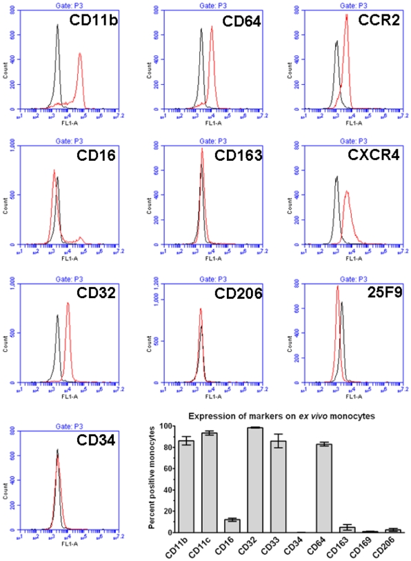
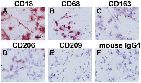
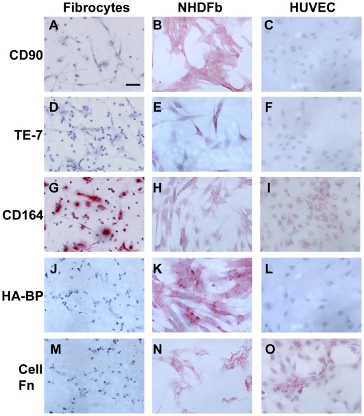
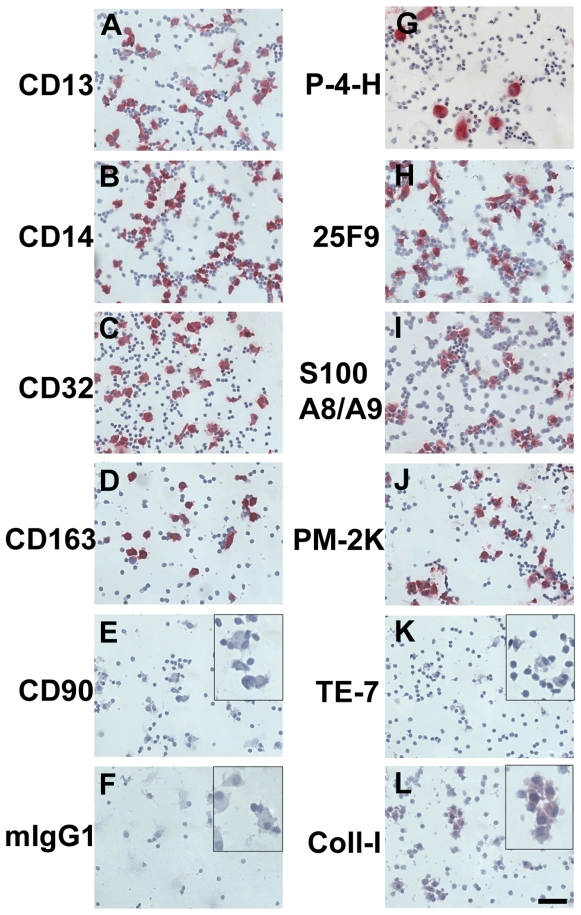
Verification
Claims the paper makes
The claims and statistics of this paper haven’t been checked yet.
Run the check — free, no account needed. It reads the open-access text and checks the sources it cites.
Claims other papers make about this one
Each is checked against this paper’s own text: does it say what the citing paper says it does?
In the citing paper
“Collectively, RhoH activity in the myeloid cell is critical in promoting macrophage terminal differentiation by the repression of the interlinked Cdc42 and Wnt intracellular signalling pathways and inflammatory effector proteins, CD11b and CD93.”
From The Role of RhoH in TCR Signalling and Its Involvement in Diseases (2021) · cites this paper as [67]
In the citing paper
“In addition, RHOH induction is demonstrated to cause the down‐regulation of CD11b and CD93 protein expression characteristic of monocytes terminally differentiating into macrophages (Figure 4C) [54, 55].”
From Bimodal expression of RHOH during myelomonocytic differentiation: Implications for the expansion of AML differentiation therapy (2021) · cites this paper as [55]
In the citing paper
“Thus this terminal phase of monocytic differentiation is characterized by repression of inflammatory effector proteins such as the β2 integrin CD11b and CD93 [54, 55].”
From Bimodal expression of RHOH during myelomonocytic differentiation: Implications for the expansion of AML differentiation therapy (2021) · cites this paper as [55]
In the citing paper
“The down‐regulation of CD11b and CD93 protein expression are individual hallmarks of the differentiation of monocytes into macrophages (Figure 4A, B) [54, 55].”
From Bimodal expression of RHOH during myelomonocytic differentiation: Implications for the expansion of AML differentiation therapy (2021) · cites this paper as [55]
In the citing paper
“These monocytes and myeloblasts were treated with phorbol ester for 48 h and terminal differentiation into macrophages confirmed by their acquisition of a strong adhesive phenotype, morphological spreading, and characteristic down‐regulation of CD11b and CD93 protein expression (Figure 4A, B, Supplemental Figure S1) [54, 55].”
From Bimodal expression of RHOH during myelomonocytic differentiation: Implications for the expansion of AML differentiation therapy (2021) · cites this paper as [55]
Self-check
Percentages, sums and reported differences re-computed from the paper’s own numbers.
The self-check needs the paper’s full text.
Citations
Cites
The chemokine system in diverse forms of macrophage activation and polarization
MANTOVANI, A., SICA, A., SOZZANI, S., ALLAVENA, P., VECCHI, A., & LOCATI, M. (2004) · Trends in Immunology · doi:10.1016/j.it.2004.09.015 · Cited by 6,534
Singer, A. J., & Clark, R. A. F. (1999) · New England Journal of Medicine · doi:10.1056/nejm199909023411006 · Cited by 6,106
Monocyte and macrophage heterogeneity
Gordon, S., & Taylor, P. R. (2005) · Nature Reviews Immunology · doi:10.1038/nri1733 · Cited by 5,190
The many faces of macrophage activation
Mosser, D. M. (2003) · Journal of Leukocyte Biology · doi:10.1189/jlb.0602325 · Cited by 1,902
Peripheral Blood Fibrocytes: Differentiation Pathway and Migration to Wound Sites
Abe, R., Donnelly, S. C., Peng, T., Bucala, R., & Metz, C. N. (2001) · The Journal of Immunology · doi:10.4049/jimmunol.166.12.7556 · Cited by 967
Reduction of Bleomycin-Induced Pulmonary Fibrosis by Serum Amyloid P
Pilling, D., Roife, D., Wang, M., Ronkainen, S. D., Crawford, J. R., Travis, E. L., & Gomer, R. H. (2007) · The Journal of Immunology · doi:10.4049/jimmunol.179.6.4035 · Cited by 230
Self-citationOlson, M. C., Scott, E. W., Hack, A. A., Su, G. H., Tenen, D. G., Singh, H., & Simon, M. C. (1995) · Immunity · doi:10.1016/1074-7613(95)90060-8 · Cited by 191
Research works printed but not linked: 79
- P Martin, 1997, Wound Healing—Aiming for Perfect Skin Regeneration., Science, 276, 75, 10.1126/science.276.5309.75 · DOI
- MF Pittenger, 1999, Multilineage potential of adult human mesenchymal stem cells., Science, 284, 143, 10.1126/science.284.5411.143 · DOI · Paper page
- DG Phinney, 2007, Concise review: mesenchymal stem/multipotent stromal cells: the state of transdifferentiation and modes of tissue repair—current views., Stem Cells, 25, 2896, 10.1634/stemcells.2007-0637 · DOI
- R Kalluri, 2003, Epithelial-mesenchymal transition and its implications for fibrosis., Journal Of Clinical Investigation, 112, 1776, 10.1172/JCI200320530 · DOI
- R Bucala, 1994, Circulating fibrocytes define a new leukocyte subpopulation that mediates tissue repair., Molecular Medicine, 1, 71, 10.1007/BF03403533 · DOI
- BN Gomperts, 2007, Fibrocytes in lung disease., Journal Of Leukocyte Biology, 82, 449, 10.1189/jlb.0906587 · DOI
- A Bellini, 2007, The role of the fibrocyte, a bone marrow-derived mesenchymal progenitor, in reactive and reparative fibroses., Lab Invest, 87, 858, 10.1038/labinvest.3700654 · DOI
- JF Wang, 2007, Fibrocytes from burn patients regulate the activities of fibroblasts., Wound Repair Regen, 15, 113, 10.1111/j.1524-475X.2006.00192.x · DOI
- SB Haudek, 2006, Bone Marrow-derived Fibroblast Precursors Mediate Ischemic Cardiomyopathy in Mice., Proceedings of the National Academy of Sciences, 103, 18284, 10.1073/pnas.0608799103 · DOI
- B Naik-Mathuria, 2008, Serum amyloid P inhibits dermal wound healing., Wound Repair and Regeneration, 16, 266, 10.1111/j.1524-475X.2008.00366.x · DOI
- L Yang, 2002, Peripheral Blood Fibrocytes from Burn Patients: Identification and Quantification of Fibrocytes in Adherent Cells Cultured from Peripheral Blood Mononuclear Cells., Laboratory Investigation, 82, 1183, 10.1097/01.LAB.0000027841.50269.61 · DOI
- D Pilling, 2003, Inhibition of fibrocyte differentiation by serum amyloid P., Journal Of Immunology, 17, 5537, 10.4049/jimmunol.171.10.5537 · DOI
- D Pilling, 2006, Aggregated IgG inhibits the differentiation of human fibrocytes., Journal Of Leukocyte Biology, 79, 1242, 10.1189/jlb.0805456 · DOI
- DD Shao, 2008, Pivotal Advance: Th-1 cytokines inhibit, and Th-2 cytokines promote fibrocyte differentiation., Journal of Leukocyte Biology, 83, 1323, 10.1189/jlb.1107782 · DOI
- J Wang, 2007, Accelerated wound healing in leukocyte-specific, protein 1-deficient mouse is associated with increased infiltration of leukocytes and fibrocytes., J Leukoc Biol, 82, 1554, 10.1189/jlb.0507306 · DOI
- RJ Phillips, 2004, Circulating fibrocytes traffic to the lungs in response to CXCL12 and mediate fibrosis., J Clin.Invest, 114, 438, 10.1172/JCI200420997 · DOI
- N Hashimoto, 2004, Bone marrow-derived progenitor cells in pulmonary fibrosis., Journal Of Clinical Investigation, 113, 243, 10.1172/JCI200418847 · DOI
- BB Moore, 2005, CCR2-Mediated Recruitment of Fibrocytes to the Alveolar Space after Fibrotic Injury., American Journal Of Pathology, 166, 675, 10.1016/S0002-9440(10)62289-4 · DOI
- BB Moore, 2006, The Role of CCL12 in the Recruitment of Fibrocytes and Lung Fibrosis., American Journal Of Respiratory Cell And Molecular Biology, 35, 175, 10.1165/rcmb.2005-0239OC · DOI
- A Andersson-Sjöland, 2008, Fibrocytes are a potential source of lung fibroblasts in idiopathic pulmonary fibrosis., The International Journal of Biochemistry & Cell Biology, 40, 2129, 10.1016/j.biocel.2008.02.012 · DOI
- L Yang, 2005, Identification of fibrocytes in postburn hypertrophic scar., Wound Repair and Regeneration, 13, 398, 10.1111/j.1067-1927.2005.130407.x · DOI
- M Salmon, 1994, The progressive differentiation of primed T cells is associated with an increasing susceptibility to apoptosis., European Journal Of Immunology, 24, 892, 10.1002/eji.1830240417 · DOI
- CD Buckley, 2000, Persistent induction of the chemokine receptor CXCR4 by TGF-beta1 on synovial T cells contributes to their accumulation within the rheumatoid synovium., J.Immunol, 165, 3423, 10.4049/jimmunol.165.6.3423 · DOI
- R García-Vicuña, 2004, CC and CXC chemokine receptors mediate migration, proliferation, and matrix metalloproteinase production by fibroblast-like synoviocytes from rheumatoid arthritis patients., Arthritis & Rheumatism, 50, 3866, 10.1002/art.20615 · DOI
- H Zhao, 2009, Tumor-promoting phenotype of CD90(hi) prostate cancer-associated fibroblasts., Prostate, 69, 991, 10.1002/pros.20946 · DOI
- Y Hosokawa, 2005, CXCL12 and CXCR4 expression by human gingival fibroblasts in periodontal disease., Clin Exp Immunol, 141, 467, 10.1111/j.1365-2249.2005.02852.x · DOI
- QH Erdag G, 2008, CD34-positive dendritic cells disappear from scars but are increased in pericicatricial tissue., Journal of Cutaneous Pathology, 35, 752, 10.1111/j.1600-0560.2007.00895.x · DOI
- BJ Nickoloff, 1991, The human progenitor cell antigen (CD34) is localized on endothelial cells, dermal dendritic cells, and perifollicular cells in formalin-fixed normal skin, and on proliferating endothelial cells and stromal spindle-shaped cells in Kaposi's sarcoma., Arch Dermatol, 127, 523, 10.1001/archderm.1991.04510010091009 · DOI
- DL Simmons, 1992, Molecular cloning of a cDNA encoding CD34, a sialomucin of human hematopoietic stem cells., Journal Of Immunology, 148, 267, 10.4049/jimmunol.148.1.267 · DOI
- J Vaage, 1990, Production of collagen type I by mouse peritoneal macrophages., J Leukoc Biol, 48, 274, 10.1002/jlb.48.3.274 · DOI
- MJ Liptay, 1993, Neointimal macrophages colocalize with extracellular matrix gene expression in human atherosclerotic pulmonary arteries., J Clin Invest, 91, 588, 10.1172/JCI116238 · DOI
- A Gratchev, 2001, Alternatively activated macrophages differentially express fibronectin and its splice variants and the extracellular matrix protein betaIG-H3., Scand J Immunol, 53, 386, 10.1046/j.1365-3083.2001.00885.x · DOI · Paper page
- JS Duffield, 2003, The inflammatory macrophage: a story of Jekyll and Hyde., Clin Sci (Lond), 104, 27, 10.1042/cs1040027 · DOI
- M Schnoor, 2008, Production of Type VI Collagen by Human Macrophages: A New Dimension in Macrophage Functional Heterogeneity., J Immunol, 180, 5707, 10.4049/jimmunol.180.8.5707 · DOI
- M Lucattelli, 2003, Collagen phagocytosis by lung alveolar macrophages in animal models of emphysema., Eur Respir J, 22, 728, 10.1183/09031936.03.00047603 · DOI
- RP Phipps, 1989, Characterization of two major populations of lung fibroblasts: distinguishing morphology and discordant display of Thy 1 and class II MHC., Am J Respir Cell Mol Biol, 1, 65, 10.1165/ajrcmb/1.1.65 · DOI
- HY Chang, 2002, Diversity, topographic differentiation, and positional memory in human fibroblasts., Proc Natl Acad Sci U S A, 99, 12877, 10.1073/pnas.162488599 · DOI
- CD Buckley, 2001, Fibroblasts regulate the switch from acute resolving to chronic persistent inflammation., Trends Immunol, 22, 199, 10.1016/S1471-4906(01)01863-4 · DOI · Paper page
- G Parsonage, 2003, Global gene expression profiles in fibroblasts from synovial, skin and lymphoid tissue reveals distinct cytokine and chemokine expression patterns., Thromb.Haemost, 90, 688, 10.1160/TH03-04-0208 · DOI
- S Halder, 2005, Generation and characterization of novel stromal specific antibodies., Cell Res, 15, 739, 10.1038/sj.cr.7290344 · DOI
- JR MacFadyen, 2005, Endosialin (TEM1, CD248) is a marker of stromal fibroblasts and is not selectively expressed on tumour endothelium., FEBS Lett, 579, 2569, 10.1016/j.febslet.2005.03.071 · DOI
- S Lax, 2007, CD248/Endosialin is dynamically expressed on a subset of stromal cells during lymphoid tissue development, splenic remodeling and repair., FEBS Lett, 581, 3550, 10.1016/j.febslet.2007.06.063 · DOI
- CJ Cabezon T, 2007, Expression of S100A4 by a variety of cell types present in the tumor microenvironment of human breast cancer., International Journal of Cancer, 121, 1433, 10.1002/ijc.22850 · DOI
- SD Ko, 1977, Fibroblast heterogeneity and prostaglandin regulation of subpopulations., Proc Natl Acad Sci U S A, 74, 3429, 10.1073/pnas.74.8.3429 · DOI
- L Peduto, 2009, Inflammation Recapitulates the Ontogeny of Lymphoid Stromal Cells., J Immunol, 182, 5789, 10.4049/jimmunol.0803974 · DOI
- F Strutz, 1995, Identification and characterization of a fibroblast marker: FSP1., J Cell Biol, 130, 393, 10.1083/jcb.130.2.393 · DOI
- JE Fonseca, 2002, Macrophage subpopulations in rheumatoid synovium: reduced CD163 expression in CD4+ T lymphocyte-rich microenvironments., Arthritis And Rheumatism, 46, 1210, 10.1002/art.10207 · DOI
- E Kunisch, 2004, Macrophage specificity of three anti-CD68 monoclonal antibodies (KP1, EBM11, and PGM1) widely used for immunohistochemistry and flow cytometry., Ann Rheum Dis, 63, 774, 10.1136/ard.2003.013029 · DOI
- T Inoue, 2005, Antibodies against macrophages that overlap in specificity with fibroblasts., Kidney Int, 67, 2488, 10.1111/j.1523-1755.2005.00358.x · DOI
- E Gottfried, 2008, Expression of CD68 in non-myeloid cell types., Scand J Immunol, 67, 453, 10.1111/j.1365-3083.2008.02091.x · DOI
- S Becker, 1987, Colony-stimulating factor-induced monocyte survival and differentiation into macrophages in serum-free cultures., J Immunol, 139, 3703, 10.4049/jimmunol.139.11.3703 · DOI
- W Brugger, 1991, Macrophage colony-stimulating factor is required for human monocyte survival and acts as a cofactor for their terminal differentiation to macrophages in vitro., J Leukoc Biol, 49, 483, 10.1002/jlb.49.5.483 · DOI
- DA Hume, 2002, The mononuclear phagocyte system revisited., Journal Of Leukocyte Biology, 72, 621, 10.1189/jlb.72.4.621 · DOI
- KJ Way, 2009, The generation and properties of human macrophage populations from hemopoietic stem cells., J Leukoc Biol, 85, 766, 10.1189/jlb.1108689 · DOI
- M Salmon, 1997, Inhibition of T cell apoptosis in the rheumatoid synovium., Journal Of Clinical Investigation, 99, 439, 10.1172/JCI119178 · DOI
- D Pilling, 1999, Interferon-β mediates stromal cell rescue of T cells from apoptosis., European Journal Of Immunology, 29, 1041, 10.1002/(SICI)1521-4141(199903)29:03<1041::AID-IMMU1041>3.0.CO;2-# · DOI
- J Olert, 2001, HOPE fixation: a novel fixing method and paraffin-embedding technique for human soft tissues., Pathol Res Pract, 197, 823, 10.1078/0344-0338-00166 · DOI
- AN Akbar, 1996, Interleukin-2 receptor common gamma-chain signaling cytokines regulate activated T cell apoptosis in response to growth factor withdrawal - selective induction of anti-apoptotic (Bcl-2, Bcl-xL) but not pro-apoptotic (Bax, Bcl-xS) gene-expression., European Journal Of Immunology, 26, 294, 10.1002/eji.1830260204 · DOI
- JM Faint, 1999, Quantitative flow cytometry for the analysis of T cell receptor Valpha chain expression., Journal Of Immunological Methods, 27, 53, 10.1016/S0022-1759(99)00027-7 · DOI
- LW Terstappen, 1990, Quantitative comparison of myeloid antigens on five lineages of mature peripheral blood cells., J Leukoc Biol, 48, 138, 10.1002/jlb.48.2.138 · DOI
- S Rosseau, 2000, Phenotypic characterization of alveolar monocyte recruitment in acute respiratory distress syndrome., Am J Physiol Lung Cell Mol Physiol, 279, L25, 10.1152/ajplung.2000.279.1.L25 · DOI
- DJ Flavell, 1987, Identification of tissue histiocytes on paraffin sections by a new monoclonal antibody., J Histochem Cytochem, 35, 1217, 10.1177/35.11.3309045 · DOI
- G Zwadlo, 1985, A monoclonal antibody to a differentiation antigen present on mature human macrophages and absent from monocytes., J Immunol, 134, 1487, 10.4049/jimmunol.134.3.1487 · DOI
- M Takeya, 1991, A new monoclonal antibody, PM-2K, specifically recognizes tissue macrophages but not blood monocytes., The Journal of pathology, 163, 315, 10.1002/path.1711630408 · DOI
- SS Bohlson, 2005, CD93 is rapidly shed from the surface of human myeloid cells and the soluble form is detected in human plasma., J Immunol, 175, 1239, 10.4049/jimmunol.175.2.1239 · DOI
- E Grage-Griebenow, 2001, Heterogeneity of human peripheral blood monocyte subsets., J.Leukoc.Biol, 69, 11, 10.1189/jlb.69.1.11 · DOI
- J Faust, 1999, Osteoclast markers accumulate on cells developing from human peripheral blood mononuclear precursors., J Cell Biochem, 72, 67, 10.1002/(SICI)1097-4644(19990101)72:1<67::AID-JCB8>3.0.CO;2-A · DOI
- SB Haudek, 2008, Fc Receptor Engagement Mediates Differentiation of Cardiac Fibroblast Precursor Cells., Proceedings of the National Academy of Sciences, 105, 10179, 10.1073/pnas.0804910105 · DOI
- B Passlick, 1989, Identification and characterization of a novel monocyte subpopulation in human peripheral blood., Blood, 74, 2527, 10.1182/blood.V74.7.2527.2527 · DOI
- M Schmidt, 2003, Identification of Circulating Fibrocytes as Precursors of Bronchial Myofibroblasts in Asthma., The Journal of Immunology, 171, 380, 10.4049/jimmunol.171.1.380 · DOI
- 2007, Leukocyte and Stromal Cell Molecules: The CD Markers: John Wiley & Sons.
- G Balciunaite, 2005, A B220+ CD117+ CD19- hematopoietic progenitor with potent lymphoid and myeloid developmental potential., Eur J Immunol, 35, 2019, 10.1002/eji.200526318 · DOI
- Y Su, 2005, Glycosylation Influences the Lectin Activities of the Macrophage Mannose Receptor., J Biol Chem, 280, 32811, 10.1074/jbc.M503457200 · DOI
- NC Henderson, 2008, Galectin-3 expression and secretion links macrophages to the promotion of renal fibrosis., Am J Pathol, 172, 288, 10.2353/ajpath.2008.070726 · DOI · Paper page
- GS Firestein, 1987, Down regulation of human monocyte differentiation antigens by interferon gamma., Cell Immunol, 104, 343, 10.1016/0008-8749(87)90036-0 · DOI
- AL Epstein, 1987, Immunohistochemical characterization of a 183 KD myeloid-specific-DNA-binding protein in B5 fixed, paraffin-embedded tissues, and bone marrow aspirates by monoclonal antibody BM-1., Blood, 70, 1124, 10.1182/blood.V70.4.1124.1124 · DOI
- FO Martinez, 2008, Macrophage activation and polarization., Front Biosci, 13, 453, 10.2741/2692 · DOI · Paper page
- L Zeng, 1996, Interspecies reactivities of anti-human macrophage monoclonal antibodies to various animal species., J Histochem Cytochem, 44, 845, 10.1177/44.8.8756757 · DOI
- O Dewald, 2004, Of mice and dogs: species-specific differences in the inflammatory response following myocardial infarction., Am J Pathol, 164, 665, 10.1016/S0002-9440(10)63154-9 · DOI
Cited by
Molecular and cellular mechanisms of liver fibrosis and its regression
Kisseleva, T., & Brenner, D. (2021) · Nature Reviews Gastroenterology & Hepatology · doi:10.1038/s41575-020-00372-7 · Cited by 2,024
Immune mechanisms in pulmonary fibrosis
Kolahian, S., Fernandez, I. E., Eickelberg, O., & Hartl, D. (2016) · American Journal of Respiratory Cell and Molecular Biology · doi:10.1165/rcmb.2016-0121tr · Cited by 349
TGF-beta driven lung fibrosis is macrophage dependent and blocked by Serum amyloid P
Murray, L. A., Chen, Q., Kramer, M. S., Hesson, D. P., Argentieri, R. L., Peng, X., Gulati, M., Homer, R. J., Russell, T., van Rooijen, N., et al. (2011) · The International Journal of Biochemistry & Cell Biology · doi:10.1016/j.biocel.2010.10.013 · Cited by 362
Nuovo, G. J., Hagood, J. S., Magro, C. M., Chin, N., Kapil, R., Davis, L., Marsh, C. B., & Folcik, V. A. (2012) · Modern Pathology · doi:10.1038/modpathol.2011.166 · Cited by 123
Tourkina, E., Bonner, M., Oates, J., Hofbauer, A., Richard, M., Znoyko, S., Visconti, R. P., Zhang, J., Hatfield, C. M., Silver, R. M., et al. (2011) · Fibrogenesis & Tissue Repair · doi:10.1186/1755-1536-4-15 · Cited by 104
The Development of Serum Amyloid P as a Possible Therapeutic
Pilling, D., & Gomer, R. H. (2018) · Frontiers in Immunology · doi:10.3389/fimmu.2018.02328 · Cited by 92
Self-citationEpithelial-mesenchymal transition in chronic liver disease: fibrogenesis or escape from death?
Pinzani, M. (2011) · Journal of Hepatology · doi:10.1016/j.jhep.2011.02.001 · Cited by 86
Frescas, D., Hall, B. M., Strom, E., Virtuoso, L. P., Gupta, M., Gleiberman, A. S., Rydkina, E., Balan, V., Vujcic, S., Chernova, O. B., et al. (2017) · Cell Cycle · doi:10.1080/15384101.2017.1339850 · Cited by 34
The Role of RhoH in TCR Signalling and Its Involvement in Diseases
Ahmad Mokhtar, A. M., Hashim, I. F., Mohd Zaini Makhtar, M., Salikin, N. H., & Amin-Nordin, S. (2021) · Cells · doi:10.3390/cells10040950 · Cited by 25
Pilling, D., Cox, N., Thomson, M. A., Karhadkar, T. R., & Gomer, R. H. (2019) · The American Journal of Pathology · doi:10.1016/j.ajpath.2019.08.005 · Cited by 17
Self-citationThe characterization of fibrocyte-like cells: A novel fibroblastic cell of the placenta
Riddell, M. R., Winkler-Lowen, B., Chakrabarti, S., Dunk, C., Davidge, S. T., & Guilbert, L. J. (2012) · Placenta · doi:10.1016/j.placenta.2011.12.012 · Cited by 14
Galiègue‐Zouitina, S., Fu, Q., Carton‐Latreche, C., Poret, N., Cheok, M., Leprêtre, F., Figeac, M., Quesnel, B., El Bouazzati, H., & Shelley, C. S. (2021) · eJHaem · doi:10.1002/jha2.128 · Cited by 7
Metadata
- Authors
- Pilling, D., Fan, T., Huang, D., Kaul, B., & Gomer, R. H.
- Venue
- PLoS ONE
- Publisher
- Public Library of Science (PLoS)
- Published
- 2009-10-16
- Volume
- 4
- Issue
- 10
- Pages
- e7475
- ISSN
- 1932-6203
- PMC
- PMC2759556
- Cited by
- 433
- References
- 7
- Language
- en
About these checks
Registry, retraction, Cites and statistics results are computed, not judged, with one exception: a printed reference that no index lists is read once by a model before it counts as a citation error. Claims are extracted by a model and kept only when their quote appears verbatim in the text; each cited source is then checked, with a model’s help, for whether it says what the claim relies on. Those verdicts can be wrong — read the claim’s quote and the cited source before relying on one.
Statistics and arithmetic are recomputed, not judged.
Confidence bands are shown only for fields calibrated on our annotated test set. None is calibrated yet, so no band is shown.
What counts as a claim ›How we measure confidence ›How we check this ›
Are you an author, or do you see a problem with this page? Request a review
