

Human Engineered Heart Tissue as a Versatile Tool in Basic Research and Preclinical Toxicology
Open accessThe DOI matches its Crossref record; 31 of the 31 references it cites were found in a registry or an index.
Abstract
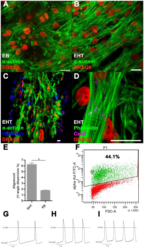
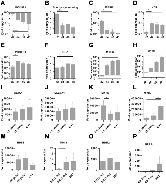
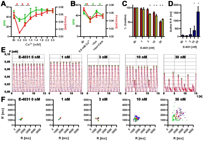
Human embryonic stem cell (hESC) progenies hold great promise as surrogates for human primary cells, particularly if the latter are not available as in the case of cardiomyocytes. However, high content experimental platforms are lacking that allow the function of hESC-derived cardiomyocytes to be studied under relatively physiological and standardized conditions. Here we describe a simple and robust protocol for the generation of fibrin-based human engineered heart tissue (hEHT) in a 24-well format using an unselected population of differentiated human embryonic stem cells containing 30-40% α-actinin-positive cardiac myocytes. Human EHTs started to show coherent contractions 5-10 days after casting, reached regular (mean 0.5 Hz) and strong (mean 100 µN) contractions for up to 8 weeks. They displayed a dense network of longitudinally oriented, interconnected and cross-striated cardiomyocytes. Spontaneous hEHT contractions were analyzed by automated video-optical recording and showed chronotropic responses to calcium and the β-adrenergic agonist isoprenaline. The proarrhythmic compounds E-4031, quinidine, procainamide, cisapride, and sertindole exerted robust, concentration-dependent and reversible decreases in relaxation velocity and irregular beating at concentrations that recapitulate findings in hERG channel assays. In conclusion this study establishes hEHT as a simple in vitro model for heart research.
Overview
The claims and statistics of this paper haven’t been checked yet.
Free: it reads the open-access text and checks the first 5 claims that cite a source we can check. Create a free account to check every claim.
Integrity signals
Registry
Cites
Statistics
Figures

Verification
Claims the paper makes
The claims and statistics of this paper haven’t been checked yet.
Free: it reads the open-access text and checks the first 5 claims that cite a source we can check. Create a free account to check every claim.
Claims other papers make about this one
Each is checked against this paper’s own text: does it say what the citing paper says it does?
The source backs part of the claim, not all of it — or backs it for a different population, condition or comparison.
In the citing paper
“but not pressure- and volume-based metrics directly representing pump performance of the heart”
From Bioengineering an electro-mechanically functional miniature ventricular heart chamber from human pluripotent stem cells (2018) · cites this paper as [16]
Experimental — accuracy not yet measuredThe cited source says what the claim attributes to it.
In the citing paper
“engineered cardiac thin films, micro-tissues and strips allow the assessment of twitch force [9,12–19]”
From Bioengineering an electro-mechanically functional miniature ventricular heart chamber from human pluripotent stem cells (2018) · cites this paper as [16]
Experimental — accuracy not yet measured
Self-check
Percentages, sums and reported differences re-computed from the paper’s own numbers.
The self-check needs the paper’s full text.
Citations
Cites
Proceedings of the National Academy of Sciences of the United States of America
Dowling, J. E., & Wald, G. (2009) · Nutrition Reviews · doi:10.1111/j.1753-4887.1981.tb06752.x · Cited by 17,893
Laflamme, M. A., Chen, K. Y., Naumova, A. V., Muskheli, V., Fugate, J. A., Dupras, S. K., Reinecke, H., Xu, C., Hassanipour, M., Police, S., et al. (2007) · Nature Biotechnology · doi:10.1038/nbt1327 · Cited by 2,206 · cited 2×
“One of the current limitations of hESC-derived cardiac myocytes is their immature and unorganized phenotype in the EB format or 2D cultures [3,5].”
BackgroundFeeder-free growth of undifferentiated human embryonic stem cells
Xu, C., Inokuma, M. S., Denham, J., Golds, K., Kundu, P., Gold, J. D., & Carpenter, M. K. (2001) · Nature Biotechnology · doi:10.1038/nbt1001-971 · Cited by 1,870 · cited 1×
“with CF1-MEF conditioned medium according to published protocols [28].”
MethodKehat, I., Kenyagin‐Karsenti, D., Snir, M., Segev, H., Amit, M., Gepstein, A., Livne, E., Binah, O., Itskovitz‐Eldor, J., & Gepstein, L. (2001) · Journal of Clinical Investigation · doi:10.1172/jci12131 · Cited by 1,814 · cited 1×
“differentiation in serum containing media yielding 8% of beating embryoid bodies (EBs) [1].”
BackgroundCai, C.-L., Liang, X., Shi, Y., Chu, P.-H., Pfaff, S. L., Chen, J., & Evans, S. (2003) · Developmental Cell · doi:10.1016/s1534-5807(03)00363-0 · Cited by 1,610 · cited 1×
“Islet-1 (ISL1) a progenitor marker of the development of the secondary heart field [21] was significantly upregulated on day 6 and 8.”
BackgroundKehat, I., Kenyagin‐Karsenti, D., Snir, M., et al. (2001) · Journal of Clinical Investigation · doi:10.1172/jci200112131 · Cited by 1,536
REDFERN, W., CARLSSON, L., DAVIS, A., LYNCH, W., MACKENZIE, I., PALETHORPE, S., SIEGL, P., STRANG, I., SULLIVAN, A., & WALLIS, R. (2003) · Cardiovascular Research · doi:10.1016/s0008-6363(02)00846-5 · Cited by 1,532 · cited 2×
“Concentrations were chosen according to published IC50 in hERG assays [23,24].”
MethodHuman cardiovascular progenitor cells develop from a KDR+ embryonic-stem-cell-derived population
Yang, L., Soonpaa, M. H., Adler, E. D., Roepke, T. K., Kattman, S. J., Kennedy, M., Henckaerts, E., Bonham, K., Abbott, G. W., Linden, R. M., et al. (2008) · Nature · doi:10.1038/nature06894 · Cited by 1,389 · cited 3×
“Cardiomyocyte differentiation was performed with a modified EB-based, three-stage, growth factor cocktail protocol [2].”
MethodInfluentialEngineered heart tissue grafts improve systolic and diastolic function in infarcted rat hearts
Zimmermann, W.-H., Melnychenko, I., Wasmeier, G., Didié, M., Naito, H., Nixdorff, U., Hess, A., Budinsky, L., Brune, K., Michaelis, B., et al. (2006) · Nature Medicine · doi:10.1038/nm1394 · Cited by 993 · cited 1×
“The principal usefulness of these techniques for in vitro testing [17,18] and cardiac repair [19] has been demonstrated.”
BackgroundSelf-citationTissue Engineering of a Differentiated Cardiac Muscle Construct
Zimmermann, W.-H., Schneiderbanger, K., Schubert, P., Didié, M., Münzel, F., Heubach, J. F., Kostin, S., Neuhuber, W. L., & Eschenhagen, T. (2002) · Circulation Research · doi:10.1161/hh0202.103644 · Cited by 917 · cited 1×
“EHTs from neonatal rat hearts develop a high degree of cellular differentiation, longitudinal orientation, intercellular coupling and force generation [15,16].”
BackgroundSelf-citation
Research works printed but not linked: 2
- [3]Mummery C Ward-van Oostwaard D Doevendans P Spijker R Brink S van den 2003 Differentiation of human embryonic stem cells to cardiomyocytes: role of coculture with visceral endoderm-like cells. Circulation 107 2733 40 12742992
- [12]FDA 2005 Guidance for Industry S7B Nonclinical Evaluation of the Potential for Delayed Ventricular Repolarization (QT Interval Prolongation) by Human Pharmaceuticals. 16237859
Cited by
Items 201–300 of 361
Daley, M. C. (2022) · ALTEX · doi:10.14573/altex.2109131 · Cited by 24
“classes of cardiotoxicity concurrently could allow for identification of the predominant toxicity (Beauchamp et al., 2015, 2020; Burridge et al., 2016; Clements et al., 2015; Forsythe et al., 2018; Schaaf et al., 2011; Chaudhari et al., 2018).”
BackgroundYe, F., Yuan, F., Li, X., Cooper, N. G. F., Tinney, J. P., & Keller, B. B. (2013) · Physiological Reports · doi:10.1002/phy2.78 · Cited by 22
“We generated ECTs from day 14.5 rat embryo ventricular cells, as previously published, and then conditioned constructs after 5 days in culture for 48 h with mechanical stretch (5%, 0.5 Hz) and/or the p38 MAPK (p38 mitogen-activated protein kinase) inhibitor BIRB796.”
MethodInfluentialMicroengineered systems with iPSC-derived cardiac and hepatic cells to evaluate drug adverse effects
Dame, K., & Ribeiro, A. J. S. (2020) · Experimental Biology and Medicine · doi:10.1177/1535370220959598 · Cited by 24
Swiatlowska, P., & Iskratsch, T. (2021) · Biophysical Reviews · doi:10.1007/s12551-021-00837-2 · Cited by 20
Construction of Defined Human Engineered Cardiac Tissues to Study Mechanisms of Cardiac Cell Therapy
Cashman, T. J., Josowitz, R., Gelb, B. D., Li, R. A., Dubois, N. C., & Costa, K. D. (2016) · Journal of Visualized Experiments · doi:10.3791/53447-v · Cited by 7
Contractile work directly modulates mitochondrial protein levels in human engineered heart tissues
Ng, R., Sewanan, L. R., Brill, A. L., Stankey, P. P., Li, X., Qyang, Y., Ehrlich, B. E., & Campbell, S. G. (2020) · American Journal of Physiology-Heart and Circulatory Physiology · doi:10.1152/ajpheart.00055.2020 · Cited by 21
Rhoden, A., Schulze, T., Pietsch, N., Christ, T., Hansen, A., & Eschenhagen, T. (2022) · American Journal of Physiology-Heart and Circulatory Physiology · doi:10.1152/ajpheart.00534.2021 · Cited by 21
Self-citationBasic Research Approaches to Evaluate Cardiac Arrhythmia in Heart Failure and Beyond
Cumberland, M. J., Riebel, L. L., Roy, A., O’Shea, C., Holmes, A. P., Denning, C. N., Kirchhof, P. F., Rodríguez, B., & Gehmlich, K. (2022) · Frontiers in Physiology · doi:10.3389/fphys.2022.806366 · Cited by 23
Regulation of ICa,L and force by PDEs in human‐induced pluripotent stem cell‐derived cardiomyocytes
Saleem, U., Ismaili, D., Mannhardt, I., Pinnschmidt, H. O., Schulze, T., Christ, T., Eschenhagen, T., & Hansen, A. (2020) · British Journal of Pharmacology · doi:10.1111/bph.15032 · Cited by 18
“Force was analysed by video-optical recording (Hansen et al., 2010; Schaaf et al., 2011).”
Self-citationSchwan, J., & Campbell, S. G. (2015) · Biomarker Insights · doi:10.4137/bmi.s23912 · Cited by 23
Rivera‐Arbeláez, J. M., Cofiño‐Fabrés, C., Schwach, V., Boonen, T., ten Den, S. A., Vermeul, K., van den Berg, A., Segerink, L. I., Ribeiro, M. C., & Passier, R. C. (2022) · PLoS ONE · doi:10.1371/journal.pone.0266834 · Cited by 24
Windt, L. M., Wiendels, M., Dostanić, M., Bellin, M., Sarro, P. M., Mastrangeli, M., Mummery, C. L., & van Meer, B. J. (2023) · Biochemical and Biophysical Research Communications · doi:10.1016/j.bbrc.2023.09.034 · Cited by 18
Human induced pluripotent stem cell-derived cardiac muscle rings for biohybrid self-beating actuator
Morita, T., Nie, M., & Takeuchi, S. (2024) · Lab on a Chip · doi:10.1039/d4lc00276h · Cited by 19
Biomaterials for pluripotent stem cell engineering: from fate determination to vascularization
Seale, N. M., & Varghese, S. (2016) · Journal of Materials Chemistry B · doi:10.1039/c5tb02658j · Cited by 16
“Biomaterials have been extensively used in cardiac tissue engineering both in vitro and in vivo(60, 61).”
MethodModulation of hepatic mitochondrial energy efficiency with age
Lionetti, L., Crescenzo, R., Mollica, M. P., Tasso, R., Barletta, A., Liverini, G., & Iossa, S. (2004) · Cellular and Molecular Life Sciences · doi:10.1007/s00018-004-4073-4 · Cited by 20
Dolatshad, N. F., Hellen, N., Jabbour, R. J., Harding, S. E., & Földes, G. (2015) · Frontiers in Cell and Developmental Biology · doi:10.3389/fcell.2015.00076 · Cited by 20
“While S1P1 couples directly to the Gi pathway, the other receptor isoforms known also as endothelial differentiation gene 3, 5 (Edg-3 and -5) stimulate Gi, Gq, and G13 pathways (Ancellin and Hla, 1999; Windh et al., 1999).”
BackgroundHuman pluripotent stem cell-derived cardiomyocyte based models for cardiotoxicity and drug discovery
Zhao, Y., Korolj, A., Feric, N. T., & Radisic, M. (2016) · Expert Opinion on Drug Safety · doi:10.1080/14740338.2016.1223624 · Cited by 19
Cardiac Myocyte–Fibroblast Interactions and the Coronary Vasculature
Bowers, S., & Baudino, T. A. (2012) · Journal of Cardiovascular Translational Research · doi:10.1007/s12265-012-9407-2 · Cited by 17
Advances in the design, generation, and application of tissue-engineered myocardial equivalents
Bernava, G., & Iop, L. (2023) · Frontiers in Bioengineering and Biotechnology · doi:10.3389/fbioe.2023.1247572 · Cited by 15
“Among other glycoconjugates with high translational power, the protein fibrin is particularly attractive for an autologous approach since it can be generated by elements, as fibrinogen, isolated from the patient’s blood (Schaaf et al., 2011; Chang et al., 2022).”
BackgroundSchwarzová, B., Stüdemann, T., Sönmez, M. I., Rössinger, J., Pan, B., Eschenhagen, T., Stenzig, J., Wiegert, J. S., Christ, T., & Weinberger, F. (2023) · Pflügers Archiv - European Journal of Physiology · doi:10.1007/s00424-023-02869-x · Cited by 15
“Cell concentration was adjusted to 10–15×10 6 cells/ml. Fibrin-based human EHTs were generated in agarose casting molds with solid silicone racks [42] (100 μl per EHT, 1 × 10 6 cells).”
MethodSelf-citationOdedra, D., Chiu, L. L. Y., Reis, L. A., Rask, F., Chiang, K., & Radisic, M. (2010) · Springer eBooks · doi:10.1007/978-3-7091-0385-2_15 · Cited by 14
Engineering Functional Tissues
Moffat, K. L., Neal, R. A., Freed, L. E., & Guilak, F. (2013) · Elsevier eBooks · doi:10.1016/b978-0-12-398358-9.00013-6 · Cited by 14
Lappi, H., Kauppila, M., Aalto‐Setälä, K., & Mörö, A. (2023) · Bioprinting · doi:10.1016/j.bprint.2023.e00313 · Cited by 13
Bioprinting approaches in cardiac tissue engineering to reproduce blood-pumping heart function
Kim, M. J., Hwang, D. G., & Jang, J. (2024) · iScience · doi:10.1016/j.isci.2024.111664 · Cited by 22
“5,16,17,39,69,134,150 These structures can be readily fabricated using mold casting methods, wherein silicone posts or nanowires embedded within the mold enable cells to remodel and compact along with these constructs, thereby enhancing cellular alignment.”
Tissue-Engineering for the Study of Cardiac Biomechanics
Ma, S. P., & Vunjak‐Novakovic, G. (2016) · Journal of Biomechanical Engineering · doi:10.1115/1.4032355 · Cited by 15
“While significant advances have been made with engineering cardiac tissues using mechanical stimulation, the results have been inconsistent, with some groups not achieving improved contractile function with cyclic stretch [62], and others demonstrating immature electrophysiology in cells subjected to passive mechanica…”
BackgroundThree-dimensional cardiac models: a pre-clinical testing platform
Groen, E., Mummery, C. L., Yiangou, L., & Davis, R. P. (2024) · Biochemical Society Transactions · doi:10.1042/bst20230444 · Cited by 14
UESUGI, K., Shima, F., Fukumoto, K. I., Hiura, A., Tsukamoto, Y., Miyagawa, S., Sawa, Y., Akagi, T., Akashi, M., & Morishima, K. (2019) · Micromachines · doi:10.3390/mi10070487 · Cited by 13
Biodegradable Materials from Natural Origin for Tissue Engineering and Stem Cells Technologies
Atwa, A., Sofy, M. R., Fakhrelden, S. M., Darwish, O., Mehany, A. B. M., Sofy, A. R., & Bakry, S. (2022) · Springer eBooks · doi:10.1007/978-3-030-83783-9_63-1 · Cited by 18
Human induced pluripotent stem cell-derived closed-loop cardiac tissue for drug assessment
Li, J., Hua, Y., Liu, Y., Qu, X., Zhang, J., Ishida, M., Yoshida, N., Tabata, A., Miyoshi, H., Shiba, M., et al. (2024) · iScience · doi:10.1016/j.isci.2024.108992 · Cited by 16
Defined Engineered Human Myocardium for Disease Modeling, Drug Screening, and Heart Repair
Tiburcy, M., Meyer, T., Satin, P.-L., & Zimmermann, W. (2022) · Methods in molecular biology · doi:10.1007/978-1-0716-2261-2_14 · Cited by 13
Self-citationLieu, D. K., Turnbull, I. C., Costa, K. D., & Li, R. A. (2012) · Drug Discovery Today Disease Models · doi:10.1016/j.ddmod.2012.06.002 · Cited by 12
“By culturing hESC-CMs in a 3D muscle strand anchored at the ends and subjected to either passive static or cyclic stretch, these CMs aligned themselves in the direction of the stretch, and the mechanically conditioned hESC-CMs showed signs of hypertrophy and formed cellular electrical couplings similar to mature CMs […”
BackgroundInduced Pluripotent Stem Cell-Derived Cardiomyocytes: From Regulatory Status to Clinical Translation
Soares, C. S. P., & Ribeiro, M. H. L. (2023) · Tissue Engineering Part B Reviews · doi:10.1089/ten.teb.2023.0080 · Cited by 10
Induced pluripotent stem cell-derived cardiomyocytes—more show than substance?
Ormrod, B., & Ehler, E. (2023) · Biophysical Reviews · doi:10.1007/s12551-023-01099-w · Cited by 11
“In one particular study, the other cell types were identified as being predominantly fibroblasts and a role for the fibroblasts in successful formation of the EHTs was postulated, likely due to their production of extracellular matrix and secretion of growth factors (Schaaf et al. 2011).”
BackgroundFrom Human Pluripotent Stem Cells to 3D Cardiac Microtissues: Progress, Applications and Challenges
Branco, M. A., Cabral, J. M. S., & Diogo, M. M. (2020) · Bioengineering · doi:10.3390/bioengineering7030092 · Cited by 11
“A different strategy relies on the development of elliptic shaped or strip-like cardiac tissues, which are anchored and stretched between two flexible pillars [89,90,93,95,97,98].”
BackgroundSpreeuwel, V. A. A., Bax, N. A. M., & Bouten, C. V. C. (2014) · OA Tissue Engineering · Cited by 12
Application of human cardiac organoids in cardiovascular disease research
Zhang, H., Qu, P., Liu, J., Cheng, P., & Lei, Q. (2025) · Frontiers in Cell and Developmental Biology · doi:10.3389/fcell.2025.1564889 · Cited by 10
Dysregulation of N-terminal acetylation causes cardiac arrhythmia and cardiomyopathy
Yoshinaga, D., Craven, I., Feng, R., Prondzynski, M., Shani, K., Tharani, Y., Mayourian, J., Milosh, J., Walker, D., Bortolin, R. H., et al. (2025) · Nature Communications · doi:10.1038/s41467-025-58539-2 · Cited by 11
Suspended Tissue Open Microfluidic Patterning (STOMP)
Haack, A. J., Brown, L. G., Goldstein, A. J., Mulimani, P. S., Berthier, J., Viswanathan, A. R., Kopyeva, I., Whitten, J. M., Lin, A., Nguyen, S. H., et al. (2025) · Advanced Science · doi:10.1002/advs.202501148 · Cited by 9
“Free-standing engineered tissues suspended between two posts are commonly generated using a casting technique where posts are placed upside down within a well plate that contains a mold where a cell-laden hydrogel precursor solution is pipetted[62].”
Trends in cardiovascular engineering: Organizing the human heart
Tulloch, N. L., & Murry, C. E. (2013) · Trends in Cardiovascular Medicine · doi:10.1016/j.tcm.2013.04.001 · Cited by 10
“…(Birla et al., 2007; Caspi et al., 2007; Dar et al., 2002; Fink et al., 2000; Liau et al., 2011; Madden et al., 2010; Marsano et al., 2010; Schaaf et al., 2011; Zimmermann et al., 2002a, 2002b, 2006) and scaffold-free approaches (Kreutziger et al., 2011; Stevens et al., 2009), often using…”
Turnbull, I. C., Lieu, D. K., Li, R. A., & Costa, K. D. (2012) · Drug Discovery Today Disease Models · doi:10.1016/j.ddmod.2012.11.001 · Cited by 9
“Considering all of these, the industry standard for screening pro-arrhythmic drugs includes hERG channel activity in HEK 293 cells, APD in dog or rabbit Purkinje fibers, and QT interval and rhythm in instrumented dogs, based on FDA and EMA recommendations for preclinical cardiotoxicity tests (35).”
MethodEngineering cardiac muscle: new ways to refurbish old hearts?
Martin, U., & Haverich, A. (2013) · European Journal of Cardio-Thoracic Surgery · doi:10.1093/ejcts/ezt490 · Cited by 9
“Miniaturized fibrin-based constructs were engineered from human ES-cell derived cardiomyocytes as a novel means for drug screening and safety pharmacology [23].”
MethodEng, G. M., Lee, B. W., Radisic, M., & Vunjak‐Novakovic, G. (2013) · Elsevier eBooks · doi:10.1016/b978-0-12-398358-9.00038-0 · Cited by 14
Organs-on-chip models for cardiovascular drug development
Zimmermann, W. (2021) · Cardiovascular Research · doi:10.1093/cvr/cvab229 · Cited by 8
Self-citationNoorani, B., Cucullo, L., Ahn, Y., Kadry, H., Bhalerao, A., Raut, S., Nozohouri, E., & Chowdhury, E. A. (2022) · Current Neuropharmacology · doi:10.2174/1570159x20666220706112711 · Cited by 9
“The system was shown to respond to low concentrations of isoprenaline, thus replicating the chronotropic effect previously observed in hESC-derived heart tissue [84, 85] and iPSC-derived cardiomyocyte monolayers at higher doses [86].”
Neuber, C., Müller, O. J., Hansen, F. C., Eder, A., Witten, A., Rühle, F., Stoll, M., Katus, H. A., Eschenhagen, T., & El‐Armouche, A. (2014) · Journal of Pharmacology and Experimental Therapeutics · doi:10.1124/jpet.113.210898 · Cited by 8
“The feasibility of the EHT system as a test bed for pharmacological (Hansen et al., 2010; Schaaf et al., 2011) and genetic interventions (ElArmouche et al., 2003, 2007; Cattin et al., 2013; Stöhr et al., 2013) has been previously proven.”
BackgroundSelf-citationEngineered cardiac micromodules for the in vitro fabrication of 3D endogenous macro-tissues
Totaro, A., Urciuolo, F., Imparato, G., & Netti, P. A. (2016) · Biofabrication · doi:10.1088/1758-5090/8/2/025014 · Cited by 10
“It has been proved that matrices having a composition as close as possible to the cardiac ECM—i.e. decellularized cardiac matrices from young rats [18] or Matrigel [16, 17]—are more effective in guiding the cardiac cell populations towards the development of functional cardiac tissue equivalents.”
BackgroundDevelopment and modeling of cardiac autonomic innervation
Patsy, M., Ge, K., Khodabukus, A., & Bursac, N. (2025) · Nature Cardiovascular Research · doi:10.1038/s44161-025-00746-7 · Cited by 7
“These tissues can be cultured as free-floating structures attached to bioinert anchoring materials 139,142,143 , suspended between polydimethylsiloxane posts 144,145 or flexible wires 146 , or cast into rings 147 .”
Gaudriault, P., Fassini, D., & Homs-Corbera, A. (2019) · Elsevier eBooks · doi:10.1016/b978-0-12-817202-5.00008-5 · Cited by 7
Functionalization of soft materials for cardiac repair and regeneration
Kuraitis, D., Hosoyama, K., Blackburn, N. J. R., Deng, C., Zhong, Z., & Suuronen, E. J. (2019) · Critical Reviews in Biotechnology · doi:10.1080/07388551.2019.1572587 · Cited by 8
Dhand, A. P., Juarros, M. A., Martin, T. G., Rodriguez‐Rivera, G. J., Hunt, D. R., Obenreder, M. C., Crosby, C. O., Meurer-Zeman, B., Mcafee, Q. W., Valle-Ayala, H., et al. (2024) · bioRxiv (Cold Spring Harbor Laboratory) · doi:10.1101/2024.10.01.616163 · Cited by 7
“Although these values are much lower than that reported for adult human ventricular cardiomyocytes, they match well with the other micropillar based systems 49,50 .”
ResultApproaches to Optimize Stem Cell-Derived Cardiomyocyte Maturation and Function
Gilbert, G. (2021) · Current Stem Cell Reports · doi:10.1007/s40778-021-00197-3 · Cited by 7
Kalkunte, N., Sohn, S., Callahan, C. J., Delambre, T., Momtahan, N., Meng, S., Brock, A., & Zoldan, J. (2026) · APL Bioengineering · doi:10.1063/5.0269538 · Cited by 6
Gowran, A., Rasponi, M., Visone, R., Nigro, P., Perrucci, G. L., Righetti, S., Zanobini, M., & Pompilio, G. (2016) · Stem Cells International · doi:10.1155/2016/4287158 · Cited by 7
“Moreover, mechanical stimulation enhanced a physiological hypertrophy and architecture of cardiac constructs as demonstrated by Tulloch and colleagues [93].”
Zhao, Y., Eng, G. M., Lee, B. W., Radisic, M., & Vunjak‐Novakovic, G. (2020) · Elsevier eBooks · doi:10.1016/b978-0-12-818422-6.00033-2 · Cited by 7
New Muscle for Old Hearts: Engineering Tissue from Pluripotent Stem Cells
Martin, U. (2015) · Human Gene Therapy · doi:10.1089/hum.2015.022 · Cited by 6
Bioengineered Cardiac Tissue Based on Human Stem Cells for Clinical Application
Avaca, M. J., & Gruh, I. (2017) · Advances in biochemical engineering, biotechnology · doi:10.1007/10_2017_24 · Cited by 6
“To support cardiomyocyte function in human EHT, primary cells have been used, including human umbilical vein endothelial cells and MSCs [152].”
MethodResultInfluentialDeveloping 3D Organized Human Cardiac Tissue within a Microfluidic Platform
Veldhuizen, J., & Nikkhah, M. (2021) · Journal of Visualized Experiments · doi:10.3791/62539 · Cited by 7
Biodegradable Materials from Natural Origin for Tissue Engineering and Stem Cells Technologies
Atwa, A., Sofy, M. R., Fakhrelden, S. M., Darwish, O., Mehany, A. B. M., Sofy, A. R., & Bakry, S. (2023) · Springer eBooks · doi:10.1007/978-3-031-09710-2_63 · Cited by 8
Trada, H., Vendra, V. K., Tinney, J. P., Yuan, F., Jackson, D. J., Walsh, K., & Keller, B. B. (2015) · BioChip Journal · doi:10.1007/s13206-015-9201-8 · Cited by 8
Spatial and Electrical Factors Regulating Cardiac Regeneration and Assembly
Pahnke, A., Montgomery, M., & Radisic, M. (2014) · Springer eBooks · doi:10.1007/978-3-319-10972-5_3 · Cited by 6
iPSCs for modeling of sarcomeric cardiomyopathies
Poetsch, M. S., & Guan, K. (2020) · Elsevier eBooks · doi:10.1016/b978-0-12-822227-0.00012-0 · Cited by 6
Designing multicellular cardiac tissue engineering technologies for clinical translation
Laskary, A. R., Hudson, J. E., & Porrello, E. R. (2025) · Seminars in Cell and Developmental Biology · doi:10.1016/j.semcdb.2025.103612 · Cited by 6
Novel xeno-free human heart matrix-derived three-dimensional scaffolds
Holt‐Casper, D., Theisen, J., Moreno, A. P., Warren, M. D., Silva, F., Grainger, D. W., Bull, D. A., & Patel, A. N. (2015) · Journal of Translational Medicine · doi:10.1186/s12967-015-0559-0 · Cited by 6
“The iPSC cardiomyocytes seeded within these same ECM scaffolds displayed electrical competence and maintained long action potentials (Figure 4a, b) consistent with human cardiomyocyte action potentials [43, 58].”
BackgroundStammzellbasierte kardiale Regeneration nach Infarkt
Reinsch, M. C., & Weinberger, F. (2017) · Herz · doi:10.1007/s00059-017-4662-2 · Cited by 5
Engineering functional tissues: in vitro culture parameters
Bara, J. J., & Guilak, F. (2020) · Elsevier eBooks · doi:10.1016/b978-0-12-818422-6.00011-3 · Cited by 7
Myocardial Tissue Engineering for Cardiac Repair
Pecha, S., & Yıldırım, Y. (2016) · Stem cells in clinical applications · doi:10.1007/978-3-319-46693-4_9 · Cited by 6
A MULTIPHASE MULTISCALE MODEL FOR NUTRIENT LIMITED TISSUE GROWTH
Holden, E., Collis, J., Brook, B. S., & O’Dea, R. D. (2018) · ANZIAM Journal · doi:10.1017/s1446181118000044 · Cited by 5
“In tissue engineering, cells, scaffolds and biologically active molecules are combined to create functional tissues for diverse uses such as organ replacement [71], toxicological research [25, 67] and cultured meat [7].”
BackgroundToward Regulatory Acceptance of MPS—Cardiac Safety Assessment as an Example—
Yamazaki, D. (2022) · YAKUGAKU ZASSHI · doi:10.1248/yakushi.22-00161-3 · Cited by 4
Biomaterials for Cardiac Tissue Engineering and Regeneration
Ruvinov, E., & Cohen, S. (2014) · Springer eBooks · doi:10.1007/978-94-017-8657-7_6 · Cited by 5
S100A4 as a Target of the E3-Ligase Asb2β and Its Effect on Engineered Heart Tissue
Braumann, S., Thottakara, T., Stücker, S., Reischmann, S., Krämer, E., Groß, J., Hirt, M. N., Doroudgar, S., Carrier, L., & Friedrich, F. W. (2018) · Frontiers in Physiology · doi:10.3389/fphys.2018.01292 · Cited by 4
“Contraction measurements were performed on day 10, 13, 15, 17, 20, and 22, as previously described (Hansen et al., 2010; Schaaf et al., 2011).”
MethodSelf-citationA Method for Contraction Force Measurement of hiPSC-Derived Engineered Cardiac Tissues
Fujiwara, Y., Deguchi, K., Miki, K., Nishimoto, T., & Yoshida, Y. (2021) · Methods in molecular biology · doi:10.1007/978-1-0716-1484-6_17 · Cited by 4
Rat engineered heart tissue: a novel tool in the safety pharmacology toolkit?
Heijman, J., & Dobrev, D. (2014) · Basic Research in Cardiology · doi:10.1007/s00395-014-0437-6 · Cited by 4
Lee, E. J., & Hitscherich, P. G. (2017) · Wiley eBooks · doi:10.1002/9783527689934.ch13 · Cited by 4
Digital light processing 3D printing enables versatile fabrication of human engineered heart tissues
Dhand, A. P., Juarros, M. A., Martin, T. G., Garay‐Sarmiento, M., Alamana, C., Hunt, D. R., Rodriguez‐Rivera, G. J., Obenreder, M. C., Crosby, C. O., Meurer-Zeman, B., et al. (2026) · Cell Biomaterials · doi:10.1016/j.celbio.2026.100405 · Cited by 9
Guiding the spatial organization of engineered heart tissues
Beeghly, G. F., Shakeri, A., Mirzapourshafiyi, F., Dikyol, C., Offen, N., Alsaafin, A., Nuber, U. A., & Radisic, M. (2026) · Advanced Drug Delivery Reviews · doi:10.1016/j.addr.2026.115907 · Cited by 3
Preclinical animal models for testing iPSC/ESC-based heart therapy
Stillitano, F., Karakikes, I., Costa, K. D., Fish, K. M., Hajjar, R. J., & Hulot, J. (2012) · Drug Discovery Today Disease Models · doi:10.1016/j.ddmod.2012.04.004 · Cited by 3
Harnessing stem cell and lineage reprogramming technology to treat cardiac fibrosis
Zeng, N., Tang, W., Wu, Y., Fan, H., Xie, S., & Cao, N. (2023) · Cell Regeneration · doi:10.1186/s13619-023-00182-7 · Cited by 5
Engineered Heart Muscle Models in Phenotypic Drug Screens
Zimmermann, W. (2020) · Handbook of experimental pharmacology · doi:10.1007/164_2020_385 · Cited by 3
Self-citationEngineering Biomaterials for Myocardial Regeneration and Repair
Ruvinov, E., & Cohen, S. (2013) · Israel Journal of Chemistry · doi:10.1002/ijch.201300050 · Cited by 3
Automated detection and analysis of depolarization events in human cardiomyocytes using MaDEC
Szymanska, A. F., Heylman, C. M., Datta, R., Gratton, E., & Nenadić, Z. (2016) · Computers in Biology and Medicine · doi:10.1016/j.compbiomed.2016.05.011 · Cited by 3
“Development of next generation human cardiac in vitro preclinical testing platforms is progressing at a rapid pace [4,9,20].”
Nano‐ and Microstructured ECM and Biomimetic Scaffolds for Cardiac Tissue Engineering
Jallerat, Q., Szymanski, J. M., & Feinberg, A. W. (2014) · Wiley eBooks · doi:10.1002/9781118843499.ch12 · Cited by 3
Design and Fabrication of Biological Wires
Miklas, J. W., Nunes, S. S., Zhang, B., & Radisic, M. (2014) · Methods in molecular biology · doi:10.1007/978-1-4939-1047-2_14 · Cited by 3
Miklas, J. W., Nunes, S. S., & Radisic, M. (2013) · Israel Journal of Chemistry · doi:10.1002/ijch.201300064 · Cited by 3
Structural Toxicity: Hypertrophy Models of Human Pluripotent Stem Cell-Derived Cardiomyocytes
Kriston-Vizi, J., Harding, S. E., & Földes, G. (2016) · Methods in pharmacology and toxicology · doi:10.1007/978-1-4939-6661-5_14 · Cited by 2
Good Cell Culture Practice (GCCP)
OECD (2018) · OECD series on testing and assessment · doi:10.1787/9789264304796-16-en · Cited by 4
Principles of Cardiovascular Tissue Engineering
Ruvinov, E., Shandalov, Y., Levenberg, S., & Cohen, S. (2014) · Elsevier eBooks · doi:10.1016/b978-0-12-420145-3.00018-3 · Cited by 6
Human-Induced Pluripotent Stem Cell-Based Modeling of Cardiac Storage Disorders
Nelson, B., Hashem, S. I., & ADLER, E. D. (2017) · Current Cardiology Reports · doi:10.1007/s11886-017-0829-y · Cited by 3
Test and reference/control items
OECD (2018) · OECD series on testing and assessment · doi:10.1787/9789264304796-11-en · Cited by 2
Seguret, M., Davidson, P. M., Robben, S., Jouve, C., Pereira, C., Cerveau, C., Berre, M. L., Ribeiro, R. S. R., & Hulot, J. (2023) · eLife · doi:10.7554/elife.87739.1 · Cited by 4
Target Selection and Validation in Drug Discovery
Jackson, C. G. (2014) · Topics in medicinal chemistry · doi:10.1007/7355_2014_59 · Cited by 1
Dong, Y., & Liu, F. (2024) · International Journal of Molecular Sciences · doi:10.3390/ijms252413396 · Cited by 1
“Nevertheless, the existing evidence indicates that these constructs may not fully replicate the functional characteristics of native heart tissue, with their performance showing variability contingent upon the duration of maturation [3,4].”
Telle, Å., Charwat, V., Charrez, B., Finsberg, H. N. T., Healy, K. E., & Wall, S. (2025) · International Journal for Numerical Methods in Biomedical Engineering · doi:10.1002/cnm.70034 · Cited by 1
Tissue engineered heart repair
Jebran, A. F., Kutschka, I., & Zimmermann, W. (2020) · Elsevier eBooks · doi:10.1016/b978-0-12-813706-2.00014-2 · Cited by 1
Self-citationSidorov, V., Sidorova, T., Samson, P. C., Reiserer, R. S., Britt, C. M., Neely, M. D., Ess, K. C., & Wikswo, J. (2024) · Bioengineering · doi:10.3390/bioengineering11030234
“Fibrin has been effectively employed to grow cardiac constructs using human embryonic stem cells [52] and neonatal cardiac cells [31,53].”
BackgroundKim, J. H., Shanmugasundaram, A., Li, L., Qu, Z., Kim, E., Lee, B., & Lee, D. (2025) · Journal of Micromechanics and Microengineering · doi:10.1088/1361-6439/adb8f9 · Cited by 1
Microfluidic Systems for Cardiac Cell Culture—Characterization
Jastrzębska, E., & Brzózka, Z. (2017) · Springer eBooks · doi:10.1007/978-3-319-70685-6_7 · Cited by 1
Cardiac Cell Culture Microtechnologies Based on Stem Cells
Kobuszewska, A., Sokołowska, P., & Jastrzębska, E. (2017) · Springer eBooks · doi:10.1007/978-3-319-70685-6_9 · Cited by 1
Model Systems for Addressing Mechanism of Arrhythmogenesis in Cardiac Repair
Zhang, X.-D., Thai, P. N., Lieu, D. K., & Chiamvimonvat, N. (2021) · Current Cardiology Reports · doi:10.1007/s11886-021-01498-z · Cited by 1
Standardisation and accreditation bodies
OECD (2018) · OECD series on testing and assessment · doi:10.1787/9789264304796-20-en · Cited by 1
Storage and retention of records and materials
OECD (2018) · OECD series on testing and assessment · doi:10.1787/9789264304796-15-en · Cited by 1
Metadata
- Authors
- Schaaf, S., Shibamiya, A., Mewe, M., Eder, A., Stöhr, A., Hirt, M. N., Rau, T., Zimmermann, W.-H., Conradi, L., Eschenhagen, T., & Hansen, A.
- Affiliations
- University Medical Center Hamburg-Eppendorf (DE)
- Corresponding author
- Hansen, Arne
- Venue
- PLoS ONE
- Publisher
- Public Library of Science (PLoS)
- Published
- 2011-10-20
- Volume
- 6
- Issue
- 10
- Pages
- e26397
- ISSN
- 1932-6203 (ISSN-L)
- PMC
- PMC3197640
- OpenAlex
- W1969832018
- Cited by
- 365
- References
- 30
- Citations by year
- 2026: 11 · 2025: 7 · 2024: 15 · 2023: 19 · 2022: 17 · 2021: 20 · 2020: 24 · 2019: 26 · 2018: 32 · 2017: 39 · 2016: 34 · 2015: 35 · 2014: 37 · 2013: 27 · 2012: 20
- FWCI
- 10.92
- Type
- journal article
- Language
- en
- Open access
- gold
- doi.org
- Locations
- PLoS ONE (publishedVersion, cc-by)
- Europe PMC (PubMed Central) (publishedVersion, cc-by)
- PubMed Central (publishedVersion, cc-by)
- PubMed (publishedVersion)
- DOAJ (DOAJ: Directory of Open Access Journals) (submittedVersion, cc-by-sa)
- Figshare (submittedVersion, cc-by)
- GRO (submittedVersion)
- Funders
- European Commission
- Deutsche Forschungsgemeinschaft (ZI 708/7-1, HA 3423/3-1)
- Deutsche Herzstiftung (F/13/10)
- Topics
- Life Sciences › Biochemistry, Genetics and Molecular Biology › Molecular Biology › Pluripotent Stem Cells Research
- Health Sciences › Medicine › Surgery › Tissue Engineering and Regenerative Medicine
- Health Sciences › Medicine › Cardiology and Cardiovascular Medicine › Cardiac electrophysiology and arrhythmias
- Keywords
- human embryonic stem cells, cardiomyocytes, cardiomyocyte contraction, spontaneous beating, proarrhythmic drugs, hERG, calcium-induced contraction, beta-adrenergic stimulation, isoproterenol, E-4031, quinidine, procainamide, cisapride, sertindole, preclinical toxicology, high-content screening
- MeSH terms
- Arrhythmias, Cardiac, Cell Differentiation, Drug Evaluation, Preclinical, Fluorescent Antibody Technique, Heart, Humans, Muscle Contraction, Myocardium, RNA, Messenger, Reproducibility of Results, Toxicity Tests, Tissue Engineering, Myocytes, Cardiac, Embryonic Stem Cells, Electrophysiological Phenomena, HEK293 Cells
About these checks
Registry, retraction, Cites and statistics results are computed, not judged, with one exception: a printed reference that no index lists is read once by a model before it counts as a citation error. Claims are extracted by a model and kept only when their quote appears verbatim in the text; each cited source is then checked, with a model’s help, for whether it says what the claim relies on. Those verdicts can be wrong — read the claim’s quote and the cited source before relying on one.
Statistics and arithmetic are recomputed, not judged.
Confidence bands are shown only for fields calibrated on our annotated test set. None is calibrated yet, so no band is shown.
What counts as a claim ›How we measure confidence ›How we check this ›
Are you an author, or do you see a problem with this page? Request a review
Data sources
Bibliographic data: Semantic Scholar, OpenAlex, Crossref.
Contains information from the Semantic Scholar Academic Graph Datasets, made available under the ODC Attribution License (ODC-BY 1.0).
Semantic Scholar’s data is described in: Kinney, R., et al. (2023). The Semantic Scholar Open Data Platform. arXiv:2301.10140.
