

Human Engineered Heart Tissue as a Versatile Tool in Basic Research and Preclinical Toxicology
Open accessThe DOI matches its Crossref record; 31 of the 31 references it cites were found in a registry or an index.
Abstract
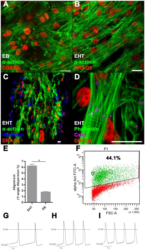
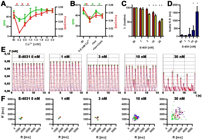
Human embryonic stem cell (hESC) progenies hold great promise as surrogates for human primary cells, particularly if the latter are not available as in the case of cardiomyocytes. However, high content experimental platforms are lacking that allow the function of hESC-derived cardiomyocytes to be studied under relatively physiological and standardized conditions. Here we describe a simple and robust protocol for the generation of fibrin-based human engineered heart tissue (hEHT) in a 24-well format using an unselected population of differentiated human embryonic stem cells containing 30-40% α-actinin-positive cardiac myocytes. Human EHTs started to show coherent contractions 5-10 days after casting, reached regular (mean 0.5 Hz) and strong (mean 100 µN) contractions for up to 8 weeks. They displayed a dense network of longitudinally oriented, interconnected and cross-striated cardiomyocytes. Spontaneous hEHT contractions were analyzed by automated video-optical recording and showed chronotropic responses to calcium and the β-adrenergic agonist isoprenaline. The proarrhythmic compounds E-4031, quinidine, procainamide, cisapride, and sertindole exerted robust, concentration-dependent and reversible decreases in relaxation velocity and irregular beating at concentrations that recapitulate findings in hERG channel assays. In conclusion this study establishes hEHT as a simple in vitro model for heart research.
Overview
The claims and statistics of this paper haven’t been checked yet.
Free: it reads the open-access text and checks the first 5 claims that cite a source we can check. Create a free account to check every claim.
Integrity signals
Registry
Cites
Statistics
Figures

Verification
Claims the paper makes
The claims and statistics of this paper haven’t been checked yet.
Free: it reads the open-access text and checks the first 5 claims that cite a source we can check. Create a free account to check every claim.
Claims other papers make about this one
Each is checked against this paper’s own text: does it say what the citing paper says it does?
The source backs part of the claim, not all of it — or backs it for a different population, condition or comparison.
In the citing paper
“but not pressure- and volume-based metrics directly representing pump performance of the heart”
From Bioengineering an electro-mechanically functional miniature ventricular heart chamber from human pluripotent stem cells (2018) · cites this paper as [16]
Experimental — accuracy not yet measuredThe cited source says what the claim attributes to it.
In the citing paper
“engineered cardiac thin films, micro-tissues and strips allow the assessment of twitch force [9,12–19]”
From Bioengineering an electro-mechanically functional miniature ventricular heart chamber from human pluripotent stem cells (2018) · cites this paper as [16]
Experimental — accuracy not yet measured
Self-check
Percentages, sums and reported differences re-computed from the paper’s own numbers.
The self-check needs the paper’s full text.
Citations
Cites
Proceedings of the National Academy of Sciences of the United States of America
Dowling, J. E., & Wald, G. (2009) · Nutrition Reviews · doi:10.1111/j.1753-4887.1981.tb06752.x · Cited by 17,893
Laflamme, M. A., Chen, K. Y., Naumova, A. V., Muskheli, V., Fugate, J. A., Dupras, S. K., Reinecke, H., Xu, C., Hassanipour, M., Police, S., et al. (2007) · Nature Biotechnology · doi:10.1038/nbt1327 · Cited by 2,206 · cited 2×
“One of the current limitations of hESC-derived cardiac myocytes is their immature and unorganized phenotype in the EB format or 2D cultures [3,5].”
BackgroundFeeder-free growth of undifferentiated human embryonic stem cells
Xu, C., Inokuma, M. S., Denham, J., Golds, K., Kundu, P., Gold, J. D., & Carpenter, M. K. (2001) · Nature Biotechnology · doi:10.1038/nbt1001-971 · Cited by 1,870 · cited 1×
“with CF1-MEF conditioned medium according to published protocols [28].”
MethodKehat, I., Kenyagin‐Karsenti, D., Snir, M., Segev, H., Amit, M., Gepstein, A., Livne, E., Binah, O., Itskovitz‐Eldor, J., & Gepstein, L. (2001) · Journal of Clinical Investigation · doi:10.1172/jci12131 · Cited by 1,814 · cited 1×
“differentiation in serum containing media yielding 8% of beating embryoid bodies (EBs) [1].”
BackgroundCai, C.-L., Liang, X., Shi, Y., Chu, P.-H., Pfaff, S. L., Chen, J., & Evans, S. (2003) · Developmental Cell · doi:10.1016/s1534-5807(03)00363-0 · Cited by 1,610 · cited 1×
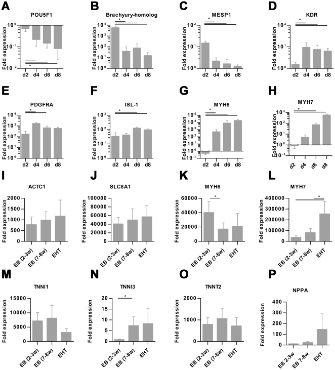
“Islet-1 (ISL1) a progenitor marker of the development of the secondary heart field [21] was significantly upregulated on day 6 and 8.”
BackgroundKehat, I., Kenyagin‐Karsenti, D., Snir, M., et al. (2001) · Journal of Clinical Investigation · doi:10.1172/jci200112131 · Cited by 1,536
REDFERN, W., CARLSSON, L., DAVIS, A., LYNCH, W., MACKENZIE, I., PALETHORPE, S., SIEGL, P., STRANG, I., SULLIVAN, A., & WALLIS, R. (2003) · Cardiovascular Research · doi:10.1016/s0008-6363(02)00846-5 · Cited by 1,532 · cited 2×
“Concentrations were chosen according to published IC50 in hERG assays [23,24].”
MethodHuman cardiovascular progenitor cells develop from a KDR+ embryonic-stem-cell-derived population
Yang, L., Soonpaa, M. H., Adler, E. D., Roepke, T. K., Kattman, S. J., Kennedy, M., Henckaerts, E., Bonham, K., Abbott, G. W., Linden, R. M., et al. (2008) · Nature · doi:10.1038/nature06894 · Cited by 1,389 · cited 3×
“Cardiomyocyte differentiation was performed with a modified EB-based, three-stage, growth factor cocktail protocol [2].”
MethodInfluentialEngineered heart tissue grafts improve systolic and diastolic function in infarcted rat hearts
Zimmermann, W.-H., Melnychenko, I., Wasmeier, G., Didié, M., Naito, H., Nixdorff, U., Hess, A., Budinsky, L., Brune, K., Michaelis, B., et al. (2006) · Nature Medicine · doi:10.1038/nm1394 · Cited by 993 · cited 1×
“The principal usefulness of these techniques for in vitro testing [17,18] and cardiac repair [19] has been demonstrated.”
BackgroundSelf-citationTissue Engineering of a Differentiated Cardiac Muscle Construct
Zimmermann, W.-H., Schneiderbanger, K., Schubert, P., Didié, M., Münzel, F., Heubach, J. F., Kostin, S., Neuhuber, W. L., & Eschenhagen, T. (2002) · Circulation Research · doi:10.1161/hh0202.103644 · Cited by 917 · cited 1×
“EHTs from neonatal rat hearts develop a high degree of cellular differentiation, longitudinal orientation, intercellular coupling and force generation [15,16].”
BackgroundSelf-citation
Research works printed but not linked: 2
- [3]Mummery C Ward-van Oostwaard D Doevendans P Spijker R Brink S van den 2003 Differentiation of human embryonic stem cells to cardiomyocytes: role of coculture with visceral endoderm-like cells. Circulation 107 2733 40 12742992
- [12]FDA 2005 Guidance for Industry S7B Nonclinical Evaluation of the Potential for Delayed Ventricular Repolarization (QT Interval Prolongation) by Human Pharmaceuticals. 16237859
Cited by
Items 101–200 of 361
Devarasetty, M., Mazzocchi, A. R., & Skardal, A. (2018) · BioDrugs · doi:10.1007/s40259-017-0258-x · Cited by 75
“Another study based around a similar model tested a variety of pro-arrhythmic compounds and demonstrated concentrationdependent and reversible changes in beating [59].”
Devalla, H. D., & Passier, R. (2018) · Science Translational Medicine · doi:10.1126/scitranslmed.aah5457 · Cited by 75
Stöhr, A., Friedrich, F. W., Flenner, F., Geertz, B., Eder, A., Schaaf, S., Hirt, M. N., Uebeler, J., Schlossarek, S., Carrier, L., et al. (2013) · Journal of Molecular and Cellular Cardiology · doi:10.1016/j.yjmcc.2013.07.011 · Cited by 72
“One such model could be an automated and miniaturized 24-well-assay based on our engineered heart tissue (EHT) technology [18,19].”
MethodSelf-citationNakane, T., Masumoto, H., Tinney, J. P., Yuan, F., Kowalski, W. J., Ye, F., LeBlanc, A. J., Sakata, R., Yamashita, J. K., & Keller, B. B. (2017) · Scientific Reports · doi:10.1038/srep45641 · Cited by 71
Mazzola, M., & Pasquale, E. D. (2020) · Frontiers in Bioengineering and Biotechnology · doi:10.3389/fbioe.2020.00455 · Cited by 78
Reconstructing the heart using iPSCs: Engineering strategies and applications
Cho, S., Lee, C., Skylar‐Scott, M. A., Heilshorn, S. C., & Wu, J. C. (2021) · Journal of Molecular and Cellular Cardiology · doi:10.1016/j.yjmcc.2021.04.006 · Cited by 79
“To date, various in vitro tissue models have been used to assess the effects of inotropic compounds at specific concentrations [131,132], and for broader systematic screening of larger panels of drugs at varying doses [77,102,133,134].”
BackgroundMiklas, J. W., Nunes, S. S., Sofla, A., Reis, L. A., Pahnke, A., Xiao, Y., Laschinger, C., & Radisic, M. (2014) · Biofabrication · doi:10.1088/1758-5082/6/2/024113 · Cited by 69
“1 mV [9] which is less negative than the resting membrane potential of comparable 7–8 week old embryoid bodies that resulted in cardiomyocytes with resting membrane potential of −60.”
BackgroundFHL2 expression and variants in hypertrophic cardiomyopathy
Friedrich, F. W., Reischmann, S., Schwalm, A., Unger, A., Ramanujam, D. P., Münch, J., Müller, O. J., Hengstenberg, C., Galve, E., Charron, P., et al. (2014) · Basic Research in Cardiology · doi:10.1007/s00395-014-0451-8 · Cited by 68
“Contraction measurements were performed on day 8, 10, 13, 14, 17, 20 and 21 as previously described [14, 21, 25, 48].”
Self-citationvan Meer, B. J., Tertoolen, L. G. J., & Mummery, C. L. (2016) · Stem Cells · doi:10.1002/stem.2403 · Cited by 68
Bielawski, K. S., Leonard, A., Bhandari, S., Murry, C., & Sniadecki, N. J. (2016) · Tissue Engineering Part C Methods · doi:10.1089/ten.tec.2016.0257 · Cited by 74
“The twitch forces and beating frequency of engineered heart tissue have been measured previously using pairs of silicone posts, where both posts deflect when a tissue undergoes a twitch contraction.(33,34) Here, we built a similar array of posts, but where one post was flexible and magnetic, while the other was rigid…”
High-Content Assessment of Cardiac Function Using Heart-on-a-Chip Devices as Drug Screening Model
Conant, G., Lai, B. F. L., Lu, R. X. Z., Korolj, A., Wang, E. Y., & Radisic, M. (2017) · Stem Cell Reviews and Reports · doi:10.1007/s12015-017-9736-2 · Cited by 75
Mayourian, J., Cashman, T. J., Ceholski, D. K., Johnson, B. V., Sachs, D. M., Kaji, D. A., Sahoo, S., Hare, J. M., Hajjar, R. J., Sobie, E. A., et al. (2017) · Circulation Research · doi:10.1161/circresaha.117.310796 · Cited by 67
“Furthermore, our model trends are consistent with our original hECT data on spontaneous beat rate variability—a proposed surrogate of arrhythmogenicity.(40,41) Most importantly, VW analyses predicted that hMSC supplementation (involving hMSC PS+HC mechanisms) did not adversely impact arrhythmogenesis and may even be a…”
ResultHarnessing organs-on-a-chip to model tissue regeneration
Tavakol, D. N., Fleischer, S., & Vunjak-Novakovic, G. (2021) · Cell Stem Cell · doi:10.1016/j.stem.2021.05.008 · Cited by 63
From engineered heart tissue to cardiac organoid
Cho, J., Lee, H., Rah, W., Chang, H., & Yoon, Y. (2022) · Theranostics · doi:10.7150/thno.67661 · Cited by 74
“Also, concentrationdependent irregular beating, and reversible decreases in relaxation velocity were induced by the proarrhythmic compounds [102].”
BackgroundMethodHuman Pluripotent Stem Cell-Derived Cardiomyocytes as Research and Therapeutic Tools
Acimovic, I., Vilotic, A., Pesl, M., Lacampagne, A., Dvorak, P., Rotrekl, V., & Meli, A. C. (2014) · BioMed Research International · doi:10.1155/2014/512831 · Cited by 66
Rupert, C. E., & Coulombe, K. L. K. (2017) · Stem Cells International · doi:10.1155/2017/7648409 · Cited by 73
“14mN/mm(2) was higher than previously reported for hESC-cardiomyocyte-only, strip-like tissues [38, 39]; was equal to recently reported values [22]; and was lower than with patterned molds where overall tissue force production was normalized by CSA, porosity, and cellular orientation [40].”
ResultBioengineering adult human heart tissue: How close are we?
Mills, R. J., & Hudson, J. E. (2019) · APL Bioengineering · doi:10.1063/1.5070106 · Cited by 68
Christoforou, N., Liau, B., Chakraborty, S., Chellappan, M. K., Bursac, N., & Leong, K. W. (2013) · PLoS ONE · doi:10.1371/journal.pone.0065963 · Cited by 66
“Currently, pharmacological studies are commonly performed on either non-cardiac cell lines or in two-dimensional (monolayer) cultures of cardiomyocytes [27,28].”
BackgroundA phenotypic in vitro model for the main determinants of human whole heart function
Stancescu, M., Molnar, P., McAleer, C. W., McLamb, W., Long, C. J., Oleaga, C., Prot, J.-M., & Hickman, J. J. (2015) · Biomaterials · doi:10.1016/j.biomaterials.2015.04.035 · Cited by 61
“Contraction force of cardiomyocytes has been measured by biopolymer microcantilevers [21], microfabricated pillars [22], videomicroscopy of flexible substrates [23,24], calcium imaging [25] and with magnetic beads [26].”
Kurokawa, Y. K., & George, S. C. (2016) · Advanced Drug Delivery Reviews · doi:10.1016/j.addr.2015.07.004 · Cited by 59
Kerscher, P., Kaczmarek, J. A., Head, S. E., Ellis, M. E., Seeto, W. J., Kim, J., Bhattacharya, S., Suppiramaniam, V. D., & Lipke, E. A. (2016) · ACS Biomaterials Science & Engineering · doi:10.1021/acsbiomaterials.6b00226 · Cited by 61
Marchianò, S., Bertero, A., & Murry, C. E. (2019) · Pediatric Cardiology · doi:10.1007/s00246-019-02165-5 · Cited by 59
Uzun, A. U., Mannhardt, I., Breckwoldt, K., Horváth, A., Johannsen, S. S., Hansen, A., Eschenhagen, T., & Christ, T. (2016) · Frontiers in Pharmacology · doi:10.3389/fphar.2016.00300 · Cited by 54
Self-citationKeung, W., Chan, P. K. W., Backeris, P. C., Lee, E. K., Wong, N., Wong, A. O. T., Wong, G. K. Y., Chan, C. W. Y., Fermini, B., Costa, K. D., et al. (2019) · Clinical Pharmacology & Therapeutics · doi:10.1002/cpt.1385 · Cited by 54
“These include 2D strain- gauge embedded muscular thin films,(28) cardiac microtissues,(29,30) cardiac microwires(31) or biowires,(32) miniaturized cardiac muscles(33) and force- generating engineered heart tissues.(34) We too have developed our version of engineered human ventricularlike cardiac tissue strip (hvCTS) m…”
BackgroundOECD (2018) · OECD series on testing and assessment · doi:10.1787/9789264304796-1-en · Cited by 54
Advances in cardiac tissue engineering and heart‐on‐a‐chip
Kieda, J., Shakeri, A., Landau, S., Wang, E. Y., Zhao, Y., Lai, B. F., Okhovatian, S., Wang, Y., Jiang, R., & Radisic, M. (2023) · Journal of Biomedical Materials Research Part A · doi:10.1002/jbm.a.37633 · Cited by 56
Ribeiro, A. J. S., Guth, B. D., Engwall, M. J., Eldridge, S. R., Foley, C. M., Guo, L., Gintant, G. A., Koerner, J. E., Parish, S. T., Pierson, J. B., et al. (2019) · Frontiers in Pharmacology · doi:10.3389/fphar.2019.00934 · Cited by 58
Saleem, U., Mannhardt, I., Braren, I., Denning, C. N., Eschenhagen, T., & Hansen, A. (2020) · Stem Cell Reports · doi:10.1016/j.stemcr.2019.12.011 · Cited by 64
Self-citationHuman cardiac tissue engineering: from pluripotent stem cells to heart repair
Jackman, C. P., Shadrin, I. Y., Carlson, A. L., & Bursac, N. (2015) · Current Opinion in Chemical Engineering · doi:10.1016/j.coche.2014.11.004 · Cited by 54
Engineered Macroscale Cardiac Constructs Elicit Human Myocardial Tissue-like Functionality
Valls-Margarit, M., Iglesias‐García, O., Guglielmo, C. D., Sarlabous, L., Tadevosyan, K., Paoli, R., Comelles, J., Blanco-Almazán, D., Jiménez‐Delgado, S., Castillo‐Fernández, Ó., et al. (2019) · Stem Cell Reports · doi:10.1016/j.stemcr.2019.05.024 · Cited by 57
“Human engineered cardiac constructs developed thus far recapitulate some of the structural complexity and electromechanical functionality of the native myocardium, leading to improved performance compared with standard 2D in vitro cultures (Ma et al., 2015; Nunes et al., 2013; Schaaf et al., 2011; Thavandiran et al.,…”
BackgroundHuman iPSC-engineered cardiac tissue platform faithfully models important cardiac physiology
de Lange, W. J., Farrell, E. T., Kreitzer, C. R., Jacobs, D. R., Lang, D., Glukhov, A. V., & Ralphe, J. C. (2021) · American Journal of Physiology-Heart and Circulatory Physiology · doi:10.1152/ajpheart.00941.2020 · Cited by 58
Strategies and Challenges to Myocardial Replacement Therapy
Feric, N. T., & Radisic, M. (2016) · Stem Cells Translational Medicine · doi:10.5966/sctm.2015-0288 · Cited by 47
Functional arrays of human pluripotent stem cell-derived cardiac microtissues
Thavandiran, N., Hale, C. M., Blit, P. H., Sandberg, M. L., McElvain, M. E., Gagliardi, M., Sun, B., Witty, A. D., Graham, G., Mcintosh, M., et al. (2020) · Scientific Reports · doi:10.1038/s41598-020-62955-3 · Cited by 56
Extracellular Vesicles in Cardiac Regeneration: Potential Applications for Tissues-on-a-Chip
Wagner, K. T., Nash, T. R., Liu, B., Vunjak‐Novakovic, G., & Radisic, M. (2020) · Trends in biotechnology · doi:10.1016/j.tibtech.2020.08.005 · Cited by 53
“Tissues suspended between or attached to flexible supports are amenable to video analysis that can determine force generation based on the deflection and physical characteristics of the supporting material [42,77,78].”
BackgroundContractile Force of Transplanted Cardiomyocytes Actively Supports Heart Function After Injury
Stüdemann, T., Rössinger, J., Manthey, C., Geertz, B., Srikantharajah, R., von Bibra, C., Shibamiya, A., Koehne, M., Wiehler, A., Wiegert, J. S., et al. (2022) · Circulation · doi:10.1161/circulationaha.122.060124 · Cited by 51
Self-citationMachine learning plus optical flow: a simple and sensitive method to detect cardioactive drugs
Lee, E. K., Kurokawa, Y. K., Tu, R., George, S. C., & Khine, M. (2015) · Scientific Reports · doi:10.1038/srep11817 · Cited by 51
Three-dimensional microengineered models of human cardiac diseases
Veldhuizen, J., Migrino, R. Q., & Nikkhah, M. (2019) · Journal of Biological Engineering · doi:10.1186/s13036-019-0155-6 · Cited by 52
Bioreactors as Engineering Support to Treat Cardiac Muscle and Vascular Disease
Massai, D., Cerino, G., Gallo, D., Pennella, F., Deriu, M. A., Rodríguez, A., Montevecchi, F. M., Bignardi, C., Audenino, A. L., & Morbiducci, U. (2013) · Journal of Healthcare Engineering · doi:10.1260/2040-2295.4.3.329 · Cited by 59
“Moreover, bioreactors, in combination with recent induced pluripotent SC technology, have been used for producing in vitro models of disease [47, 48].”
Effects of Geometry on the Mechanics and Alignment of Three-Dimensional Engineered Microtissues
Bose, P., Eyckmans, J., Nguyen, T. D., Chen, C. S., & Reich, D. H. (2018) · ACS Biomaterials Science & Engineering · doi:10.1021/acsbiomaterials.8b01183 · Cited by 51
Wei, X., Zhuang, L., Li, H., He, C., Wan, H., Hu, N., & Wang, P. (2020) · Small · doi:10.1002/smll.202005828 · Cited by 50
Calmodulinopathy: A Novel, Life-Threatening Clinical Entity Affecting the Young
Kotta, M., Sala, L., Ghidoni, A., Badone, B., Ronchi, C., Parati, G., Zaza, A., & Crotti, L. (2018) · Frontiers in Cardiovascular Medicine · doi:10.3389/fcvm.2018.00175 · Cited by 46
Cutts, J., Nikkhah, M., & Brafman, D. A. (2015) · Biomarker Insights · doi:10.4137/bmi.s20313 · Cited by 50
Chen, A., Lee, E., Tu, R., Santiago, K. C., Grosberg, A., Fowlkes, C. C., & Khine, M. (2013) · Biomaterials · doi:10.1016/j.biomaterials.2013.10.007 · Cited by 51
Jacob, F., Yonis, A. Y., Cuello, F., Luther, P. K., Schulze, T., Eder, A., Streichert, T., Mannhardt, I., Hirt, M. N., Schaaf, S., et al. (2016) · PLoS ONE · doi:10.1371/journal.pone.0145937 · Cited by 48
Self-citationLiguori, G. R., Jeronimus, B. F., Liguori, T. T. A., Moreira, L. F. P., & Harmsen, M. C. (2017) · Tissue Engineering Part C Methods · doi:10.1089/ten.tec.2017.0189 · Cited by 50
“71 The use of these models was already shown not only to pathophysiology 79 and pharmacological testing 78,80,81 but also for gene therapy.”
Malandraki-Miller, S., Lopez, C. A., Al-Siddiqi, H., & Carr, C. A. (2018) · Frontiers in Cardiovascular Medicine · doi:10.3389/fcvm.2018.00119 · Cited by 53
Massai, D., Pisani, G., Isu, G., Rodríguez, A., Cerino, G., Galluzzi, R., Pisanu, A., Tonoli, A., Bignardi, C., Audenino, A. L., et al. (2020) · Frontiers in Bioengineering and Biotechnology · doi:10.3389/fbioe.2020.00733 · Cited by 46
Topological and electrical control of cardiac differentiation and assembly
Thavandiran, N., Nunes, S. S., Xiao, Y., & Radisic, M. (2013) · Stem Cell Research & Therapy · doi:10.1186/scrt162 · Cited by 49
“Th e Eschenhagen and Zimmerman groups have investigated and reported on the role and benefi cial eff ects of mechanical stimulation in cardiac cells [31,48-50].”
BackgroundTechniques for the induction of human pluripotent stem cell differentiation towards cardiomyocytes
Lewandowski, J., Kolanowski, T. J., & Kurpisz, M. K. (2016) · Journal of Tissue Engineering and Regenerative Medicine · doi:10.1002/term.2117 · Cited by 48
“The new convenient ways of cardiomyocyte generations holds also a great promise for developmental studies and drug screening (Schaaf et al., 2011).”
BackgroundGeneration of Strip-Format Fibrin-Based Engineered Heart Tissue (EHT)
Schaaf, S., Eder, A., Vollert, I., Stoehr, A., Hansen, A., & Eschenhagen, T. (2014) · Methods in molecular biology · doi:10.1007/978-1-4939-1047-2_11 · Cited by 47
Self-citationA Multimaterial Microphysiological Platform Enabled by Rapid Casting of Elastic Microwires
Zhao, Y., Wang, E. Y., Huyer, L. D., Liao, Y., Yeager, K. J., Vunjak‐Novakovic, G., Radisic, M., & Zhang, B. (2019) · Advanced Healthcare Materials · doi:10.1002/adhm.201801187 · Cited by 46
Hartung, S., Schwanke, K., Haase, A., David, R., Franz, W., Martin, U., & Zweigerdt, R. (2012) · Stem Cells and Development · doi:10.1089/scd.2012.0351 · Cited by 47
“They also represent a human cell source for the development of more predictive drug discovery and drug safety assays [1].”
Yıldırım, Z., Swanson, K., Wu, X., Zou, J., & Wu, J. C. (2024) · The Annual Review of Pharmacology and Toxicology · doi:10.1146/annurev-pharmtox-022724-095035 · Cited by 49
A Miniaturized EHT Platform for Accurate Measurements of Tissue Contractile Properties
Dostanic, M., Windt, L. M., Stein, J. M., van Meer, B. J., Bellin, M., Orlova, V., Mastrangeli, M., Mummery, C. L., & Sarro, P. M. (2020) · Journal of Microelectromechanical Systems · doi:10.1109/jmems.2020.3011196 · Cited by 41
“Some of the existing EHT platforms [14], [19], [20] use more than 500,000 cells per tissue, which could confound large scale”
MethodMaturation of human cardiac organoids enables complex disease modeling and drug discovery
Pocock, M. W., Reid, J. D., Robinson, H., Charitakis, N., Krycer, J. R., Foster, S. R., Fitzsimmons, R. L., Lor, M., Devilée, L., Batho, C. A. P., et al. (2025) · Nature Cardiovascular Research · doi:10.1038/s44161-025-00669-3 · Cited by 45
Bioengineering Approaches to Mature Human Pluripotent Stem Cell-Derived Cardiomyocytes
Sun, X., & Nunes, S. S. (2017) · Frontiers in Cell and Developmental Biology · doi:10.3389/fcell.2017.00019 · Cited by 46
“Electrical stimulation also significantly increased the conduction velocity of biowires from ∼11.5 to 18.5 cm/s. Biowires exposed to electrical stimulation also showed increased Ca2+ transient amplitudes vs. unstimulated controls. hPSC-CMs in biowires exhibited improved hERG current and inward rectifier current (Ik1)…”
BackgroundInfluentialVascularisation to improve translational potential of tissue engineering systems for cardiac repair
Dilley, R. J., & Morrison, W. A. (2014) · The International Journal of Biochemistry & Cell Biology · doi:10.1016/j.biocel.2014.10.020 · Cited by 42
Ruvinov, E., Sapir, Y., & Cohen, S. (2012) · Synthesis lectures on tissue engineering · doi:10.2200/s00437ed1v01y201207tis009 · Cited by 37
Modeling myocardial growth and hypertrophy in engineered heart muscle
Tiburcy, M., & Zimmermann, W. (2013) · Trends in Cardiovascular Medicine · doi:10.1016/j.tcm.2013.05.003 · Cited by 37
Self-citationAutomated Contraction Analysis of Human Engineered Heart Tissue for Cardiac Drug Safety Screening
Mannhardt, I., Saleem, U., Benzin, A., Schulze, T., Klampe, B., Eschenhagen, T., & Hansen, A. (2017) · Journal of Visualized Experiments · doi:10.3791/55461 · Cited by 44
Self-citation2D and 3D Self-Assembling Nanofiber Hydrogels for Cardiomyocyte Culture
Ikonen, L., Kerkelä, E., Metselaar, G., Stuart, M. C. A., de Jong, M. R., & Aalto‐Setälä, K. (2012) · BioMed Research International · doi:10.1155/2013/285678 · Cited by 37
“Human engineered heart tissue (hEHT) has also been developed from hESC-CMs and �brinogen; it forms a dense network that responds to chronotropic compounds [42].”
BackgroundVisone, R., Ugolini, G. S., Cruz‐Moreira, D., Marzorati, S., Piazza, S., Pesenti, E. A., Redaelli, A. C. L., Moretti, M., Occhetta, P., & Rasponi, M. (2021) · Biofabrication · doi:10.1088/1758-5090/abe4c4 · Cited by 44
“Recent studies indeed demonstrated the fundamental role of spatially organized 3D tissue models [10] and mechanical [11–14] or electromechanical [15] stimulations, designed ad hoc to achieve superior maturation of h-iPSC-CMs leading to more physiological responses to known compounds [10, 16].”
BackgroundLopez, C. A., Al-Siddiqi, H., Purnama, U., Iftekhar, S., Bruyneel, A. A. N., Kerr, M. J., Nazir, R., Fialho, M. D. L. S., Malandraki-Miller, S., Alonaizan, R., et al. (2021) · Scientific Reports · doi:10.1038/s41598-021-87186-y · Cited by 38
Sewanan, L. R., & Campbell, S. G. (2019) · The Journal of Physiology · doi:10.1113/jp276753 · Cited by 39
“…neonatal rat (Zimmermann et al. 2000) models that were readily adapted for pluripotent stem cell-derived cardiomyocyte technology to make intact 3D muscle structures whose force and contractility can be measured through various direct or indirect methods (Schaaf et al. 2011; Tulloch et al. 2011).”
BackgroundMechanobiology Assays with Applications in Cardiomyocyte Biology and Cardiotoxicity
Blair, C. A., & Pruitt, B. L. (2020) · Advanced Healthcare Materials · doi:10.1002/adhm.201901656 · Cited by 37
Engineering and Assessing Cardiac Tissue Complexity
Tadevosyan, K., Iglesias‐García, O., Mazo, M. M., Prósper, F., & Raya, Á. (2021) · International Journal of Molecular Sciences · doi:10.3390/ijms22031479 · Cited by 36
“4 n/a Human ESC-CM No No 14 Gene/protein expression, patch clamp Force of contraction [37]”
BackgroundMethodThe harder the climb the better the view: The impact of substrate stiffness on cardiomyocyte fate
Querceto, S., Santoro, R., Gowran, A., Grandinetti, B., Pompilio, G., Regnier, M., Tesi, C., Poggesi, C., Ferrantini, C., & Pioner, J. M. (2022) · Journal of Molecular and Cellular Cardiology · doi:10.1016/j.yjmcc.2022.02.001 · Cited by 39
“Studies have shown that hiPSC-CMs used to create collagen I-based engineered heart tissues (EHTs) were subjected to different mechanical stress and electrical pacing conditions.”
BackgroundInfluentialEffects of proarrhythmic drugs on relaxation time and beating pattern in rat engineered heart tissue
Eder, A., Hansen, A., Uebeler, J., Schulze, T., Neuber, C., Schaaf, S., Yuan, L., Christ, T., Vos, M. A., & Eschenhagen, T. (2014) · Basic Research in Cardiology · doi:10.1007/s00395-014-0436-7 · Cited by 33
Self-citationBioengineering heart tissue for in vitro testing
Cimetta, E., Godier-Furnémont, A., & Vunjak-Novakovic, G. (2013) · Current Opinion in Biotechnology · doi:10.1016/j.copbio.2013.07.002 · Cited by 32
“Analogous approaches were followed to generate 3D miniaturized, force-generating human heart muscle from hESC-CM [46 ,47].”
Modeling the Human Scarred Heart In Vitro: Toward New Tissue Engineered Models
Deddens, J. C., Sadeghi, A. H., Hjortnaes, J., Laake, L. W. V., Buijsrogge, M. P., Doevendans, P. A., Khademhosseini, A., & Sluijter, J. P. G. (2016) · Advanced Healthcare Materials · doi:10.1002/adhm.201600571 · Cited by 33
Breckwoldt, K., Weinberger, F., & Eschenhagen, T. (2015) · Biochimica et Biophysica Acta (BBA) - Molecular Cell Research · doi:10.1016/j.bbamcr.2015.11.010 · Cited by 37
“EHTs from hPSC cardiomyocytes show good cardiac tissue development in vitro, spontaneous contractions, reactivity to sympathetic (isoprenaline) and parasympathetic (carbachol) signals, reaction to changes in extracellular calcium-concentrations and follow the Frank–Starling law [146, 147,151].”
BackgroundSelf-citationMicro-Arrayed Human Embryonic Stem Cells-Derived Cardiomyocytes for In Vitro Functional Assay
Serena, E., Cimetta, E., Zatti, S., Zaglia, T., Zagallo, M., Keller, G. M., & Elvassore, N. (2012) · PLoS ONE · doi:10.1371/journal.pone.0048483 · Cited by 34
“Despite the originality of these works, they all test animal derived cardiomyocytes and only few in vitro cardiac models were developed based on human embryonic stem cell-derived cardiomyocytes (hCMs) [15],[16],[17],[18].”
BackgroundBiomechanical regulation of in vitro cardiogenesis for tissue-engineered heart repair
Zimmermann, W. (2013) · Stem Cell Research & Therapy · doi:10.1186/scrt348 · Cited by 31
“Proof of concept for a wide range of research applications of tissue-engineered heart muscle has been presented; this includes applications in drug screening [12], target validation [13], and disease modeling [14].”
BackgroundSelf-citationPutame, G., Gabetti, S., Carbonaro, D., Meglio, F. D., Romano, V., Sacco, A. M., Belviso, I., Serino, G., Bignardi, C., Morbiducci, U., et al. (2020) · Medical Engineering & Physics · doi:10.1016/j.medengphy.2020.07.018 · Cited by 36
Maturation of Human Stem Cell-derived Cardiomyocytes in Biowires Using Electrical Stimulation
Sun, X., & Nunes, S. S. (2017) · Journal of Visualized Experiments · doi:10.3791/55373 · Cited by 32
Gouadon, E., Moore‐Morris, T., Smit, N. W., Chatenoud, L., Coronel, R., Harding, S. E., Jourdon, P., Lambert, V., Rücker‐Martin, C., & Pucéat, M. (2015) · Stem Cells · doi:10.1002/stem.2205 · Cited by 29
Fibrosis in tissue engineering and regenerative medicine: treat or trigger?
Fernández‐Colino, A., Iop, L., Ferreira, M. S. V., & Mela, P. (2019) · Advanced Drug Delivery Reviews · doi:10.1016/j.addr.2019.07.007 · Cited by 30
Vuorenpää, H., Penttinen, K., Heinonen, T., Pekkanen-Mattila, M., Sarkanen, J., Ylikomi, T. J., & Aalto‐Setälä, K. (2017) · Cytotechnology · doi:10.1007/s10616-017-0088-1 · Cited by 33
“In addition to the increased time in culture, electrical stimulation (Chan et al. 2013) and 3D culture environment (Schaaf et al. 2011; van Spreeuwel et al. 2014; Garzoni et al. 2009; Valarmathi et al. 2010; Pontes Soares et al. 2012) have been utilized in the production of more mature hPSC-CMs.”
Metabolic Regulation of Human Pluripotent Stem Cell-Derived Cardiomyocyte Maturation
Batho, C. A. P., Mills, R. J., & Hudson, J. E. (2020) · Current Cardiology Reports · doi:10.1007/s11886-020-01303-3 · Cited by 30
Stem cell toxicology: a powerful tool to assess pollution effects on human health
Yao, X., Yin, N., & Faiola, F. (2016) · National Science Review · doi:10.1093/nsr/nww089 · Cited by 27
3D engineered cardiac tissue models of human heart disease: learning more from our mice
Ralphe, J. C., & de Lange, W. J. (2013) · Trends in Cardiovascular Medicine · doi:10.1016/j.tcm.2012.08.007 · Cited by 26
“In 2011, the successful formation of functional ECT from human embryonic stem cells in a fibrin-based matrix was reported (Schaaf et al., 2011).”
Feaster, T. K., Casciola, M., Narkar, A., & Blinova, K. (2021) · Physiological Reports · doi:10.14814/phy2.15085 · Cited by 29
“5 mM] calcium is comparable with the EC50 value for calcium in hiPSC- CMs (Mannhardt et al., 2016, 2017; Schaaf et al., 2011).”
MethodElectrical and Mechanical Strategies to Enable Cardiac Repair and Regeneration
Cao, H., Kang, B. J., Lee, C.-A., Shung, K. K., & Hsiai, T. K. (2015) · IEEE Reviews in Biomedical Engineering · doi:10.1109/rbme.2015.2431681 · Cited by 26
“12 mN/mm2 in comparison with 40–80 mN/mm2 in the intact heart muscle [15], [67], [68].”
Myocardial tissue engineering for cardiac repair
Pecha, S., Eschenhagen, T., & Reichenspurner, H. C. (2016) · The Journal of Heart and Lung Transplantation · doi:10.1016/j.healun.2015.12.007 · Cited by 29
Self-citationCardiac macrotissues-on-a-plate models for phenotypic drug screens
Meyer, T., Tiburcy, M., & Zimmermann, W. (2019) · Advanced Drug Delivery Reviews · doi:10.1016/j.addr.2019.03.002 · Cited by 32
Self-citationSalazar, B., Cashion, A., Dennis, R. G., & Birla, R. K. (2015) · Cardiovascular Engineering and Technology · doi:10.1007/s13239-015-0236-8 · Cited by 29
Kinase inhibitor screening using artificial neural networks and engineered cardiac biowires
Conant, G., Ahadian, S., Zhao, Y., & Radisic, M. (2017) · Scientific Reports · doi:10.1038/s41598-017-12048-5 · Cited by 25
Farouz, Y., Chen, Y., Terzic, A., & Menasché, P. (2014) · Stem Cells · doi:10.1002/stem.1929 · Cited by 27
Ribeiro, M. C., Rivera‐Arbeláez, J. M., Cofiño‐Fabrés, C., Schwach, V., Slaats, R. H., ten Den, S. A., Vermeul, K., van den Berg, A., Pérez-Pomares, J. M., Segerink, L. I., et al. (2022) · Journal of Personalized Medicine · doi:10.3390/jpm12020214 · Cited by 26
“The currently available EHT models or platforms which use a comparable cylindrical flexible cantilever in a similar format produce a lower number of EHTs (1 per custom-made bioreactor and 24 in 24-well plates) [17,18] or do not fit in a commercial culture plate.”
MethodInfluentialHuman Stem Cell-Derived Cardiac Model of Chronic Drug Exposure
Nunes, S. S., Feric, N. T., Pahnke, A., Miklas, J. W., Li, M., Coles, J. G., Gagliardi, M., Keller, G. M., & Radisic, M. (2016) · ACS Biomaterials Science & Engineering · doi:10.1021/acsbiomaterials.5b00496 · Cited by 28
The Rapidly Evolving Concept of Whole Heart Engineering
Iop, L., Sasso, E. D., Menabò, R., Lisa, F. D., & Gerosa, G. (2017) · Stem Cells International · doi:10.1155/2017/8920940 · Cited by 22
“Constant electrical stimulation proved to exert similar effects in terms of cell maturation in tissue-engineered heart constructs [98, 99], as well as in repopulated hearts [7, 27, 33].”
BackgroundMurphy, J. F., Mayourian, J., Stillitano, F., Munawar, S., Broughton, K. M., Agulló-Pascual, E., Sussman, M. A., Hajjar, R. J., Costa, K. D., & Turnbull, I. C. (2019) · Stem Cell Research & Therapy · doi:10.1186/s13287-019-1486-4 · Cited by 25
Seguret, M., Davidson, P. M., Robben, S., Jouve, C., Pereira, C., Lelong, Q., Deshayes, L., Cerveau, C., Berre, M. L., Ribeiro, R. S. R., et al. (2024) · eLife · doi:10.7554/elife.87739 · Cited by 25
Biophysical stimulation for in vitro engineering of functional cardiac tissues
Korolj, A., Wang, E. Y., Civitarese, R., & Radisic, M. (2017) · Clinical Science · doi:10.1042/cs20170055 · Cited by 25
Regenerative Medicine/Cardiac Cell Therapy: Pluripotent Stem Cells
Duran, A., Reidell, O., Stachelscheid, H., Klose, K., Gossen, M., Falk, V., Roell, W., & Stamm, C. (2017) · The Thoracic and Cardiovascular Surgeon · doi:10.1055/s-0037-1608761 · Cited by 23
Pang, J. K. S., Ho, B. X., Chan, W., & Soh, B.-S. (2021) · Frontiers in Cell and Developmental Biology · doi:10.3389/fcell.2021.788955 · Cited by 25
“Hydrogels and decellularized extracellular matrix have been developed into bioinks that serve as a scaffold for individual cell types to be printed on (Lepilina et al., 2006; Caspi et al., 2007; Sekine et al., 2008;Weeke-Klimp et al., 2010).”
BackgroundArefin, A., Mendoza, M., Dame, K., Garcia, M. I., Strauss, D. G., & Ribeiro, A. J. S. (2023) · Frontiers in Pharmacology · doi:10.3389/fphar.2023.1212092 · Cited by 21
“Engineered heart tissues (EHTs) are three-dimensional (3D) in vitro cellular platforms containing cardiomyocytes (Schaaf et al., 2011) differentiated from human pluripotent stem cells (hPSC-cardiomyocytes) (Yamanaka, 2007; Burridge et al., 2012) and have demonstrated potential to predict cardiac…”
InfluentialMedicinal Chemistry Approaches to Heart Regeneration
Schade, D., & Plowright, A. T. (2015) · Journal of Medicinal Chemistry · doi:10.1021/acs.jmedchem.5b00446 · Cited by 26
Goßmann, M., Linder, P., Thomas, U., Juhász, K., Lemme, M., George, M., Fertig, N., Dragicevic, E., & Stoelzle‐Feix, S. (2020) · Journal of Pharmacological and Toxicological Methods · doi:10.1016/j.vascn.2020.106892 · Cited by 21
Disease modeling and functional screening using engineered heart tissue
Mills, R. J., Voges, H. K., Porrello, E. R., & Hudson, J. E. (2017) · Current Opinion in Physiology · doi:10.1016/j.cophys.2017.08.003 · Cited by 20
Metadata
- Authors
- Schaaf, S., Shibamiya, A., Mewe, M., Eder, A., Stöhr, A., Hirt, M. N., Rau, T., Zimmermann, W.-H., Conradi, L., Eschenhagen, T., & Hansen, A.
- Affiliations
- University Medical Center Hamburg-Eppendorf (DE)
- Corresponding author
- Hansen, Arne
- Venue
- PLoS ONE
- Publisher
- Public Library of Science (PLoS)
- Published
- 2011-10-20
- Volume
- 6
- Issue
- 10
- Pages
- e26397
- ISSN
- 1932-6203 (ISSN-L)
- PMC
- PMC3197640
- OpenAlex
- W1969832018
- Cited by
- 365
- References
- 30
- Citations by year
- 2026: 11 · 2025: 7 · 2024: 15 · 2023: 19 · 2022: 17 · 2021: 20 · 2020: 24 · 2019: 26 · 2018: 32 · 2017: 39 · 2016: 34 · 2015: 35 · 2014: 37 · 2013: 27 · 2012: 20
- FWCI
- 10.92
- Type
- journal article
- Language
- en
- Open access
- gold
- doi.org
- Locations
- PLoS ONE (publishedVersion, cc-by)
- Europe PMC (PubMed Central) (publishedVersion, cc-by)
- PubMed Central (publishedVersion, cc-by)
- PubMed (publishedVersion)
- DOAJ (DOAJ: Directory of Open Access Journals) (submittedVersion, cc-by-sa)
- Figshare (submittedVersion, cc-by)
- GRO (submittedVersion)
- Funders
- European Commission
- Deutsche Forschungsgemeinschaft (ZI 708/7-1, HA 3423/3-1)
- Deutsche Herzstiftung (F/13/10)
- Topics
- Life Sciences › Biochemistry, Genetics and Molecular Biology › Molecular Biology › Pluripotent Stem Cells Research
- Health Sciences › Medicine › Surgery › Tissue Engineering and Regenerative Medicine
- Health Sciences › Medicine › Cardiology and Cardiovascular Medicine › Cardiac electrophysiology and arrhythmias
- Keywords
- human embryonic stem cells, cardiomyocytes, cardiomyocyte contraction, spontaneous beating, proarrhythmic drugs, hERG, calcium-induced contraction, beta-adrenergic stimulation, isoproterenol, E-4031, quinidine, procainamide, cisapride, sertindole, preclinical toxicology, high-content screening
- MeSH terms
- Arrhythmias, Cardiac, Cell Differentiation, Drug Evaluation, Preclinical, Fluorescent Antibody Technique, Heart, Humans, Muscle Contraction, Myocardium, RNA, Messenger, Reproducibility of Results, Toxicity Tests, Tissue Engineering, Myocytes, Cardiac, Embryonic Stem Cells, Electrophysiological Phenomena, HEK293 Cells
About these checks
Registry, retraction, Cites and statistics results are computed, not judged, with one exception: a printed reference that no index lists is read once by a model before it counts as a citation error. Claims are extracted by a model and kept only when their quote appears verbatim in the text; each cited source is then checked, with a model’s help, for whether it says what the claim relies on. Those verdicts can be wrong — read the claim’s quote and the cited source before relying on one.
Statistics and arithmetic are recomputed, not judged.
Confidence bands are shown only for fields calibrated on our annotated test set. None is calibrated yet, so no band is shown.
What counts as a claim ›How we measure confidence ›How we check this ›
Are you an author, or do you see a problem with this page? Request a review
Data sources
Bibliographic data: Semantic Scholar, OpenAlex, Crossref.
Contains information from the Semantic Scholar Academic Graph Datasets, made available under the ODC Attribution License (ODC-BY 1.0).
Semantic Scholar’s data is described in: Kinney, R., et al. (2023). The Semantic Scholar Open Data Platform. arXiv:2301.10140.
