

Structural Brain Changes in Chronic Pain Reflect Probably Neither Damage Nor Atrophy
Open accessThe DOI matches its Crossref record; 50 of the 51 references it cites were found in a registry or an index.
Abstract
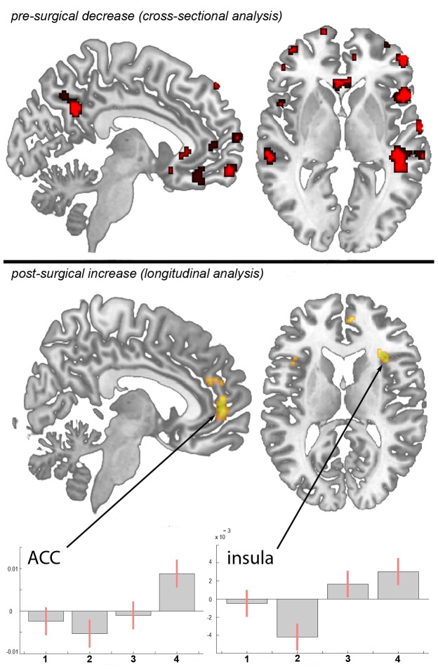
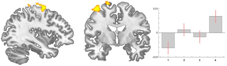
Chronic pain appears to be associated with brain gray matter reduction in areas ascribable to the transmission of pain. The morphological processes underlying these structural changes, probably following functional reorganisation and central plasticity in the brain, remain unclear. The pain in hip osteoarthritis is one of the few chronic pain syndromes which are principally curable. We investigated 20 patients with chronic pain due to unilateral coxarthrosis (mean age 63.25±9.46 (SD) years, 10 female) before hip joint endoprosthetic surgery (pain state) and monitored brain structural changes up to 1 year after surgery: 6-8 weeks, 12-18 weeks and 10-14 month when completely pain free. Patients with chronic pain due to unilateral coxarthrosis had significantly less gray matter compared to controls in the anterior cingulate cortex (ACC), insular cortex and operculum, dorsolateral prefrontal cortex (DLPFC) and orbitofrontal cortex. These regions function as multi-integrative structures during the experience and the anticipation of pain. When the patients were pain free after recovery from endoprosthetic surgery, a gray matter increase in nearly the same areas was found. We also found a progressive increase of brain gray matter in the premotor cortex and the supplementary motor area (SMA). We conclude that gray matter abnormalities in chronic pain are not the cause, but secondary to the disease and are at least in part due to changes in motor function and bodily integration.
Overview
The claims and statistics of this paper haven’t been checked yet.
Free: it reads the open-access text and checks the first 5 claims that cite a source we can check. Create a free account to check every claim.
Integrity signals
Registry
Cites
Statistics
Figures

Verification
Claims the paper makes
The claims and statistics of this paper haven’t been checked yet.
Free: it reads the open-access text and checks the first 5 claims that cite a source we can check. Create a free account to check every claim.
Claims other papers make about this one
Each is checked against this paper’s own text: does it say what the citing paper says it does?
The source backs part of the claim, not all of it — or backs it for a different population, condition or comparison.
In the citing paper
“some chronic pains are associated with decreased left DLPFC gray matter”
From The dorsolateral prefrontal cortex in acute and chronic pain (2017) · cites this paper as [79]
Experimental — accuracy not yet measuredThe cited source says what the claim attributes to it.
In the citing paper
“There is growing evidence that cLBP, like other chronic pain conditions, can progress beyond a symptomatic state to a complex condition unto itself,109 involving persistent anatomical and functional changes in the central nervous system9, 93, 100 in addition to structural changes in the back (e.g., degenerative spinal changes, atrophy or asymmetry of para-spinal muscles).10, 11, 14”
From Report of the NIH Task Force on research standards for chronic low back pain. (2014) · cites this paper as [93]
Self-check
Percentages, sums and reported differences re-computed from the paper’s own numbers.
The self-check needs the paper’s full text.
Citations
Cites
An inventory for measuring depression
BECK, A. T. (1961) · Archives of General Psychiatry · doi:10.1001/archpsyc.1961.01710120031004 · Cited by 38,032 · cited 6×
“Adding the BDI scores as a covariate did not change the results.”
MethodInfluentialVoxel-Based Morphometry—The Methods
Ashburner, J., & Friston, K. J. (2000) · NeuroImage · doi:10.1006/nimg.2000.0582 · Cited by 8,676 · cited 2×
“For the pre-processing steps, we used an optimized protocol [22,23] and a scanner- and study-specific gray matter template [17].”
MethodA Voxel-Based Morphometric Study of Ageing in 465 Normal Adult Human Brains
Good, C. D., Johnsrude, I. S., Ashburner, J., Henson, R. N. A., Friston, K. J., & Frackowiak, R. S. J. (2001) · NeuroImage · doi:10.1006/nimg.2001.0786 · Cited by 4,561
Neuronal plasticity: Increasing the gain in pain
Woolf, C. J., & Salter, M. W. (2000) · Science · doi:10.1126/science.288.5472.1765 · Cited by 3,781 · cited 1×
“Evidence of functional and structural reorganization in chronic pain patients support the idea that chronic pain should not only be conceptualized as an altered functional state, but also as a consequence of functional and structural brain plasticity [1,2,3,4,5,6].”
BackgroundThe estimation of a preference-based measure of health from the SF-36
Brazier, J., Roberts, J., & Deverill, M. (2002) · Journal of Health Economics · doi:10.1016/s0167-6296(01)00130-8 · Cited by 3,026 · cited 2×
“In the short form health survey (SF-36), which consists of the summary measures of a Physical Health Score and a Mental Health Score [29], the patients improved significantly in the Physical Health score from scan I to scan II (t(17) = 24.”
MethodInfluentialChanges in grey matter induced by training
Draganski, B., Gaser, C., Busch, V., Schuierer, G., Bogdahn, U., & May, A. (2004) · Nature · doi:10.1038/427311a · Cited by 2,373 · cited 4×
“Of course we cannot exclude that these changes are at least partly irreversible although that is unlikely, given the findings of exercise specific structural plasticity and reorganisation [4,12,30,33,34].”
BackgroundInfluentialSelf-citationChronic back pain is associated with decreased prefrontal and thalamic gray matter density
Apkarian, A. V., Sosa, Y., Sonty, S., Levy, R. M., Harden, R. N., Parrish, T. B., & Gitelman, D. R. (2004) · The Journal of Neuroscience · doi:10.1523/jneurosci.2541-04.2004 · Cited by 1,434 · cited 4×
“These findings are often discussed as cellular atrophy, reinforcing the idea of damage or loss of brain gray matter [7,8,9].”
BackgroundInfluentialPhantom limb pain: a case of maladaptive CNS plasticity?
Flor, H., Nikolajsen, L., & Staehelin Jensen, T. (2006) · Nature Reviews Neuroscience · doi:10.1038/nrn1991 · Cited by 905 · cited 1×
“Evidence of functional and structural reorganization in chronic pain patients support the idea that chronic pain should not only be conceptualized as an altered functional state, but also as a consequence of functional and structural brain plasticity [1,2,3,4,5,6].”
BackgroundTowards a theory of chronic pain
Apkarian, A. V., Baliki, M. N., & Geha, P. Y. (2009) · Progress in Neurobiology · doi:10.1016/j.pneurobio.2008.09.018 · Cited by 876 · cited 3×
“In fact, researchers found a correlation between brain gray matter decreases and duration of pain [6,10].”
BackgroundSF-36, Fragebogen zum Gesundheitszustand
Bullinger, M., & Kirchberger, I. (1998) · Cited by 835
Franke, K., Ziegler, G., Klöppel, S., & Gaser, C. (2010) · NeuroImage · doi:10.1016/j.neuroimage.2010.01.005 · Cited by 794 · cited 1×
“But the duration of pain is also linked to the patient’s age, and the age dependent global, but also regionally specific decline of gray matter is well documented [11].”
BackgroundBaliki, M. N., Chialvo, D. R., Geha, P. Y., Levy, R. M., Harden, R. N., Parrish, T. B., & Apkarian, A. V. (2006) · The Journal of Neuroscience · doi:10.1523/jneurosci.3576-06.2006 · Cited by 756 · cited 2×
“001 (uncorrected) across the whole brain because of our strong a priory hypothesis, which is based on 9 independent studies and cohorts showing decreases in gray matter in chronic pain patients [7,8,9,15,24,25,26,27,28], that gray matter increases will appear in the same (for pain processing relevant) regions as in ou…”
BackgroundExtensive reorganization of primary somatosensory cortex in chronic back pain patients
Flor, H., Braun, C., Elbert, T., & Birbaumer, N. (1997) · Neuroscience Letters · doi:10.1016/s0304-3940(97)13441-3 · Cited by 713 · cited 1×
“Nevertheless, (functional) motor cortex reorganization in patients with pain syndromes is well documented [35,36,37,38].”
BackgroundTraining-Induced Brain Structure Changes in the Elderly
Boyke, J., Driemeyer, J., Gaser, C., Büchel, C., & May, A. (2008) · The Journal of Neuroscience · doi:10.1523/jneurosci.0742-08.2008 · Cited by 681
Self-citationChronic pain may change the structure of the brain
May, A. (2008) · Pain · doi:10.1016/j.pain.2008.02.034 · Cited by 652
Self-citationEffective treatment of chronic low back pain in humans reverses abnormal brain anatomy and function
Seminowicz, D. A., Wideman, T. H., Naso, L., Hatami-Khoroushahi, Z., Fallatah, S., Ware, M. A., Jarzem, P., Bushnell, M. C., Shir, Y., Ouellet, J. A., et al. (2011) · The Journal of Neuroscience · doi:10.1523/jneurosci.5280-10.2011 · Cited by 622 · cited 1×
“Two recent pilot studies focussed on hip replacement therapy in osteoarthritis patients, the only chronic pain syndrome which is principally curable with total hip replacement [17,46] and these data are flanked by a very recent study in chronic low back pain patients [47].”
BackgroundAccelerated Brain Gray Matter Loss in Fibromyalgia Patients: Premature Aging of the Brain?
Kuchinad, A., Schweinhardt, P., Seminowicz, D. A., Wood, P. B., Chizh, B. A., & Bushnell, M. C. (2007) · The Journal of Neuroscience · doi:10.1523/jneurosci.0098-07.2007 · Cited by 612 · cited 4×
“These findings are often discussed as cellular atrophy, reinforcing the idea of damage or loss of brain gray matter [7,8,9].”
BackgroundInfluentialAdkins, D. L., Boychuk, J., Remple, M. S., & Kleim, J. A. (2006) · Journal of Applied Physiology · doi:10.1152/japplphysiol.00515.2006 · Cited by 593 · cited 2×
“A recent study demonstrated that a specific motor experience can alter the structure of the brain [13].”
BackgroundEffect of sensory discrimination training on cortical reorganisation and phantom limb pain
Flor, H., Denke, C., Schaefer, M., & Grüsser, S. (2001) · The Lancet · doi:10.1016/s0140-6736(00)04890-x · Cited by 560 · cited 1×
“Nevertheless, (functional) motor cortex reorganization in patients with pain syndromes is well documented [35,36,37,38].”
BackgroundGeha, P. Y., Baliki, M. N., Harden, R. N., Bauer, W. R., Parrish, T. B., & Apkarian, A. V. (2008) · Neuron · doi:10.1016/j.neuron.2008.08.022 · Cited by 557 · cited 4×
“In fact, a recent study in neuropathic pain patients pointed out abnormalities in brain regions that encompass emotional, autonomic, and pain perception, implying that they play a critical role in the global clinical picture of chronic pain [28].”
BackgroundInfluentialBrain gray matter decrease in chronic pain is the consequence and not the cause of pain
Rodriguez-Raecke, R., Niemeier, A., Ihle, K., Ruether, W., & May, A. (2009) · The Journal of Neuroscience · doi:10.1523/jneurosci.3687-09.2009 · Cited by 527
Self-citationNon-invasive brain stimulation techniques for chronic pain
O'Connell, N. E., Wand, B. M., Marston, L., Spencer, S., & DeSouza, L. H. (2014) · Cochrane Database of Systematic Reviews · doi:10.1002/14651858.cd008208.pub3 · Cited by 488
Tsao, H., Galea, M. P., & Hodges, P. W. (2008) · Brain · doi:10.1093/brain/awn154 · Cited by 457 · cited 1×
“showed reorganisation in the motor cortex of patients with chronic low back pain that seem to be back pain-specific [44] and Puri et al.”
BackgroundHow neuroimaging studies have challenged us to rethink: Is chronic pain a disease?
Tracey, I., & Bushnell, M. C. (2009) · The Journal of Pain · doi:10.1016/j.jpain.2009.09.001 · Cited by 455 · cited 1×
“In fact, researchers found a correlation between brain gray matter decreases and duration of pain [6,10].”
BackgroundChanges in Gray Matter Induced by Learning—Revisited
Driemeyer, J., Boyke, J., Gaser, C., Büchel, C., & May, A. (2008) · PLoS ONE · doi:10.1371/journal.pone.0002669 · Cited by 440
Self-citationTraining-induced structural changes in the adult human brain
Draganski, B., & May, A. (2008) · Behavioural Brain Research · doi:10.1016/j.bbr.2008.02.015 · Cited by 437 · cited 3×
“Of course we cannot exclude that these changes are at least partly irreversible although that is unlikely, given the findings of exercise specific structural plasticity and reorganisation [4,12,30,33,34].”
BackgroundSelf-citationGray matter decrease in patients with chronic tension type headache
Schmidt-Wilcke, T., Leinisch, E., Straube, A., Kämpfe, N., Draganski, B., Diener, H. C., Bogdahn, U., & May, A. (2005) · Neurology · doi:10.1212/01.wnl.0000183067.94400.80 · Cited by 429 · cited 1×
“No study so far has shown effects of pain medication on brain morphology but several papers found that changes in brain structure in chronic pain patients are neither solely explained by pain related inactivity [15], nor by pain medication [7,9,49].”
BackgroundSelf-citationStructural Brain Alterations following 5 Days of Intervention: Dynamic Aspects of Neuroplasticity
May, A., Hajak, G., Gänßbauer, S., Steffens, T., Langguth, B., Kleinjung, T., & Eichhammer, P. (2007) · Cerebral Cortex · doi:10.1093/cercor/bhj138 · Cited by 344 · cited 2×
“Of course we cannot exclude that these changes are at least partly irreversible although that is unlikely, given the findings of exercise specific structural plasticity and reorganisation [4,12,30,33,34].”
BackgroundSelf-citationThe motor system shows adaptive changes in complex regional pain syndrome
Maihofner, C., Baron, R., DeCol, R., Binder, A., Birklein, F., Deuschl, G., Handwerker, H. O., & Schattschneider, J. (2007) · Brain · doi:10.1093/brain/awm131 · Cited by 343 · cited 1×
“Nevertheless, (functional) motor cortex reorganization in patients with pain syndromes is well documented [35,36,37,38].”
BackgroundBrain Gray Matter Changes in Migraine Patients With T2-Visible Lesions: A 3-T MRI Study
Rocca, M. A., Ceccarelli, A., Falini, A., Colombo, B., Tortorella, P., Bernasconi, L., Comi, G., Scotti, G., & Filippi, M. (2006) · Stroke · doi:10.1161/01.str.0000226589.00599.4d · Cited by 328 · cited 3×
“These findings are often discussed as cellular atrophy, reinforcing the idea of damage or loss of brain gray matter [7,8,9].”
BackgroundPankow, G. (1980) · PubMed · Cited by 307
Gwilym, S. E., Filippini, N., Douaud, G., Carr, A. J., & Tracey, I. (2010) · Arthritis & Rheumatism · doi:10.1002/art.27585 · Cited by 302 · cited 1×
“Two recent pilot studies focussed on hip replacement therapy in osteoarthritis patients, the only chronic pain syndrome which is principally curable with total hip replacement [17,46] and these data are flanked by a very recent study in chronic low back pain patients [47].”
BackgroundLutz, J., Jäger, L., de Quervain, D., Krauseneck, T., Padberg, F., Wichnalek, M., Beyer, A., Stahl, R., Zirngibl, B., Morhard, D., et al. (2008) · Arthritis & Rheumatism · doi:10.1002/art.24070 · Cited by 285 · cited 3×
“[25] found an increase of brain gray matter in the somatosensory and midcingulate cortex in healthy volunteers that experienced painful stimulation in a daily protocol for eight consecutive days.”
BackgroundChronic pain following total hip arthroplasty: a nationwide questionnaire study
Nikolajsen, L., Brandsborg, B., Lucht, U., Jensen, T. S., & Kehlet, H. (2006) · Acta Anaesthesiologica Scandinavica · doi:10.1111/j.1399-6576.2006.00976.x · Cited by 279 · cited 1×
“However, the pain in hip osteoarthritis (OA) is one of the few chronic pain syndrome which is principally curable, as 88% of these patients are regularly free of pain following total hip replacement (THR) surgery [16].”
BackgroundInfluentialWrigley, P. J., Gustin, S. M., Macey, P. M., Nash, P. G., Gandevia, S. C., Macefield, V. G., Siddall, P. J., & Henderson, L. A. (2009) · Cerebral Cortex · doi:10.1093/cercor/bhn072 · Cited by 238 · cited 2×
“001 (uncorrected) across the whole brain because of our strong a priory hypothesis, which is based on 9 independent studies and cohorts showing decreases in gray matter in chronic pain patients [7,8,9,15,24,25,26,27,28], that gray matter increases will appear in the same (for pain processing relevant) regions as in ou…”
BackgroundFontaine, D., Hamani, C., & Lozano, A. (2009) · Journal of Neurosurgery · doi:10.3171/2008.6.17602 · Cited by 233 · cited 1×
“Moreover, the motor cortex is one target in therapeutic approaches in medically intractable chronic pain patients using direct brain stimulation [39,40], transcranial direct current stimulation [41], and repetitive transcranial magnetic Table 2.”
BackgroundChanges in brain gray matter due to repetitive painful stimulation
Teutsch, S., Herken, W., Bingel, U., Schoell, E., & May, A. (2008) · NeuroImage · doi:10.1016/j.neuroimage.2008.05.044 · Cited by 200
Self-citationDecrease of thalamic gray matter following limb amputation
Draganski, B., Moser, T., Lummel, N., Gänssbauer, S., Bogdahn, U., Haas, F., & May, A. (2006) · NeuroImage · doi:10.1016/j.neuroimage.2006.01.018 · Cited by 185
Self-citationAntal, A., Brepohl, N., Poreisz, C., Boros, K., Csifcsak, G., & Paulus, W. (2008) · The Clinical Journal of Pain · doi:10.1097/ajp.0b013e318157233b · Cited by 179 · cited 1×
“Moreover, the motor cortex is one target in therapeutic approaches in medically intractable chronic pain patients using direct brain stimulation [39,40], transcranial direct current stimulation [41], and repetitive transcranial magnetic Table 2.”
BackgroundMagnetic resonance-based morphometry: a window into structural plasticity of the brain
May, A., & Gaser, C. (2006) · Current Opinion in Neurology · doi:10.1097/01.wco.0000236622.91495.21 · Cited by 171 · cited 1×
“It is more than likely that behavioural changes, such as deprivation or enhancement of social contacts, agility, physical training and life style changes are sufficient to shape the brain [6,12,28,48].”
BackgroundSelf-citationIntracranial Neurostimulation for Pain Control: A Review
Deer, T. R. (2010) · Pain Physician · doi:10.36076/ppj.2010/13/157 · Cited by 171
Low-frequency rTMS of the vertex in the prophylactic treatment of migraine
Teepker, M., Hötzel, J., Timmesfeld, N., Reis, J., Mylius, V., Haag, A., Oertel, W., Rosenow, F., & Schepelmann, K. (2009) · Cephalalgia · doi:10.1111/j.1468-2982.2009.01911.x · Cited by 151
Intracranial neurostimulation for pain control: a review.
Levy, R. M., Deer, T. R., & Henderson, J. M. (2010) · PubMed · Cited by 150
Cortical changes in complex regional pain syndrome (CRPS)
Swart, C. M. A. K., Stins, J. F., & Beek, P. J. (2009) · European Journal of Pain · doi:10.1016/j.ejpain.2008.11.010 · Cited by 103 · cited 1×
“Nevertheless, (functional) motor cortex reorganization in patients with pain syndromes is well documented [35,36,37,38].”
BackgroundMorphing voxels: the hype around structural imaging of headache patients
May, A. (2009) · Brain · doi:10.1093/brain/awp116 · Cited by 91
Self-citationO'Connell, N. E., Wand, B. M., Marston, L., Spencer, S., & DeSouza, L. (2011) · PubMed · Cited by 64
(1995) · PubMed · Cited by 62
Altered Regional Brain Morphology in Patients With Chronic Facial Pain
Schmidt‐Wilcke, T., Hierlmeier, S., & Leinisch, E. (2010) · Headache: The Journal of Head and Face Pain · doi:10.1111/j.1526-4610.2010.01637.x · Cited by 57 · cited 2×
“001 (uncorrected) across the whole brain because of our strong a priory hypothesis, which is based on 9 independent studies and cohorts showing decreases in gray matter in chronic pain patients [7,8,9,15,24,25,26,27,28], that gray matter increases will appear in the same (for pain processing relevant) regions as in ou…”
BackgroundPuri, B., Agour, M., Gunatilake, K., Fernando, K., Gurusinghe, A., & Treasaden, I. (2010) · Journal of International Medical Research · doi:10.1177/147323001003800429 · Cited by 22 · cited 1×
“observed a reduction in left supplemental motor area gray matter in fibromyalgia sufferers [45].”
BackgroundPractice Makes Cortex: Figure 1.
Duerden, E. G., & Laverdure-Dupont, D. (2008) · Journal of Neuroscience · doi:10.1523/jneurosci.2650-08.2008 · Cited by 18 · cited 1×
“Whatever the source is, for our interpretation of such findings it is important to see these morphometric findings in the light of a wealth of morphometric studies in exercise dependant plasticity, given that regionally specific structural brain changes have been repeatedly shown following cognitive and physical exerc…”
Background
Research works printed but not linked: 2
- PJ Wrigley, 2008, Anatomical Changes in Human Motor Cortex and Motor Pathways following Complete Thoracic Spinal Cord Injury, Cereb Cortex, 19, 224, 10.1093/cercor/bhn072 · DOI · Paper page
- A May, 2009, Morphing voxels: the hype around structural imaging of headache patients, Brain 132(Pt, 6), 1419, 10.1093/brain/awp116 · DOI
Cited by
Role of the Prefrontal Cortex in Pain Processing
Ong, W.-Y., Stohler, C. S., & Herr, D. R. (2019) · Molecular Neurobiology · doi:10.1007/s12035-018-1130-9 · Cited by 766
Report of the NIH Task Force on research standards for chronic low back pain.
Deyo, R. A., Dworkin, S. F., Amtmann, D., Andersson, G., Borenstein, D., Carragee, E., Carrino, J., Chou, R., Cook, K., DeLitto, A., et al. (2014) · The Journal of Pain · doi:10.1016/j.jpain.2014.03.005 · Cited by 533
Deyo, R. A., Von Korff, M., & Duhrkoop, D. (2015) · BMJ · doi:10.1136/bmj.g6380 · Cited by 507
The dorsolateral prefrontal cortex in acute and chronic pain
Seminowicz, D. A., & Moayedi, M. (2017) · The Journal of Pain · doi:10.1016/j.jpain.2017.03.008 · Cited by 502
Complex regional pain syndrome
Bruehl, S. (2015) · BMJ · doi:10.1136/bmj.h2730 · Cited by 362
Metadata
- Authors
- Rodriguez-Raecke, R., Niemeier, A., Ihle, K., Ruether, W., & May, A.
- Affiliations
- University Medical Center Hamburg-Eppendorf (DE)
- Corresponding author
- May, Arne
- ORCID
- May, Arne
- Venue
- PLoS ONE
- Publisher
- Public Library of Science (PLoS)
- Published
- 2013-02-06
- Volume
- 8
- Issue
- 2
- Pages
- e54475
- ISSN
- 1932-6203 (ISSN-L)
- PMC
- PMC3566164
- OpenAlex
- W2010535784
- Cited by
- 141
- References
- 51
- Citations by year
- 2026: 9 · 2025: 5 · 2024: 5 · 2023: 6 · 2022: 14 · 2021: 11 · 2020: 11 · 2019: 13 · 2018: 14 · 2017: 10 · 2016: 7 · 2015: 14 · 2014: 16 · 2013: 5
- FWCI
- 9.75
- Type
- journal article
- Language
- en
- Open access
- gold
- doi.org
- Locations
- PLoS ONE (publishedVersion, cc-by)
- Europe PMC (PubMed Central) (publishedVersion, other-oa)
- PubMed Central (publishedVersion, cc-by)
- PubMed (publishedVersion)
- DOAJ (DOAJ: Directory of Open Access Journals) (submittedVersion, cc-by-sa)
- Funders
- Deutsche Forschungsgemeinschaft
- Bundesministerium für Bildung und Forschung (371 57 01)
- Topics
- Health Sciences › Medicine › Physiology (medical) › Pain Mechanisms and Treatments
- Health Sciences › Medicine › Anesthesiology and Pain Medicine › Pain Management and Treatment
- Life Sciences › Neuroscience › Cognitive Neuroscience › Pain Management and Placebo Effect
- Keywords
- chronic pain, hip osteoarthritis, gray matter reduction, brain structural changes, anterior cingulate cortex, insula, dorsolateral prefrontal cortex, orbitofrontal cortex, supplementary motor area, premotor cortex, central plasticity, pain anticipation, motor function
- MeSH terms
- Atrophy, Behavior, Brain, Brain Mapping, Cross-Sectional Studies, Female, Humans, Longitudinal Studies, Male, Middle Aged, Motor Cortex, Osteoarthritis, Hip, Prefrontal Cortex, Chronic Pain
About these checks
Registry, retraction, Cites and statistics results are computed, not judged, with one exception: a printed reference that no index lists is read once by a model before it counts as a citation error. Claims are extracted by a model and kept only when their quote appears verbatim in the text; each cited source is then checked, with a model’s help, for whether it says what the claim relies on. Those verdicts can be wrong — read the claim’s quote and the cited source before relying on one.
Statistics and arithmetic are recomputed, not judged.
Confidence bands are shown only for fields calibrated on our annotated test set. None is calibrated yet, so no band is shown.
What counts as a claim ›How we measure confidence ›How we check this ›
Are you an author, or do you see a problem with this page? Request a review
Data sources
Bibliographic data: Semantic Scholar, OpenAlex, Crossref.
Contains information from the Semantic Scholar Academic Graph Datasets, made available under the ODC Attribution License (ODC-BY 1.0).
Semantic Scholar’s data is described in: Kinney, R., et al. (2023). The Semantic Scholar Open Data Platform. arXiv:2301.10140.
