

The DOI matches its Crossref record; 19 of the 19 references it cites were found in a registry or an index.
Single-Subject Grey Matter Graphs in Alzheimer's Disease
Open accessAbstract
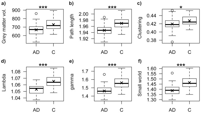
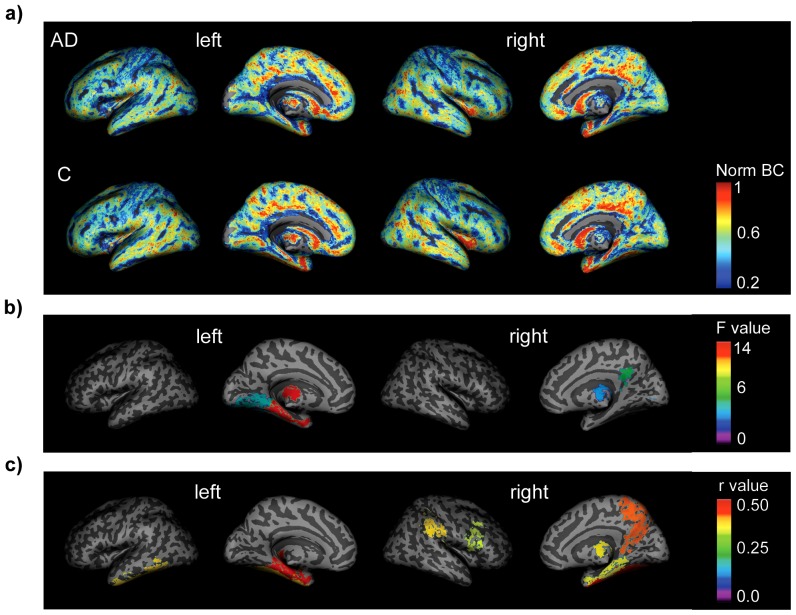
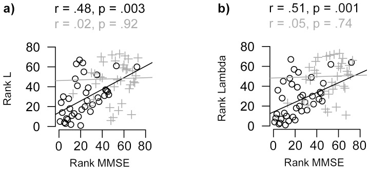
Coordinated patterns of cortical morphology have been described as structural graphs and previous research has demonstrated that properties of such graphs are altered in Alzheimer's disease (AD). However, it remains unknown how these alterations are related to cognitive deficits in individuals, as such graphs are restricted to group-level analysis. In the present study we investigated this question in single-subject grey matter networks. This new method extracts large-scale structural graphs where nodes represent small cortical regions that are connected by edges when they show statistical similarity. Using this method, unweighted and undirected networks were extracted from T1 weighted structural magnetic resonance imaging scans of 38 AD patients (19 female, average age 72±4 years) and 38 controls (19 females, average age 72±4 years). Group comparisons of standard graph properties were performed after correcting for grey matter volumetric measurements and were correlated to scores of general cognitive functioning. AD networks were characterised by a more random topology as indicated by a decreased small world coefficient (p = 3.53×10(-5)), decreased normalized clustering coefficient (p = 7.25×10(-6)) and decreased normalized path length (p = 1.91×10(-7)). Reduced normalized path length explained significantly (p = 0.004) more variance in measurements of general cognitive decline (32%) in comparison to volumetric measurements (9%). Altered path length of the parahippocampal gyrus, hippocampus, fusiform gyrus and precuneus showed the strongest relationship with cognitive decline. The present results suggest that single-subject grey matter graphs provide a concise quantification of cortical structure that has clinical value, which might be of particular importance for disease prognosis. These findings contribute to a better understanding of structural alterations and cognitive dysfunction in AD.
Overview
The claims and statistics of this paper haven’t been checked yet.
Run the check — free, no account needed. It reads the open-access text and checks the sources it cites.
Integrity signals
Registry
Cites
Statistics
Figures
Verification
Claims the paper makes
The claims and statistics of this paper haven’t been checked yet.
Run the check — free, no account needed. It reads the open-access text and checks the sources it cites.
Claims other papers make about this one
Each is checked against this paper’s own text: does it say what the citing paper says it does?
The cited source says what the claim attributes to it.
In the citing paper
“However, some studies have observed conflicting results, including lower characteristic path length in DTI (Daianu et al., 2013), gray matter (Tijms, Moller et al., 2013), fMRI (Sanz-Arigita et al., 2010), EEG (de Haan et al., 2009), and magneto-encephalogram (MEG; Stam et al., 2009) connectomes in patients with AD.”
From Functional and structural connectome properties in the 5XFAD transgenic mouse model of Alzheimer’s disease (2018) · cites this paper as Tijms, Moller et al., 2013
Self-check
Percentages, sums and reported differences re-computed from the paper’s own numbers.
The self-check needs the paper’s full text.
Citations
Cites
Kok, J. S., Berg, I. J., & Scherder, E. J. A. (2013) · Dementia and Geriatric Cognitive Disorders Extra · doi:10.1159/000353441 · Cited by 20,589
Neuropathological stageing of Alzheimer-related changes.
Braak, H., & Braak, E. (1991) · Acta Neuropathologica · doi:10.1007/bf00308809 · Cited by 16,123
Mapping the structural core of human cerebral cortex
Hagmann, P., Cammoun, L., Gigandet, X., Meuli, R., Honey, C. J., Wedeen, V. J., & Sporns, O. (2008) · PLoS Biology · doi:10.1371/journal.pbio.0060159 · Cited by 4,173
A PROPOSED MECHANISM OF EMOTION
PAPEZ, J. W. (1937) · Archives of Neurology And Psychiatry · doi:10.1001/archneurpsyc.1937.02260220069003 · Cited by 3,563
The economy of brain network organization
Bullmore, E., & Sporns, O. (2012) · Nature Reviews Neuroscience · doi:10.1038/nrn3214 · Cited by 3,284
The Human Connectome: A Structural Description of the Human Brain
Sporns, O., Tononi, G., & Kötter, R. (2005) · PLoS Computational Biology · doi:10.1371/journal.pcbi.0010042 · Cited by 3,260
Buckner, R. L., Sepulcre, J., Talukdar, T., Krienen, F. M., Liu, H., Hedden, T., Andrews-Hanna, J. R., Sperling, R. A., & Johnson, K. A. (2009) · The Journal of Neuroscience · doi:10.1523/jneurosci.5062-08.2009 · Cited by 2,862
Changes in grey matter induced by training
Draganski, B., Gaser, C., Busch, V., Schuierer, G., Bogdahn, U., & May, A. (2004) · Nature · doi:10.1038/427311a · Cited by 2,373
Buckner, R. L., Snyder, A. Z., Shannon, B. J., LaRossa, G., Sachs, R., Fotenos, A. F., Sheline, Y. I., Klunk, W. E., Mathis, C. A., Morris, J. C., et al. (2005) · The Journal of Neuroscience · doi:10.1523/jneurosci.2177-05.2005 · Cited by 2,140
Jack, C. R., Albert, M. S., Knopman, D. S., McKhann, G. M., Sperling, R. A., Carrillo, M. C., Thies, B., & Phelps, C. H. (2011) · Alzheimer's & Dementia · doi:10.1016/j.jalz.2011.03.004 · Cited by 2,042
Research works printed but not linked: 76
- JA Hardy, 1992, Alzheimer's disease: the amyloid cascade hypothesis, Science, 256, 184, 10.1126/science.1566067 · DOI · Paper page
- MC De Lacoste, 1993, The role of cortical connectivity in Alzheimer's disease pathogenesis: a review and model system, Neurobiol Aging, 14, 1, 10.1016/0197-4580(93)90015-4 · DOI
- X Delbeuck, 2003, Alzheimer's disease as a disconnection syndrome?, Neuropsychol Rev, 13, 79, 10.1023/A:1023832305702 · DOI
- RA Sperling, 2009, Amyloid deposition is associated with impaired default network function in older persons without dementia, Neuron, 63, 178, 10.1016/j.neuron.2009.07.003 · DOI · Paper page
- A Takashima, 2009, Amyloid-β, tau, and dementia, J Alzheimers Dis, 17, 729, 10.3233/JAD-2009-1090 · DOI
- GB Frisoni, 2002, Detection of grey matter loss in mild Alzheimer's disease with voxel based morphometry, J Neurol Neurosurg Psychiatry, 73, 657, 10.1136/jnnp.73.6.657 · DOI
- G Karas, 2004, Global and local gray matter loss in mild cognitive impairment and Alzheimer's disease, Neuroimage, 23, 708, 10.1016/j.neuroimage.2004.07.006 · DOI · Paper page
- MH Rosenbloom, 2011, Distinct clinical and metabolic deficits in PCA and AD are not related to amyloid distribution, Neurology, 76, 1789, 10.1212/WNL.0b013e31821cccad · DOI
- A Shiino, 2006, Four subgroups of Alzheimer's disease based on patterns of atrophy using VBM and a unique pattern for early onset disease, Neuroimage, 33, 17, 10.1016/j.neuroimage.2006.06.010 · DOI
- GR Ridgway, 2012, Early-onset Alzheimer disease clinical variants: Multivariate analyses of cortical thickness, Neurology, 79, 80, 10.1212/WNL.0b013e31825dce28 · DOI
- C Gratton, 2012, Focal brain lesions to critical locations cause widespread disruption of the modular organization of the brain, J Cogn Neurosci, 24, 1275, 10.1162/jocn_a_00222 · DOI
- O Sporns, 2004, Organization, development and function of complex brain networks, Trends Cogn Sci, 8, 418, 10.1016/j.tics.2004.07.008 · DOI · Paper page
- CJ Stam, 2012, The organization of physiological brain networks, Clin Neurophysiol, 123, 1067, 10.1016/j.clinph.2012.01.011 · DOI
- Y He, 2007, Small-world anatomical networks in the human brain revealed by cortical thickness from MRI, Cereb Cortex, 17, 2407, 10.1093/cercor/bhl149 · DOI
- D Bassett, 2008, Hierarchical organization of human cortical networks in health and schizophrenia, J Neurosci, 28, 9239, 10.1523/JNEUROSCI.1929-08.2008 · DOI
- ZJ Chen, 2008, Revealing modular architecture of human brain structural networks by using cortical thickness from MRI, Cereb Cortex, 18, 2374, 10.1093/cercor/bhn003 · DOI · Paper page
- G Sanabria-Diaz, 2010, Surface area and cortical thickness descriptors reveal different attributes of the structural human brain networks, Neuroimage, 50, 1497, 10.1016/j.neuroimage.2010.01.028 · DOI
- BM Tijms, 2012, Similarity-based extraction of individual networks from gray matter MRI scans, Cereb Cortex, 22, 1530, 10.1093/cercor/bhr221 · DOI
- A Mechelli, 2005, Structural covariance in the human cortex, J Neurosci, 25, 8303, 10.1523/JNEUROSCI.0357-05.2005 · DOI · Paper page
- J Lerch, 2006, Mapping anatomical correlations across cerebral cortex (MACACC) using cortical thickness from MRI, Neuroimage, 31, 993, 10.1016/j.neuroimage.2006.01.042 · DOI
- Y He, 2008, Structural insights into aberrant topological patterns of large-scale cortical networks in Alzheimer's disease, J Neurosci, 28, 4756, 10.1523/JNEUROSCI.0141-08.2008 · DOI · Paper page
- Y Li, 2012, Discriminant analysis of longitudinal cortical thickness changes in Alzheimer's disease using dynamic and network features, Neurobiol Aging 33, 427
- Z Yao, 2010, Abnormal cortical networks in mild cognitive impairment and Alzheimer's disease, PLoS Comp Biol, 6, e1001006, 10.1371/journal.pcbi.1001006 · DOI
- C Stam, 2009, Graph theoretical analysis of magnetoencephalographic functional connectivity in Alzheimer's disease, Brain, 132, 213, 10.1093/brain/awn262 · DOI
- W de Haan, 2009, Functional neural network analysis in frontotemporal dementia and Alzheimer's disease using EEG and graph theory, BMC Neurosci, 10, 101, 10.1186/1471-2202-10-101 · DOI
- EC Mormino, 2011, Relationships between beta-amyloid and functional connectivity in different components of the default mode network in aging, Cereb Cortex, 21, 2399, 10.1093/cercor/bhr025 · DOI
- W de Haan, 2012, Activity dependent degeneration explains hub vulnerability in Alzheimer's disease, PLoS Comp Biol, 8, e1002582, 10.1371/journal.pcbi.1002582 · DOI
- GM Mckhann, 2011, The diagnosis of dementia due to Alzheimer's disease: Recommendations from the National Institute on Aging-Alzheimer’s Association workgroups on diagnostic guidelines for Alzheimer's disease, Alzheimers Dement, 7, 263, 10.1016/j.jalz.2011.03.005 · DOI · Paper page
- MF Folstein, 1975, “Mini-mental state.” A practical method for grading the cognitive state of patients for the clinician, J Psychiatr Res, 12, 189
- M Jenkinson, 2001, A global optimisation method for robust affine registration of brain images, Med Image Anal, 5, 143, 10.1016/S1361-8415(01)00036-6 · DOI
- C Nikou, 1999, Robust voxel similarity metrics for the registration of dissimilar single and multimodal images, Pattern Recognition, 32, 1351, 10.1016/S0031-3203(98)00167-8 · DOI
- J Weese, 1999, Gray-value based registration of CT and MR images by maximization of local correlation, Medical Image Computing and Computer-Assisted Intervention–MICCAI', 99, 656, 10.1007/10704282_71 · DOI
- T VanCourt, 2005, Three-dimensional template correlation: object recognition in 3D voxel data, Computer Architecture for Machine Perception, 2005 CAMP 2005 Proceedings Seventh International Workshop on, 153, 10.1109/CAMP.2005.52 · DOI
- G Penney, 1998, A comparison of similarity measures for use in 2D-3D medical image registration, Lect Notes Comput Sc, 1496, 1153, 10.1007/BFb0056305 · DOI
- A Barrat, 2004, The architecture of complex weighted networks, Proc Natl Acad Sci U S A, 101, 3747, 10.1073/pnas.0400087101 · DOI
- M Rubinov, 2010, Complex network measures of brain connectivity: uses and interpretations, Neuroimage, 52, 1059, 10.1016/j.neuroimage.2009.10.003 · DOI · Paper page
- D Watts, 1998, Collective dynamics of “small-world” networks, Nature, 393, 440, 10.1038/30918 · DOI · Paper page
- L Freeman, 1977, A set of measures of centrality based on betweenness, Sociometry, 40, 35, 10.2307/3033543 · DOI
- MD Humphries, 2008, Network “Small-World-Ness”: A quantitative method for determining canonical network equivalence, PLoS ONE, 3, e0002051, 10.1371/journal.pone.0002051 · DOI · Paper page
- S Maslov, 2002, Specificity and stability in topology of protein networks, Science, 296, 910, 10.1126/science.1065103 · DOI · Paper page
- AL Barabasi, 1999, Emergence of scaling in random networks, Science, 286, 509, 10.1126/science.286.5439.509 · DOI · Paper page
- D Bassett, 2006, Small-world brain networks, Neuroscientist, 12, 512, 10.1177/1073858406293182 · DOI · Paper page
- M Kaiser, 2011, A tutorial in connectome analysis: topological and spatial features of brain networks, Neuroimage, 57, 892, 10.1016/j.neuroimage.2011.05.025 · DOI · Paper page
- QK Telesford, 2011, The brain as a complex system: using network science as a tool for understanding the brain, Brain Connect, 1, 295, 10.1089/brain.2011.0055 · DOI
- Conover W, Iman RL (1981) Rank transformations as a bridge between parametric and nonparametric statistics. American Statistician: 124–129. · DOI · Paper page
- WJ Conover, 1982, Analysis of covariance using the rank transformation, Biometrics, 38, 715, 10.2307/2530051 · DOI
- A Zalesky, 2010, Whole-brain anatomical networks: does the choice of nodes matter?, Neuroimage, 50, 970, 10.1016/j.neuroimage.2009.12.027 · DOI · Paper page
- N Tzourio-Mazoyer, 2002, Automated anatomical labeling of activations in SPM using a macroscopic anatomical parcellation of the MNI MRI single-subject brain, Neuroimage, 15, 273, 10.1006/nimg.2001.0978 · DOI · Paper page
- Y Benjamini, 1995, Controlling the false discovery rate: a practical and powerful approach to multiple testing, Journal of the Royal Statistical Society Series B (Methodological), 57, 289, 10.1111/j.2517-6161.1995.tb02031.x · DOI · Paper page
- Y Benjamini, 2001, The control of the false discovery rate in multiple testing under dependency, The Annals of Statistics, 29, 1165, 10.1214/aos/1013699998 · DOI · Paper page
- T Andrews, 1997, Correlated size variations in human visual cortex, lateral geniculate nucleus, and optic tract, J Neurosci, 17, 2859, 10.1523/JNEUROSCI.17-08-02859.1997 · DOI
- M Raichle, 2001, A default mode of brain function, Proc Natl Acad Sci U S A, 98, 676, 10.1073/pnas.98.2.676 · DOI · Paper page
- MD Greicius, 2004, Default-mode network activity distinguishes Alzheimer's disease from healthy aging: evidence from functional MRI, Proc Natl Acad Sci U S A, 101, 4637, 10.1073/pnas.0308627101 · DOI · Paper page
- SARB Rombouts, 2005, Altered resting state networks in mild cognitive impairment and mild Alzheimer's disease: An fMRI study, Hum Brain Mapp, 26, 231, 10.1002/hbm.20160 · DOI · Paper page
- A Burggren, 2002, Specificity of brain activation patterns in people at genetic risk for Alzheimer disease, American Journal of Geriatric Psych, 10, 44, 10.1097/00019442-200201000-00006 · DOI
- E Reiman, 2004, Functional brain abnormalities in young adults at genetic risk for late-onset Alzheimer's dementia, Proc Natl Acad Sci U S A, 101, 284, 10.1073/pnas.2635903100 · DOI · Paper page
- DC Van Essen, 1997, A tension-based theory of morphogenesis and compact wiring in the central nervous system, Nature, 385, 313, 10.1038/385313a0 · DOI
- C Hilgetag, 2004, Clustered organization of cortical connectivity, Neuroinformatics, 2, 353, 10.1385/NI:2:3:353 · DOI
- G Gong, 2012, Convergence and divergence of thickness correlations with diffusion connections across the human cerebral cortex, Neuroimage, 59, 1239, 10.1016/j.neuroimage.2011.08.017 · DOI
- I Ferrer, 1995, Transforming growth factor alpha immunoreactivity in the developing and adult brain, Neuroscience, 66, 189, 10.1016/0306-4522(94)00584-R · DOI
- K Kiuchi, 2009, Abnormalities of the uncinate fasciculus and posterior cingulate fasciculus in mild cognitive impairment and early Alzheimer's disease: A diffusion tensor tractography study, Brain Research, 1287, 184, 10.1016/j.brainres.2009.06.052 · DOI
- G Gong, 2009, Age- and gender-related differences in the cortical anatomical network, J Neurosci, 29, 15684, 10.1523/JNEUROSCI.2308-09.2009 · DOI
- M Greicius, 2003, Functional connectivity in the resting brain: a network analysis of the default mode hypothesis, Proc Natl Acad Sci U S A, 100, 253, 10.1073/pnas.0135058100 · DOI · Paper page
- AR Laird, 2009, Investigating the functional heterogeneity of the default mode network using coordinate-based meta-analytic modeling, J Neurosci, 29, 14496, 10.1523/JNEUROSCI.4004-09.2009 · DOI
- W De Haan, 2012, Disrupted modular brain dynamics reflect cognitive dysfunction in Alzheimer's disease, Neuroimage, 59, 3085, 10.1016/j.neuroimage.2011.11.055 · DOI
- DS Bassett, 2011, Conserved and variable architecture of human white matter connectivity, Neuroimage, 54, 1262, 10.1016/j.neuroimage.2010.09.006 · DOI
- A Fornito, 2010, Network scaling effects in graph analytic studies of human resting-state FMRI data, Front Syst Neurosci, 4, 22
- J Hänggi, 2011, Globally Altered Structural Brain Network Topology in Grapheme-Color Synesthesia, J Neurosci, 31, 5816, 10.1523/JNEUROSCI.0964-10.2011 · DOI
- T Paus, 1996, Human cingulate and paracingulate sulci: pattern, variability, asymmetry, and probabilistic map, Cereb Cortex, 6, 207, 10.1093/cercor/6.2.207 · DOI
- M Pievani, 2011, Functional network disruption in the degenerative dementias, Lancet Neurol, 10, 829, 10.1016/S1474-4422(11)70158-2 · DOI · Paper page
- Dai D, He H, Vogelstein J, Hou Z (2011) Network-based classification using cortical thickness of AD patients. Machine Learning in Medical Imaging: 193–200. · DOI
- L Zhou, 2011, Hierarchical anatomical brain networks for MCI prediction: Revisiting volumetric measures, PLoS ONE, 6, e21935, 10.1371/journal.pone.0021935 · DOI
- CJ Stam, 2007, Small-world networks and functional connectivity in Alzheimer's disease, Cereb Cortex, 17, 92, 10.1093/cercor/bhj127 · DOI
- C-Y Lo, 2010, Diffusion tensor tractography reveals abnormal topological organization in structural cortical networks in Alzheimer's disease, J Neurosci, 30, 16876, 10.1523/JNEUROSCI.4136-10.2010 · DOI
- N Langer, 2011, Functional brain network efficiency predicts intelligence, Hum Brain Mapp, 33, 1393, 10.1002/hbm.21297 · DOI
- AJ Mitchell, 2008, The clinical significance of subjective memory complaints in the diagnosis of mild cognitive impairment and dementia: a meta-analysis, Int J Geriat Psychiatry, 23, 1191, 10.1002/gps.2053 · DOI
Cited by
Sorted by citations
Items 101–117 of 117
Vermunt, L., Sutphen, C. L., Dicks, E., de Leeuw, D. M., Allegri, R., Berman, S., Cash, D. M., Chhatwal, J. P., Cruchaga, C., Day, G. S., et al. (2024) · Brain Communications · doi:10.1093/braincomms/fcae357 · Cited by 6
“40 Finally, we calculated the small-world metric for each network with the brain connectivity toolbox (https://sites.google.com/site/bctnet/ 41 ) modified for large sized networks.”
MethodResultSelf-citationProtas, H. D., Ghisays, V., Goradia, D. D., Bauer, R., Devadas, V., Chen, K., Reiman, E. M., & Su, Y. (2023) · Frontiers in Neuroscience · doi:10.3389/fnins.2023.1089134 · Cited by 4
Lorenzini, L., Ingala, S., Wottschel, V., Wink, A. M., Mutsaerts, H. J. M. M., Haller, S., Blennow, K., O'Brien, J. T., Frisoni, B. G., Chételat, G., et al. (2022) · Aging Brain · doi:10.1016/j.nbas.2022.100054 · Cited by 3
“have been observed in relation with Abdeposition in patients with AD [63,27,55].”
BackgroundSelf-citationChen, G., Wu, C., Liu, Y., Fang, Z., Luo, L., Lai, X., Wang, W., & Dong, L. (2022) · Brain Imaging and Behavior · doi:10.1007/s11682-022-00709-7 · Cited by 3
Sharma, R., & Joshi, S. D. (2023) · Annals of Neurosciences · doi:10.1177/09727531231186503 · Cited by 3
Connectivity-Driven Parcellation Methods for the Human Cerebral Cortex
Arslan, S. (2017) · arXiv.org · doi:10.48550/arxiv.1802.06772 · Cited by 2
“It has been common practice in recent neuroscience studies to explore several neurological [247, 77] and neuro-developmental disorders [107, 145, 60, 78] from a network perspective.”
BackgroundThe bi-phasic behaviour of grey matter networks after the first demyelinating attack
Collorone, S., Pontillo, G., Foster, M. A., Prados, F., Kanber, B., Yiannakas, M. C., Burke, A., Ogunbowale, L., Davagnanam, I., Wheeler‐Kingshott, C. A. M. G., et al. (2025) · Brain Communications · doi:10.1093/braincomms/fcaf367 · Cited by 1
“Notably, the analysis of single-subject grey matter networks in advanced multiple sclerosis, 13 like studies using the same methodology in dementia, 40 showed that cognitively impaired patients tend towards random network topology.”
Self-citationSingle-subject gray matter networks in patients with early- to late-stage Parkinson’s disease
Yoko, S., Matsuda, H., Maikusa, N., Ota, M., Kimura, Y., Maki, H., Kagaya, R., Imokawa, T., Mukai, Y., Takahashi, Y., et al. (2025) · Neurological Research · doi:10.1080/01616412.2025.2561116 · Cited by 1
Structural Similarity Networks Reveal Brain Vulnerability in DementiaPreprint
Montagnese, M., Ebneabbasi, A., García-San-Martín, N., Pecci-Terroba, C., Romero-García, R., Morgan, S. E., Cole, J. H., Seidlitz, J., Rittman, T., & Bethlehem, R. A. I. (2025) · medRxiv · doi:10.1101/2025.06.10.25328978
“Our findings contribute to a growing body of work suggesting that Alzheimer’s disease may reflect not only regional atrophy, but also disruptions in the coordinated structural organisation of the cortex 48,49 .”
Detecting Amyloid Positivity Using Morphometric Magnetic Resonance Imaging
Pereira, H. R., Initiative, A. D. N., Diogo, V. S., Prata, D. P., & Ferreira, H. A. (2024) · Journal of Alzheimer s Disease · doi:10.3233/jad-240366 · Cited by 3
“11 , 18 , 21 Given that, MRI morphometric GT metrics have been surprisingly little explored as a potential biomarker for A (cid:2) positivity, especially given its clinical usefulness potential (non-invasiveness, low cost, and high accessibility compared to PET/CSF exams), at least as a preliminary…”
Quantitative Assessment of Mesoscale Cellular Order and Organization in the Mouse Hippocampus
Hein, K., Romero-Limon, H., Möckel, C., Karasinsky, A., Möllmert, S., Kayser, J., Zaccone, A., Guck, J., & Toda, T. (2026) · bioRxiv · doi:10.64898/2026.07.06.736467
“…altered graph properties of the neural network, as well as changes in the density of hippocampal tissue have been linked to cognitive decline, mem-55 ory deficits and neurological diseases such as Alzheimer’s disease (Allen et al., 2007; Wang et al., 2006; Tijms et al., 2013; Berron et al., 2021).”
MVMGNN;Multi-View Masked Graph Neural Network for Alzheimer's Disease Diagnosis using Structural MRI
Yao, N., Wang, Z., Sun, D., Han, C., Li, Y., Nan, J.-F., Zhu, F., Zhao, C., & Zhou, W. (2026) · arXiv.org · doi:10.48550/arxiv.2607.09788
de Leeuw, D. M., Duits, F. H., Dicks, E., Vromen, E. M., Teunissen, C. E., Barkhof, F., van der Flier, W. M., Visser, P. J., & Tijms, B. M. (2026) · Brain Communications · doi:10.1093/braincomms/fcag257
“43 For each individual’s scan, a network was constructed from native space GM segmentations.”
Self-citationMarie, D., Kokkinou, D., Junker-Tschopp, C., Kliegel, M., Allali, G., Guevara, A. B., Frisoni, G., & James, C. E. (2026) · bioRxiv · doi:10.64898/2026.06.19.733422
“These findings align with graph-theoretical and connectomics evidence of disrupted hub organization and efficiency in MCI/AD (Sala-Llonch et al., 2015; Tijms et al., 2013; Yu et al., 2021).”
Rubido, N., Batziou, V., Fuad, M., & Vuksanović, V. (2025) · doi:10.48550/arxiv.2505.03458
“Both MCI and AD affect local de Haan et al. (2012); Heringa et al. (2014) and small-world network metrics Tijms et al. (2013); Seo et al. (2013) through changes in clustering (i.e., segregation) Brier et al. (2014) and global efficiency (i.e., integration) Lo et al. (2010); Yao et al. (2010);…”
Dehkordi, A. M. G., Kashaninia, A., Farokhi, F., & Dabanloo, N. J. (2022) · Engineering Analysis with Boundary Elements · doi:10.1016/j.enganabound.2022.10.037
Yoko, S., Sone, D., Maikusa, N., Kimura, Y., Suzuki, F., Fujii, H., Sato, N., & Matsuda, H. (2020) · eNeurologicalSci · doi:10.1016/j.ensci.2021.100343
Metadata
- Authors
- Tijms, B. M., Möller, C., Vrenken, H., Wink, A. M., de Haan, W., van der Flier, W. M., Stam, C. J., Scheltens, P., & Barkhof, F.
- Affiliations
- Amsterdam UMC Location Vrije Universiteit Amsterdam (NL)
- Corresponding author
- Tijms, Betty M.
- ORCID
- Venue
- PLoS ONE
- Publisher
- Public Library of Science (PLoS)
- Published
- 2013-03-11
- Volume
- 8
- Issue
- 3
- Pages
- e58921
- ISSN
- 1932-6203 (ISSN-L)
- PMC
- PMC3594199
- OpenAlex
- W2058087702
- Cited by
- 132
- References
- 102
- Citations by year
- 2026: 2 · 2025: 6 · 2024: 12 · 2023: 8 · 2022: 11 · 2021: 15 · 2020: 11 · 2019: 6 · 2018: 18 · 2017: 11 · 2016: 10 · 2015: 12 · 2014: 7 · 2013: 3
- FWCI
- 5.85
- Type
- journal article
- Language
- en
- Update notices
- correction (2013-09-09)
- Open access
- gold
- doi.org
- Locations
- PLoS ONE (publishedVersion, cc-by)
- Europe PMC (PubMed Central) (publishedVersion, cc-by)
- PubMed Central (publishedVersion, cc-by)
- PubMed (publishedVersion)
- DOAJ (DOAJ: Directory of Open Access Journals) (submittedVersion, cc-by-sa)
- UCL Discovery (University College London) (submittedVersion)
- Pure Amsterdam UMC (submittedVersion)
- Data Archiving and Networked Services (DANS) (submittedVersion, other-oa)
- Data Archiving and Networked Services (DANS) (submittedVersion)
- Funders
- Stichting Dioraphte
- Alzheimer Nederland
- Topics
- Life Sciences › Neuroscience › Cognitive Neuroscience › Functional Brain Connectivity Studies
- Health Sciences › Medicine › Psychiatry and Mental health › Dementia and Cognitive Impairment Research
- Life Sciences › Neuroscience › Cellular and Molecular Neuroscience › Alzheimer's disease research and treatments
- Keywords
- Alzheimer's disease, structural graph, small-world property, path length, clustering coefficient, cognitive decline, cortical morphology, hippocampus, parahippocampal gyrus, precuneus, fusiform gyrus, gray matter volume, disease prognosis
- MeSH terms
- Aged, Alzheimer Disease, Brain Mapping, Electroencephalography, Female, Humans, Magnetic Resonance Imaging, Male, Psychiatric Status Rating Scales, Severity of Illness Index, Case-Control Studies, Neuroimaging
About these checks
Registry, retraction, Cites and statistics results are computed, not judged, with one exception: a printed reference that no index lists is read once by a model before it counts as a citation error. Claims are extracted by a model and kept only when their quote appears verbatim in the text; each cited source is then checked, with a model’s help, for whether it says what the claim relies on. Those verdicts can be wrong — read the claim’s quote and the cited source before relying on one.
Statistics and arithmetic are recomputed, not judged.
Confidence bands are shown only for fields calibrated on our annotated test set. None is calibrated yet, so no band is shown.
What counts as a claim ›How we measure confidence ›How we check this ›
Are you an author, or do you see a problem with this page? Request a review
