

The DOI matches its Crossref record; 74 of the 74 references it cites were found in a registry or an index.
Microgravity Induces Pelvic Bone Loss through Osteoclastic Activity, Osteocytic Osteolysis, and Osteoblastic Cell Cycle Inhibition by CDKN1a/p21
Open accessAbstract
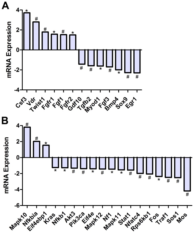
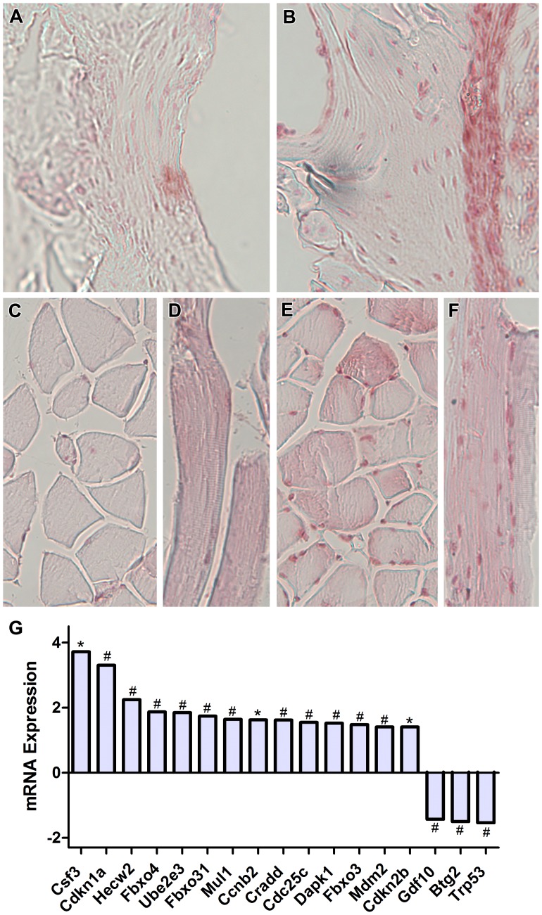
Bone is a dynamically remodeled tissue that requires gravity-mediated mechanical stimulation for maintenance of mineral content and structure. Homeostasis in bone occurs through a balance in the activities and signaling of osteoclasts, osteoblasts, and osteocytes, as well as proliferation and differentiation of their stem cell progenitors. Microgravity and unloading are known to cause osteoclast-mediated bone resorption; however, we hypothesize that osteocytic osteolysis, and cell cycle arrest during osteogenesis may also contribute to bone loss in space. To test this possibility, we exposed 16-week-old female C57BL/6J mice (n = 8) to microgravity for 15-days on the STS-131 space shuttle mission. Analysis of the pelvis by µCT shows decreases in bone volume fraction (BV/TV) of 6.29%, and bone thickness of 11.91%. TRAP-positive osteoclast-covered trabecular bone surfaces also increased in microgravity by 170% (p = 0.004), indicating osteoclastic bone degeneration. High-resolution X-ray nanoCT studies revealed signs of lacunar osteolysis, including increases in cross-sectional area (+17%, p = 0.022), perimeter (+14%, p = 0.008), and canalicular diameter (+6%, p = 0.037). Expression of matrix metalloproteinases (MMP) 1, 3, and 10 in bone, as measured by RT-qPCR, was also up-regulated in microgravity (+12.94, +2.98 and +16.85 fold respectively, p<0.01), with MMP10 localized to osteocytes, and consistent with induction of osteocytic osteolysis. Furthermore, expression of CDKN1a/p21 in bone increased 3.31 fold (p<0.01), and was localized to osteoblasts, possibly inhibiting the cell cycle during tissue regeneration as well as conferring apoptosis resistance to these cells. Finally the apoptosis inducer Trp53 was down-regulated by -1.54 fold (p<0.01), possibly associated with the quiescent survival-promoting function of CDKN1a/p21. In conclusion, our findings identify the pelvic and femoral region of the mouse skeleton as an active site of rapid bone loss in microgravity, and indicate that this loss is not limited to osteoclastic degradation. Therefore, this study offers new evidence for microgravity-induced osteocytic osteolysis, and CDKN1a/p21-mediated osteogenic cell cycle arrest.
Overview
The claims and statistics of this paper haven’t been checked yet.
Run the check — free, no account needed. It reads the open-access text and checks the sources it cites.
Integrity signals
Registry
Cites
Statistics
Figures
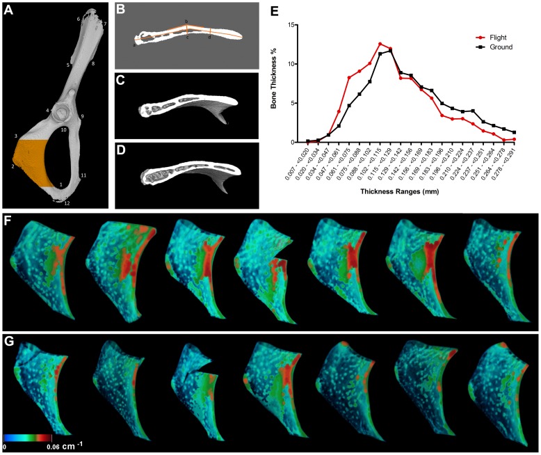
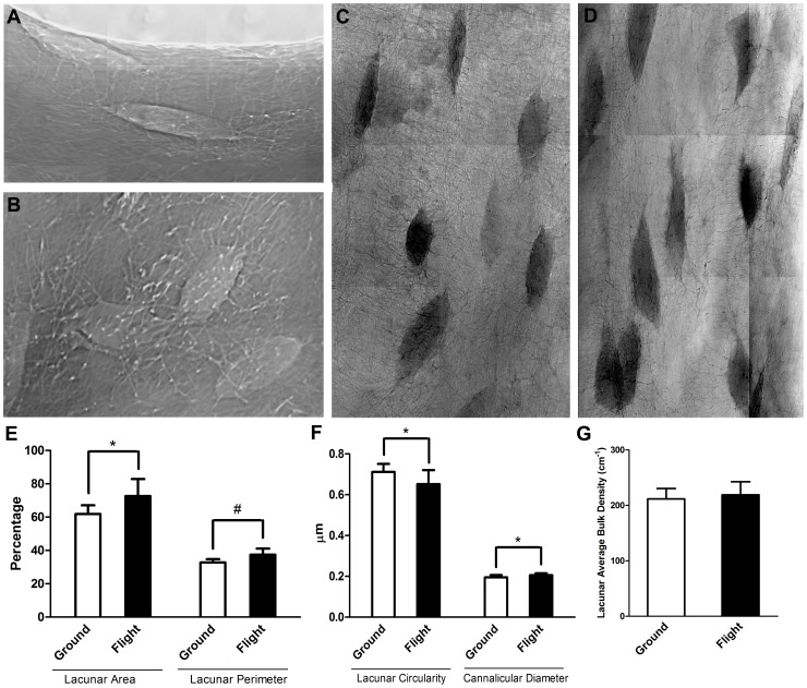
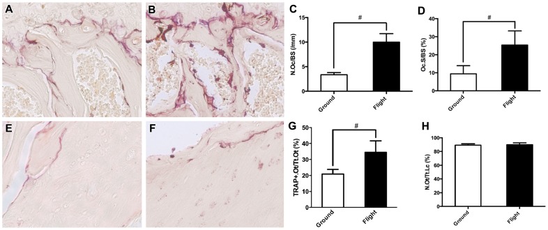
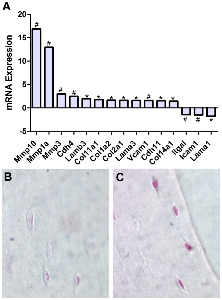
Verification
Claims the paper makes
The claims and statistics of this paper haven’t been checked yet.
Run the check — free, no account needed. It reads the open-access text and checks the sources it cites.
Claims other papers make about this one
Each is checked against this paper’s own text: does it say what the citing paper says it does?
The cited source doesn’t support the claim as written.
In the citing paper
“They also documented osteocyte apoptosis as reflected by increases in lacunar and canalicular diameters [60].”
From Osteoclasts and Microgravity (2020) · cites this paper as [60]
Checked by a reasoning model — may be wrong.
Experimental — accuracy not yet measuredThe source backs part of the claim, not all of it — or backs it for a different population, condition or comparison.
In the citing paper
“They found that microgravity reduced the bone volume by 6.3% and bone thickness by 11.9% and increased the number of bone-surface TRAP-positive osteoclasts by 170% compared to that of ground controls, indicating enhanced osteoclast-mediated bone resorption.”
From Osteoclasts and Microgravity (2020) · cites this paper as [60]
Experimental — accuracy not yet measuredThe source backs part of the claim, not all of it — or backs it for a different population, condition or comparison.
In the citing paper
“conditions of microgravity and simulated microgravity decrease the mineral content and cortical and trabecular microstructures of bone”
From Osteoclasts and Microgravity (2020) · cites this paper as [60]
Experimental — accuracy not yet measuredThe source backs part of the claim, not all of it — or backs it for a different population, condition or comparison.
In the citing paper
“decrease osteoblastogenesis and osteoblast secretion of OPG”
From Osteoclasts and Microgravity (2020) · cites this paper as [60]
Experimental — accuracy not yet measuredThe source backs part of the claim, not all of it — or backs it for a different population, condition or comparison.
In the citing paper
“increase osteoclast differentiation, fusion, and expression of regulatory and osteoclast-specific genes”
From Osteoclasts and Microgravity (2020) · cites this paper as [60]
Experimental — accuracy not yet measured
The check wasn’t confident either way, so it gives no verdict — never disputed.
In the citing paper
“increase osteoclast secretion of sclerostin”
From Osteoclasts and Microgravity (2020) · cites this paper as [60]
Experimental — accuracy not yet measured
Self-check
Percentages, sums and reported differences re-computed from the paper’s own numbers.
The self-check needs the paper’s full text.
Citations
Cites
Vogelstein, B. (1992) · Cell · doi:10.1016/0092-8674(92)90421-8 · Cited by 1,849
Cellular and molecular mechanisms of bone remodeling
Raggatt, L. J., & Partridge, N. C. (2010) · Journal of Biological Chemistry · doi:10.1074/jbc.r109.041087 · Cited by 1,402
Correlation of Terminal Cell Cycle Arrest of Skeletal Muscle with Induction of p21 by MyoD
Halevy, O., Novitch, B. G., Spicer, D. B., Skapek, S. X., Rhee, J., Hannon, G. J., Beach, D., & Lassar, A. B. (1995) · Science · doi:10.1126/science.7863327 · Cited by 1,139
Matrix metalloproteinases: a tail of a frog that became a prince
Brinckerhoff, C., & Matrisian, L. M. (2002) · Nature Reviews Molecular Cell Biology · doi:10.1038/nrm763 · Cited by 1,112
Vico, L., Collet, P., Guignandon, A., Lafage-Proust, M.-H., Thomas, T., Rehailia, M., & Alexandre, C. (2000) · The Lancet · doi:10.1016/s0140-6736(00)02217-0 · Cited by 821
Matrix metalloproteinases (MMPs) in health and disease: an overview
Malemud, C. (2006) · Frontiers in Bioscience · doi:10.2741/1915 · Cited by 705
The cell biology of bone metabolism
Datta, H. K., Ng, W. F., Walker, J. A., Tuck, S. P., & Varanasi, S. S. (2008) · Journal of Clinical Pathology · doi:10.1136/jcp.2007.048868 · Cited by 614
Yoshihara, Y., Nakamura, H., Obata, K., Yamada, H., Hayakawa, T., Fujikawa, K., & Okada, Y. (2000) · Annals of the Rheumatic Diseases · doi:10.1136/ard.59.6.455 · Cited by 566
Mechanosensation and transduction in osteocytes
Klein-Nulend, J., Bakker, A. D., Bacabac, R. G., Vatsa, A., & Weinbaum, S. (2013) · Bone · doi:10.1016/j.bone.2012.10.013 · Cited by 528
Ge, C., Xiao, G., Jiang, D., & Franceschi, R. T. (2007) · The Journal of Cell Biology · doi:10.1083/jcb.200610046 · Cited by 510
Aguirre, J. I., Plotkin, L. I., Stewart, S. A., Weinstein, R. S., Parfitt, A. M., Manolagas, S. C., & Bellido, T. (2006) · Journal of Bone and Mineral Research · doi:10.1359/jbmr.060107 · Cited by 494
Demonstration of osteocytic perilacunar/canalicular remodeling in mice during lactation
Qing, H., Ardeshirpour, L., Divieti Pajevic, P., Dusevich, V., Jähn, K., Kato, S., Wysolmerski, J., & Bonewald, L. F. (2012) · Journal of Bone and Mineral Research · doi:10.1002/jbmr.1567 · Cited by 489
Functional and structural adaptations of skeletal muscle to microgravity
Fitts, R. H., Riley, D. R., & Widrick, J. J. (2001) · Journal of Experimental Biology · doi:10.1242/jeb.204.18.3201 · Cited by 478
Osteocytes as Dynamic Multifunctional Cells
Bonewald, L. F. (2007) · Annals of the New York Academy of Sciences · doi:10.1196/annals.1402.018 · Cited by 457
Exercise in space: human skeletal muscle after 6 months aboard the International Space Station
Trappe, S., Costill, D., Gallagher, P., Creer, A., Peters, J. R., Evans, H., Riley, D. A., & Fitts, R. H. (2009) · Journal of Applied Physiology · doi:10.1152/japplphysiol.91578.2008 · Cited by 454
Role of Transforming Growth Factor-Beta in Bone Remodeling
Bonewald, L. F., & Mundy, G. R. (1990) · Clinical Orthopaedics and Related Research · doi:10.1097/00003086-199001000-00036 · Cited by 431
Murakami, S., Kan, M., McKeehan, W. L., & Crombrugghe, B. D. (2000) · Proceedings of the National Academy of Sciences · doi:10.1073/pnas.97.3.1113 · Cited by 377
Hayman, A. R., Jones, S. J., BOYDE, A., Foster, D. B., Colledge, W. H., Carlton, M. B. L., Evans, M. J., & Cox, T. M. (1996) · Development · doi:10.1242/dev.122.10.3151 · Cited by 373
Phosphorylation-Dependent Ubiquitination of Cyclin D1 by the SCFFBX4-αB Crystallin Complex
Lin, D. I., Barbash, O., Kumar, K. G. S., Weber, J. D., Harper, J. W., Klein-Szanto, A. J. P., Rustgi, A., Fuchs, S. Y., & Diehl, J. A. (2006) · Molecular Cell · doi:10.1016/j.molcel.2006.09.007 · Cited by 349
Mechanosensation and transduction in osteocytes
Bonewald, L. F. (2006) · BoneKEy-Osteovision · doi:10.1138/20060233 · Cited by 283
Zehnder, Y., L�thi, M., Michel, D., Knecht, H., Perrelet, R., Neto, I. C., KRAENZLIN, M. E., Z�ch, G., & Lippuner, K. (2004) · Osteoporosis International · doi:10.1007/s00198-003-1529-6 · Cited by 281
Wang, X., Kua, H.-Y., Hu, Y., Guo, K., Zeng, Q., Wu, Q., Ng, H., Karsenty, G., Crombrugghe, B. D., Yeh, J. K., et al. (2005) · The Journal of Cell Biology · doi:10.1083/jcb.200507106 · Cited by 254
Effects of Microgravity on Osteoblast Growth Activation
Hughes‐Fulford, M., & Lewis, M. L. (1996) · Experimental Cell Research · doi:10.1006/excr.1996.0116 · Cited by 250
Do osteocytes contribute to bone mineral homeostasis? Osteocytic osteolysis revisited
Teti, A. M., & Zallone, A. Z. (2008) · Bone · doi:10.1016/j.bone.2008.09.017 · Cited by 245
Holmbeck, K., Bianco, P., Pidoux, I., Inoué, S., Billinghurst, R. C., Wu, W. K. K., Chrysovergis, K., Yamada, S. S., Birkedal‐Hansen, H., & Poole, A. R. (2004) · Journal of Cell Science · doi:10.1242/jcs.01581 · Cited by 231
Effects of spaceflight on trabecular bone in rats
Jee, W. S. S., Wronski, T. J., Morey, E. R., & Kimmel, D. B. (1983) · American Journal of Physiology-Regulatory, Integrative and Comparative Physiology · doi:10.1152/ajpregu.1983.244.3.r310 · Cited by 225
Gius, D. R., Cao, X., Rauscher, F. J., Cohén, D., Curran, T., & Sukhatme, V. P. (1990) · Molecular and Cellular Biology · doi:10.1128/mcb.10.8.4243 · Cited by 214
Filvaroff, E. H., Erlebacher, A., Ye, J., Gitelman, S. E., Lotz, J. C., Heilman, M., & Derynck, R. (1999) · Development · doi:10.1242/dev.126.19.4267 · Cited by 212
Caiozzo, V. J., Baker, M. J., Herrick, R. E., Tao, M., & Baldwin, K. M. (1994) · Journal of Applied Physiology · doi:10.1152/jappl.1994.76.4.1764 · Cited by 208
Lack of p21 expression links cell cycle control and appendage regeneration in mice
Bedelbaeva, K., Snyder, A., Gourevitch, D., Clark, L., Zhang, X.-M., Leferovich, J., Cheverud, J. M., Lieberman, P., & Heber-Katz, E. (2010) · Proceedings of the National Academy of Sciences · doi:10.1073/pnas.1000830107 · Cited by 207
Hill, P. (1998) · British Journal of Orthodontics · doi:10.1093/ortho/25.2.101 · Cited by 206
Srivastava, S., Weitzmann, M. N., Kimble, R. B., Rizzo, M., Zahner, M., Milbrandt, J., Ross, F. P., & Pacifici, R. (1998) · Journal of Clinical Investigation · doi:10.1172/jci4561 · Cited by 197
Nabavi, N., Khandani, A., Camirand, A., & Harrison, R. E. (2011) · Bone · doi:10.1016/j.bone.2011.07.036 · Cited by 174
Dai, Z., Wang, R., Ling, S., Wan, Y., & Li, Y. H. (2007) · Cell Proliferation · doi:10.1111/j.1365-2184.2007.00461.x · Cited by 165
Hill, P. A., Murphy, G., Docherty, A., Hembry, R. M., Millican, T. A., Reynolds, J. J., & Meikle, M. C. (1994) · Journal of Cell Science · doi:10.1242/jcs.107.11.3055 · Cited by 164
Osteocyte Remodeling of the Perilacunar and Pericanalicular Matrix
Qing, H., & Bonewald, L. F. (2009) · International Journal of Oral Science · doi:10.4248/ijos.09019 · Cited by 152
Space flight and oxidative stress
Stein, T. P. (2002) · Nutrition · doi:10.1016/s0899-9007(02)00938-3 · Cited by 152
Function of the cytoskeleton in gravisensing during spaceflight
Hughes‐Fulford, M. (2003) · Advances in Space Research · doi:10.1016/s0273-1177(03)90399-1 · Cited by 149
Effect of spaceflight on periosteal bone formation in rats
Wronski, T. J., & Morey, E. R. (1983) · American Journal of Physiology-Regulatory, Integrative and Comparative Physiology · doi:10.1152/ajpregu.1983.244.3.r305 · Cited by 145
Vico, L., Chappard, D., Alexandre, C., Palle, S., MINAIRE, P. M., Riffat, G., Morukov, B. V., & Rakhmanov, S. (1987) · PubMed · Cited by 144
Barbash, O., Zamfirova, P., Lin, D. I., Chen, X., Yang, K., Nakagawa, H., Lu, F., Rustgi, A. K., & Diehl, J. A. (2008) · Cancer Cell · doi:10.1016/j.ccr.2008.05.017 · Cited by 140
Cardiovascular function and basics of physiology in microgravity
AUBERT, A. E., Beckers, F., & Verheyden, B. (2005) · Acta Cardiologica · doi:10.2143/ac.60.2.2005024 · Cited by 129
Microgravity during spaceflight directly affects in vitro osteoclastogenesis and bone resorption
Tamma, R., Colaianni, G., Camerino, C., Di Benedetto, A., Greco, G., Strippoli, M., Vergari, R., Grano, A., Mancini, L., Mori, G., et al. (2009) · The FASEB Journal · doi:10.1096/fj.08-127951 · Cited by 121
Microgravity: the immune response and bone
Zayzafoon, M., Meyers, V. E., & McDonald, J. M. (2005) · Immunological Reviews · doi:10.1111/j.0105-2896.2005.00330.x · Cited by 117
Bone resorption is induced on the second day of bed rest: results of a controlled crossover trial
Baecker, N., Tomic, A., Mika, C., Gotzmann, A., Platen, P., Gerzer, R., & Heer, M. A. (2003) · Journal of Applied Physiology · doi:10.1152/japplphysiol.00264.2003 · Cited by 103
Tavella, S., Ruggiu, A., Giuliani, A., Brun, F., Canciani, B., Mănescu, A., Marozzi, K., Cilli, M., Costa, D., Liu, Y., et al. (2012) · PLoS ONE · doi:10.1371/journal.pone.0033179 · Cited by 95
Cavolina, J. M., Evans, G. L., Harris, S. A., Zhang, M., Westerlind, K. C., & Turner, R. T. (1997) · Endocrinology · doi:10.1210/endo.138.4.5040 · Cited by 94
Effects of Long-Term Space Flight on Erythrocytes and Oxidative Stress of Rodents
Rizzo, A. M., Corsetto, P. A., Montorfano, G., Milani, S., Zava, S., Tavella, S., Cancedda, R., & Berra, B. (2012) · PLoS ONE · doi:10.1371/journal.pone.0032361 · Cited by 91
The effect of microgravity on morphology and gene expression of osteoblasts in vitro
Carmeliet, G., & Bouillon, R. (1999) · The FASEB Journal · doi:10.1096/fasebj.13.9001.s129 · Cited by 91
Westerlind, K. C., & Turner, R. T. (1995) · Journal of Bone and Mineral Research · doi:10.1002/jbmr.5650100603 · Cited by 88
Calcium and bone metabolism during space flight
Smith, S. M., & Heer, M. A. (2002) · Nutrition · doi:10.1016/s0899-9007(02)00895-x · Cited by 85
Vico, L., Новиков, В. Е., Very, J. M., & Alexandre, C. (1991) · PubMed · Cited by 82
Alterations in protein metabolism during space flight and inactivity
Ferrando, A. A., Paddon‐Jones, D., & Wolfe, R. R. (2002) · Nutrition · doi:10.1016/s0899-9007(02)00930-9 · Cited by 81
Chang, S., Chang, T., Peng, H., Yeh, Y. T., Lee, D. Y., Yeh, C. R., Zhou, J., Cheng, C. K., Chang, C. A., & Chiu, J. (2009) · Molecular Endocrinology · doi:10.1210/me.2009-0143 · Cited by 69
The Canalicular Structure of Compact Bone in the Rat at Different Ages
OKADA, S., Yoshida, S., Ashrafi, S. H., & Schraufnagel, D. E. (2002) · Microscopy and Microanalysis · doi:10.1017/s1431927601020037 · Cited by 67
The scarless heart and the MRL mouse
Heber‐Katz, E., Leferovich, J. M., Bedelbaeva, K., Gourevitch, D., & Clark, L. D. (2004) · Philosophical Transactions of the Royal Society B Biological Sciences · doi:10.1098/rstb.2004.1468 · Cited by 59
Cardiovascular and Fluid Volume Control in Humans in Space
Norsk, P. (2005) · Current Pharmaceutical Biotechnology · doi:10.2174/1389201054553734 · Cited by 57
Bajotto, G., & Shimomura, Y. (2006) · Journal of Nutritional Science and Vitaminology · doi:10.3177/jnsv.52.233 · Cited by 54
Siu, P. M., Pistilli, E. E., Murlasits, Z., & Alway, S. E. (2005) · Journal of Applied Physiology · doi:10.1152/japplphysiol.01012.2005 · Cited by 53
The effects of TGF-beta on bone.
Mundy, G. R. (1991) · PubMed · Cited by 51
The effect of simulated microgravity on osteoblasts is independent of the induction of apoptosis
Bucaro, M. A., Zahm, A. M., Risbud, M. V., AYYASWAMY, P. S., Mukundakrishnan, K., Steinbeck, M. J., Shapiro, I. M., & Adams, C. S. (2007) · Journal of Cellular Biochemistry · doi:10.1002/jcb.21310 · Cited by 50
Sakai, A., Sakata, T., Tanaka, S., Okazaki, R., Kunugita, N., Norimura, T., & Nakamura, T. (2002) · Journal of Bone and Mineral Research · doi:10.1359/jbmr.2002.17.1.119 · Cited by 48
Nakano, Y., Toyosawa, S., & Takano, Y. (2004) · Journal of Histochemistry & Cytochemistry · doi:10.1369/jhc.4a6378.2004 · Cited by 47
Osteobiology, strain, and microgravity. Part II: Studies at the tissue level
Vico, L., Hinsenkamp, M., Jones, D. M., Marie, P. J., Zallone, A. Z., & Cancedda, R. (2001) · Calcified Tissue International · doi:10.1007/bf02684996 · Cited by 41
Effects of parity on pelvic size and shape dimorphism in Mus
Schutz, H., Donovan, E. R., & Hayes, J. P. (2009) · Journal of Morphology · doi:10.1002/jmor.10723 · Cited by 40
p53 control of bone remodeling
Liu, H., & Li, B. (2010) · Journal of Cellular Biochemistry · doi:10.1002/jcb.22749 · Cited by 39
Zhang, R., Ran, H., Ma, J., Bai, Y., & Lin, L. (2011) · Journal of Physiology and Biochemistry · doi:10.1007/s13105-011-0123-1 · Cited by 35
Effects of microgravity on bone and calcium homeostasis
Zérath, E. (1998) · Advances in Space Research · doi:10.1016/s0273-1177(98)00026-x · Cited by 34
Hydrogen therapy may reduce the risks related to radiation-induced oxidative stress in space flight
Schoenfeld, M. P., Ansari, R. R., Zakrajsek, J. F., Billiar, T. R., Toyoda, Y., Wink, D. A., & Nakao, A. (2010) · Medical Hypotheses · doi:10.1016/j.mehy.2010.08.046 · Cited by 34
Convertino, V. A. (2005) · PubMed · Cited by 29
[Bone loss and bone metabolism in astronauts during long-duration space flight].
Ohshima, H. (2006) · PubMed · Cited by 19
FBXO31: A New Player in the Ever-Expanding DNA Damage Response Orchestra
Shiloh, Y. (2009) · Science Signaling · doi:10.1126/scisignal.296pe73 · Cited by 15
[Effect of space flight on rat skeletal bones (an optical light and electron microscopic study)].
Vs, I., La, T., & Gorokhova, G. P. (1977) · PubMed · Cited by 15
Cardiovascular Consequences of Weightlessness Promote Advances in Clinical and Trauma Care
Cooke, W. H., & Convertino, V. A. (2005) · Current Pharmaceutical Biotechnology · doi:10.2174/1389201054553671 · Cited by 12
Cited by
Sorted by citations
Robling, A. G., & Bonewald, L. F. (2020) · Annual Review of Physiology · doi:10.1146/annurev-physiol-021119-034332 · Cited by 539
The impact of microgravity on bone in humans
Grimm, D. G., Grosse, J., Wehland, M., Mann, V., Reseland, J. E., Sundaresan, A., & Corydon, T. J. (2016) · Bone · doi:10.1016/j.bone.2015.12.057 · Cited by 283
Skeletal changes during and after spaceflight
Vico, L., & Hargens, A. (2018) · Nature Reviews Rheumatology · doi:10.1038/nrrheum.2018.37 · Cited by 236
Update on the effects of microgravity on the musculoskeletal system
Juhl, O. J., Buettmann, E. G., Friedman, M. A., DeNapoli, R. C., Hoppock, G. A., & Donahue, H. J. (2021) · npj Microgravity · doi:10.1038/s41526-021-00158-4 · Cited by 204
The effects of microgravity on bone structure and function
Man, J., Graham, T., Squires-Donelly, G., & Laslett, A. L. (2022) · npj Microgravity · doi:10.1038/s41526-022-00194-8 · Cited by 175
Metadata
- Authors
- Blaber, E. A., Dvorochkin, N., Lee, C., Alwood, J. S., Yousuf, R., Pianetta, P., Globus, R. K., Burns, B. P., & Almeida, E. A. C.
- Affiliations
- Ames Research Center (US)
- SLAC National Accelerator Laboratory (US)
- UNSW Sydney (AU)
- Corresponding author
- Almeida, Eduardo A. C.
- Venue
- PLoS ONE
- Publisher
- Public Library of Science (PLoS)
- Published
- 2013-04-18
- Volume
- 8
- Issue
- 4
- Pages
- e61372
- ISSN
- 1932-6203 (ISSN-L)
- PMC
- PMC3630201
- OpenAlex
- W2142491318
- Cited by
- 196
- References
- 74
- Citations by year
- 2026: 15 · 2025: 17 · 2024: 10 · 2023: 9 · 2022: 9 · 2021: 21 · 2020: 21 · 2019: 16 · 2018: 19 · 2017: 11 · 2016: 15 · 2015: 12 · 2014: 19 · 2013: 2
- FWCI
- 11.14
- Type
- journal article
- Language
- en
- Open access
- gold
- doi.org
- Licence
- Locations
- PLoS ONE (publishedVersion, public-domain)
- Europe PMC (PubMed Central) (publishedVersion, other-oa)
- PubMed Central (publishedVersion, public-domain)
- PubMed (publishedVersion)
- DOAJ (DOAJ: Directory of Open Access Journals) (submittedVersion, cc-by-sa)
- Funders
- National Aeronautics and Space Administration
- Topics
- Health Sciences › Medicine › Physiology (medical) › Spaceflight effects on biology
- Life Sciences › Biochemistry, Genetics and Molecular Biology › Molecular Biology › Bone Metabolism and Diseases
- Health Sciences › Medicine › Orthopedics and Sports Medicine › Bone health and osteoporosis research
- Keywords
- microgravity, spaceflight, bone loss, osteoclasts, osteoblasts, CDKN1A/p21, Trp53, matrix metalloproteinases, osteocytes, bone resorption, trabecular bone, bone unloading, female mice, bone homeostasis
- MeSH terms
- Animals, Bone and Bones, Bone Resorption, Female, Mice, Inbred C57BL, Osteoclasts, Osteocytes, Osteolysis, Pelvic Bones, Weightlessness, Cyclin-Dependent Kinase Inhibitor p21, Mice, X-Ray Microtomography, Cell Cycle Checkpoints
About these checks
Registry, retraction, Cites and statistics results are computed, not judged, with one exception: a printed reference that no index lists is read once by a model before it counts as a citation error. Claims are extracted by a model and kept only when their quote appears verbatim in the text; each cited source is then checked, with a model’s help, for whether it says what the claim relies on. Those verdicts can be wrong — read the claim’s quote and the cited source before relying on one.
Statistics and arithmetic are recomputed, not judged.
Confidence bands are shown only for fields calibrated on our annotated test set. None is calibrated yet, so no band is shown.
What counts as a claim ›How we measure confidence ›How we check this ›
Are you an author, or do you see a problem with this page? Request a review
