

The DOI matches its Crossref record; 69 of the 70 references it cites were found in a registry or an index.
Targeting the Intrinsically Disordered Structural Ensemble of α-Synuclein by Small Molecules as a Potential Therapeutic Strategy for Parkinson’s Disease
Open accessAbstract
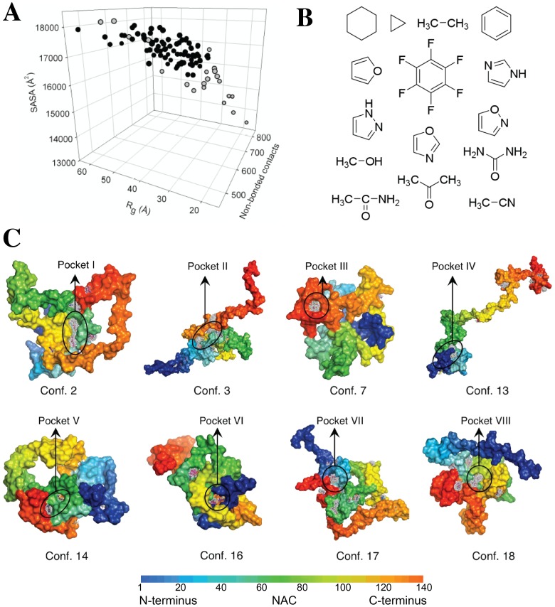
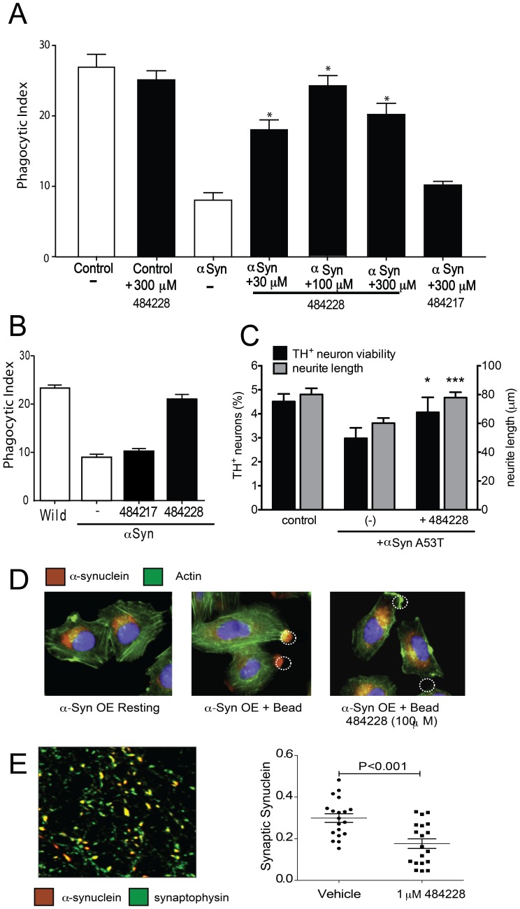
The misfolding of intrinsically disordered proteins such as α-synuclein, tau and the Aβ peptide has been associated with many highly debilitating neurodegenerative syndromes including Parkinson's and Alzheimer's diseases. Therapeutic targeting of the monomeric state of such intrinsically disordered proteins by small molecules has, however, been a major challenge because of their heterogeneous conformational properties. We show here that a combination of computational and experimental techniques has led to the identification of a drug-like phenyl-sulfonamide compound (ELN484228), that targets α-synuclein, a key protein in Parkinson's disease. We found that this compound has substantial biological activity in cellular models of α-synuclein-mediated dysfunction, including rescue of α-synuclein-induced disruption of vesicle trafficking and dopaminergic neuronal loss and neurite retraction most likely by reducing the amount of α-synuclein targeted to sites of vesicle mobilization such as the synapse in neurons or the site of bead engulfment in microglial cells. These results indicate that targeting α-synuclein by small molecules represents a promising approach to the development of therapeutic treatments of Parkinson's disease and related conditions.
Overview
The claims and statistics of this paper haven’t been checked yet.
Run the check — free, no account needed. It reads the open-access text and checks the sources it cites.
Integrity signals
Registry
Cites
Statistics
Figures
Verification
Claims the paper makes
The claims and statistics of this paper haven’t been checked yet.
Run the check — free, no account needed. It reads the open-access text and checks the sources it cites.
Claims other papers make about this one
Each is checked against this paper’s own text: does it say what the citing paper says it does?
In the citing paper
“Targeting the intrinsically disordered structural ensemble of α-synuclein by small molecules as a potential therapeutic strategy for Parkinson’s disease has been shown to be promising.429”
From Protein Ensembles: How Does Nature Harness Thermodynamic Fluctuations for Life? The Diverse Functional Roles of Conformational Ensembles in the Cell (2016) · cites this paper as [429]
Self-check
Percentages, sums and reported differences re-computed from the paper’s own numbers.
The self-check needs the paper’s full text.
Citations
Cites
Spillantini, M. G., Schmidt, M. L., Lee, V. M.-Y., Trojanowski, J. Q., Jakes, R., & Goedert, M. (1997) · Nature · doi:10.1038/42166 · Cited by 8,382
Mutation in the α-Synuclein Gene Identified in Families with Parkinson's Disease
Polymeropoulos, M. H., Lavedan, C., Leroy, E., Ide, S. E., Dehejia, A., Dutra, A., Pike, B., Root, H., Rubenstein, J., Boyer, R., et al. (1997) · Science · doi:10.1126/science.276.5321.2045 · Cited by 8,240
Protein Misfolding, Functional Amyloid, and Human Disease
Chiti, F., & Dobson, C. M. (2006) · Annual Review of Biochemistry · doi:10.1146/annurev.biochem.75.101304.123901 · Cited by 6,497
Self-citationProtein folding and misfolding
Dobson, C. M. (2003) · Nature · doi:10.1038/nature02261 · Cited by 4,791
Self-citationCox’s regression model for counting processes: A large sample study
Andersen, P. K., & Gill, R. D. (1982) · The Annals of Statistics · doi:10.1214/aos/1176345976 · Cited by 4,419
α-Synuclein Locus Triplication Causes Parkinson's Disease
Singleton, A. B., Farrer, M., Johnson, J., Singleton, A., Hague, S., Kachergus, J., Hulihan, M., Peuralinna, T., Dutra, A., Nussbaum, R., et al. (2003) · Science · doi:10.1126/science.1090278 · Cited by 4,383
Intrinsically unstructured proteins and their functions
Dyson, H. J., & Wright, P. E. (2005) · Nature Reviews Molecular Cell Biology · doi:10.1038/nrm1589 · Cited by 3,871
DrugBank: a knowledgebase for drugs, drug actions and drug targets
Wishart, D. S., Knox, C., Guo, A. C., Cheng, D., Shrivastava, S., Tzur, D., Gautam, B., & Hassanali, M. (2007) · Nucleic Acids Research · doi:10.1093/nar/gkm958 · Cited by 2,896
The new mutation, E46K, of α‐synuclein causes parkinson and Lewy body dementia
Zarranz, J. J., Alegre, J., Gómez‐Esteban, J. C., Lezcano, E., Ros, R., Ampuero, I., Vidal, L., Hoenicka, J., Rodriguez, O., Atarés, B., et al. (2004) · Annals of Neurology · doi:10.1002/ana.10795 · Cited by 2,737
A Hot Spot of Binding Energy in a Hormone-Receptor Interface
Clackson, T., & Wells, J. A. (1995) · Science · doi:10.1126/science.7529940 · Cited by 1,987
Cited by
Sorted by citations
The amyloid state and its association with protein misfolding diseases.
Knowles, T. P. J., Vendruscolo, M., & Dobson, C. M. (2014) · Nature Reviews Molecular Cell Biology · doi:10.1038/nrm3810 · Cited by 2,382
Self-citationSavelieff, M. G., Nam, G., Kang, J., Lee, H. J., Lee, M., & Lim, M. H. (2018) · Chemical Reviews · doi:10.1021/acs.chemrev.8b00138 · Cited by 612
Wei, G., Xi, W., Nussinov, R., & Ma, B. (2016) · Chemical Reviews · doi:10.1021/acs.chemrev.5b00562 · Cited by 436
Historical Perspective: Models of Parkinson’s Disease
Chia, S. J., Tan, E., & Chao, Y. (2020) · International Journal of Molecular Sciences · doi:10.3390/ijms21072464 · Cited by 438
Alpha-synuclein biology in Lewy body diseases
Kim, W. S., Kågedal, K., & Halliday, G. M. (2014) · Alzheimer s Research & Therapy · doi:10.1186/s13195-014-0073-2 · Cited by 397
Metadata
- Authors
- Tóth, G., Gardai, S. J., Zago, W., Bertoncini, C. W., Cremades, N., Roy, S. L., Tambe, M. A., Rochet, J.-C., Galvagnion, C., Skibinski, G., Finkbeiner, S., Bova, M., Regnstrom, K., Chiou, S.-S., Johnston, J., Callaway, K., Anderson, J. P., Jobling, M. F., Buell, A. K., Yednock, T. A., Knowles, T. P. J., Vendruscolo, M., Christodoulou, J., Dobson, C. M., Schenk, D., & McConlogue, L.
- Affiliations
- University of Cambridge (GB)
- Elan Pharmaceuticals, Inc. (Ireland) (IE)
- Purdue University West Lafayette (US)
- Gladstone Institutes (US)
- University College London (GB)
- Corresponding author
- Tóth, Gergely
- Vendruscolo, Michele
- Dobson, Christopher M.
- Schenk, Dale
- McConlogue, Lisa
- ORCID
- Venue
- PLoS ONE
- Publisher
- Public Library of Science (PLoS)
- Published
- 2014-02-14
- Volume
- 9
- Issue
- 2
- Pages
- e87133
- ISSN
- 1932-6203 (ISSN-L)
- PMC
- PMC3925190
- OpenAlex
- W2145653859
- Cited by
- 151
- References
- 70
- Citations by year
- 2026: 5 · 2025: 9 · 2024: 7 · 2023: 11 · 2022: 9 · 2021: 12 · 2020: 16 · 2019: 7 · 2018: 19 · 2017: 16 · 2016: 18 · 2015: 12 · 2014: 10
- FWCI
- 12.10
- Type
- journal article
- Language
- en
- Update notices
- correction (2014-05-27)
- Open access
- gold
- doi.org
- Locations
- PLoS ONE (publishedVersion, cc-by)
- Europe PMC (PubMed Central) (publishedVersion, other-oa)
- PubMed Central (publishedVersion, cc-by)
- PubMed (publishedVersion)
- LA Referencia (Red Federada de Repositorios Institucionales de Publicaciones Científicas) (publishedVersion, other-oa)
- Americanae (AECID Library) (submittedVersion, cc-by-nc-sa)
- DOAJ (DOAJ: Directory of Open Access Journals) (submittedVersion)
- eScholarship (California Digital Library) (submittedVersion)
- Figshare (submittedVersion, cc-by)
- Greater South Information System
- Funders
- Wellcome Trust (097806)
- University of Cambridge
- Ministerio de Ciencia e Innovación
- National Institutes of Health
- Medical Research Council (MC_G1000734)
- Biotechnology and Biological Sciences Research Council (BB/H003843/1)
- National Institute of Neurological Disorders and Stroke (R01NS039074, R01NS083390)
- Topics
- Health Sciences › Medicine › Clinical Neurology › Parkinson's Disease Mechanisms and Treatments
- Life Sciences › Neuroscience › Cellular and Molecular Neuroscience › Alzheimer's disease research and treatments
- Life Sciences › Biochemistry, Genetics and Molecular Biology › Molecular Biology › Protein Structure and Dynamics
- Keywords
- alpha-synuclein, Parkinson's disease, intrinsically disordered proteins, small molecule drug candidates, protein misfolding, vesicle trafficking, dopaminergic neurons, neurite retraction, microglia, synapses, drug repurposing
- MeSH terms
- Animals, Binding Sites, Humans, Models, Biological, Models, Molecular, Nerve Degeneration, Parkinson Disease, Phagocytes, Synapses, Mice, alpha-Synuclein, Small Molecule Libraries, Molecular Targeted Therapy, Dopaminergic Neurons, Intrinsically Disordered Proteins
About these checks
Registry, retraction, Cites and statistics results are computed, not judged, with one exception: a printed reference that no index lists is read once by a model before it counts as a citation error. Claims are extracted by a model and kept only when their quote appears verbatim in the text; each cited source is then checked, with a model’s help, for whether it says what the claim relies on. Those verdicts can be wrong — read the claim’s quote and the cited source before relying on one.
Statistics and arithmetic are recomputed, not judged.
Confidence bands are shown only for fields calibrated on our annotated test set. None is calibrated yet, so no band is shown.
What counts as a claim ›How we measure confidence ›How we check this ›
Are you an author, or do you see a problem with this page? Request a review
