

The DOI matches its Crossref record; 64 of the 64 references it cites were found in a registry or an index.
Key Regulatory Role of Dermal Fibroblasts in Pigmentation as Demonstrated Using a Reconstructed Skin Model: Impact of Photo-Aging
Open accessAbstract
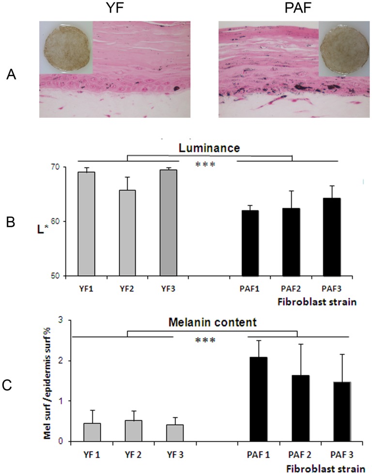
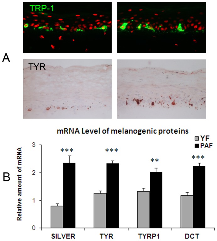
To study cutaneous pigmentation in a physiological context, we have previously developed a functional pigmented reconstructed skin model composed of a melanocyte-containing epidermis grown on a dermal equivalent comprising living fibroblasts. The present studies, using the same model, aimed to demonstrate that dermal fibroblasts influence skin pigmentation up to the macroscopic level. The proof of principle was performed with pigmented skins differing only in the fibroblast component. First, the in vitro system was reconstructed with or without fibroblasts in order to test the global influence of the presence of this cell type. We then assessed the impact of the origin of the fibroblast strain on the degree of pigmentation using fetal versus adult fibroblasts. In both experiments, impressive variation in skin pigmentation at the macroscopic level was observed and confirmed by quantitative parameters related to skin color, melanin content and melanocyte numbers. These data confirmed the responsiveness of the model and demonstrated that dermal fibroblasts do indeed impact the degree of skin pigmentation. We then hypothesized that a physiological state associated with pigmentary alterations such as photo-aging could be linked to dermal fibroblasts modifications that accumulate over time. Pigmentation of skin reconstructed using young unexposed fibroblasts (n = 3) was compared to that of tissues containing natural photo-aged fibroblasts (n = 3) which express a senescent phenotype. A stimulation of pigmentation in the presence of the natural photo-aged fibroblasts was revealed by a significant increase in the skin color (decrease in Luminance) and an increase in both epidermal melanin content and melanogenic gene expression, thus confirming our hypothesis. Altogether, these data demonstrate that the level of pigmentation of the skin model is influenced by dermal fibroblasts and that natural photo-aged fibroblasts can contribute to the hyperpigmentation that is associated with photo-aging.
Overview
The claims and statistics of this paper haven’t been checked yet.
Run the check — free, no account needed. It reads the open-access text and checks the sources it cites.
Integrity signals
Registry
Cites
Statistics
Figures
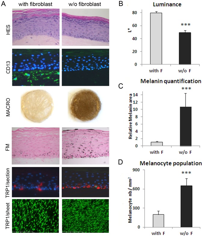
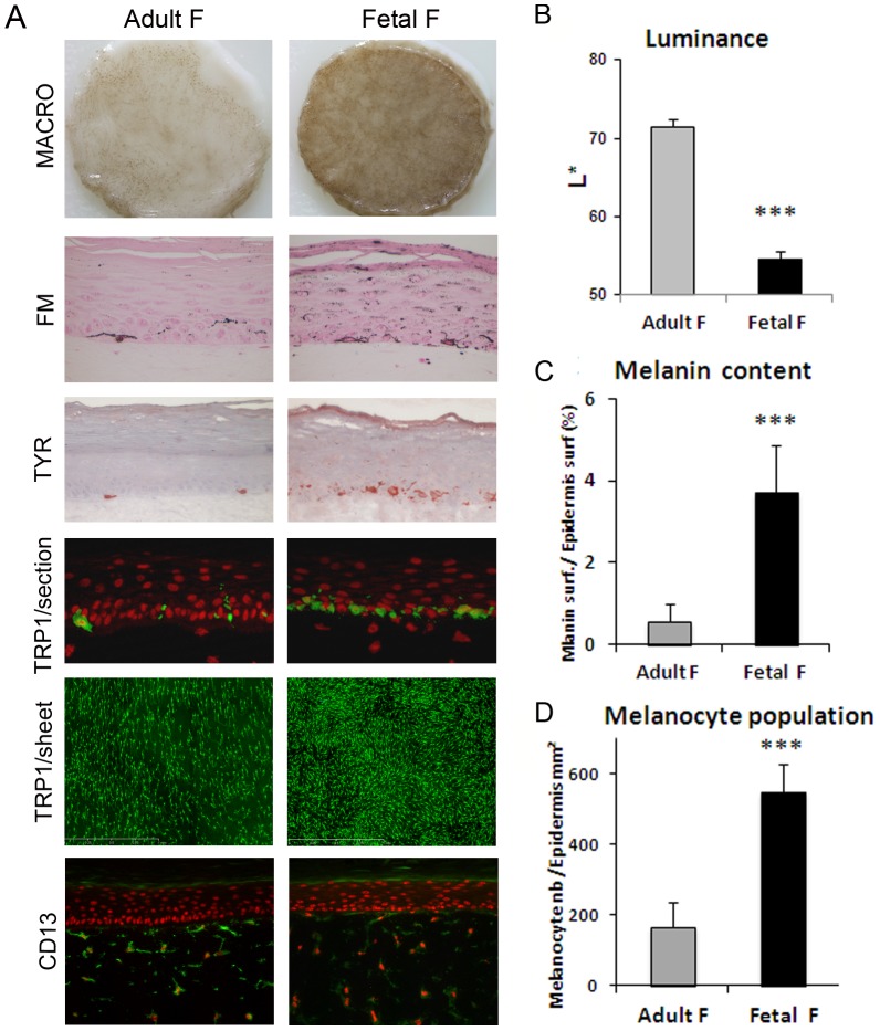
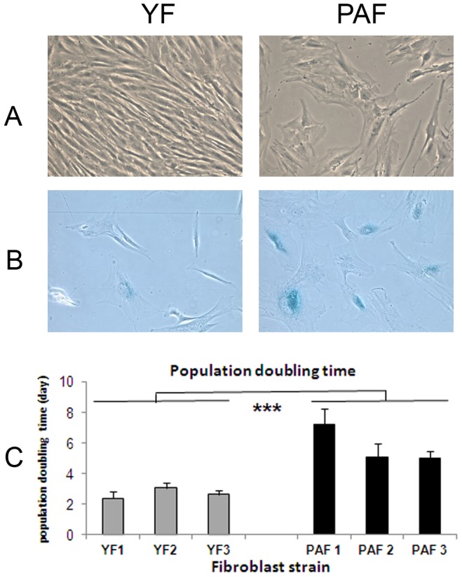
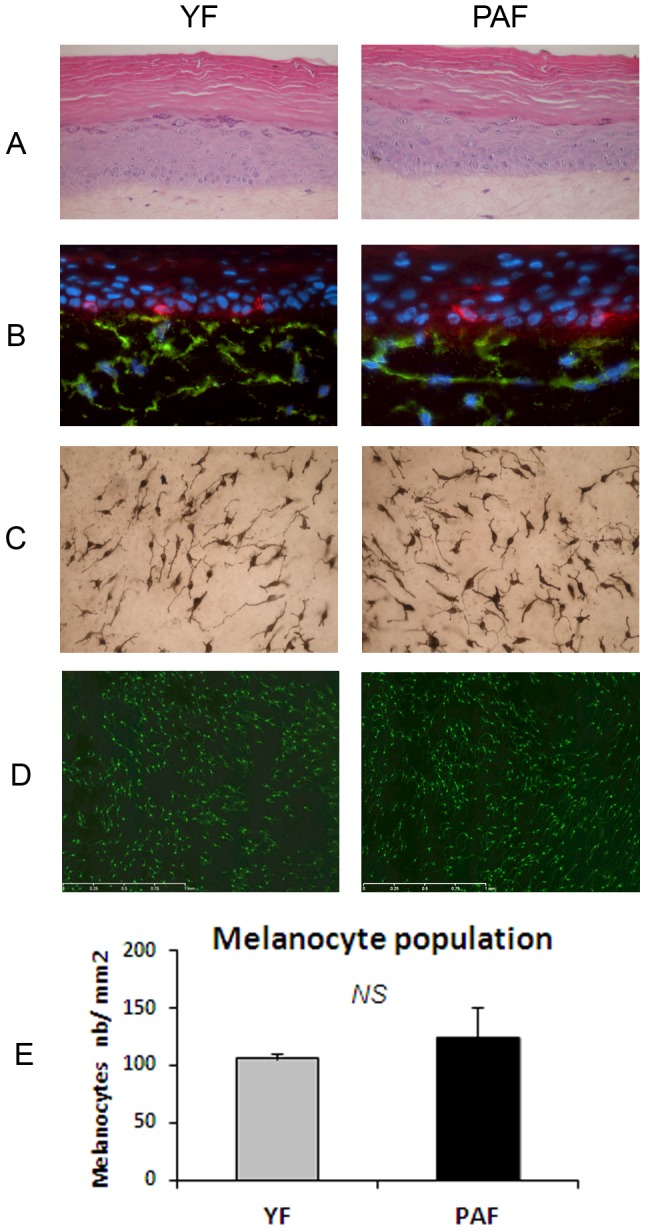
Verification
Claims the paper makes
The claims and statistics of this paper haven’t been checked yet.
Run the check — free, no account needed. It reads the open-access text and checks the sources it cites.
Claims other papers make about this one
Each is checked against this paper’s own text: does it say what the citing paper says it does?
In the citing paper
“Further, aging skin fibroblasts cause pigmentation and dark spots by promoting the transcription of the melanin gene [28].”
From Diet and Skin Aging—From the Perspective of Food Nutrition (2020) · cites this paper as [28]
Self-check
Percentages, sums and reported differences re-computed from the paper’s own numbers.
The self-check needs the paper’s full text.
Citations
Cites
Oncogenic ras provokes premature cell senescence associated with accumulation of p53 and p16INK4a
Serrano, M., Lin, A. W., McCurrach, M. E., Beach, D., & Lowe, S. W. (1997) · Cell · doi:10.1016/s0092-8674(00)81902-9 · Cited by 5,218
Rheinwatd, J. G., & Green, H. (1975) · Cell · doi:10.1016/s0092-8674(75)80001-8 · Cited by 4,581
Melanin pigmentation in mammalian skin and its hormonal regulation
Slominski, A., Tobin, D. J., Shibahara, S., & Wortsman, J. (2004) · Physiological Reviews · doi:10.1152/physrev.00044.2003 · Cited by 2,015
Slominski, A., Wortsman, J., Luger, T., Paus, R., & Solomon, S. (2000) · Physiological Reviews · doi:10.1152/physrev.2000.80.3.979 · Cited by 776
Methods to Detect Biomarkers of Cellular Senescence
Itahana, K., Campisi, J., & Dimri, G. P. (2007) · Methods in molecular biology · doi:10.1007/978-1-59745-361-5_3 · Cited by 535
Solar UV irradiation and dermal photoaging
Wlaschek, M., Tantcheva-Poór, I., Naderi, L., Ma, W., Schneider, L. A., Razi-Wolf, Z., Schüller, J., & Scharffetter-Kochanek, K. (2001) · Journal of Photochemistry and Photobiology B Biology · doi:10.1016/s1011-1344(01)00201-9 · Cited by 509
Slominski, A. T., Zmijewski, M. A., Skobowiat, C., Zbytek, B., Slominski, R. M., & Steketee, J. D. (2012) · Advances in anatomy, embryology and cell biology · doi:10.1007/978-3-642-19683-6 · Cited by 505
Physiological factors that regulate skin pigmentation
Yamaguchi, Y., & Hearing, V. J. (2009) · BioFactors · doi:10.1002/biof.29 · Cited by 495
Slominski (2012) · doi:10.1007/978-3-642-19683-6_1 · Cited by 461
L‐tyrosine and L‐dihydroxyphenylalanine as hormone‐like regulators of melanocyte functions
Slominski, A. T., Zmijewski, M. A., & Pawelek, J. M. (2011) · Pigment Cell & Melanoma Research · doi:10.1111/j.1755-148x.2011.00898.x · Cited by 449
Research works printed but not linked: 4
- Lavker R.M. (1995) Cutaneous aging: chronologic versus photoaging. in: Gilchrest B.A. (ed): Photodamage. Cambridge USA, Blackwell Science Inc, pp 123–135.
- SJ Hedley, 2002, Fibroblasts play a regulatory role in the control of pigmentation in reconstructed human skin from skin types I and II, Pigment Cell Res, 15, 49, 10.1034/j.1600-0749.2002.00067.x · DOI
- G Imokawa, 1998, Biological characterization of human fibroblast-derived mitogenic factors for human melanocytes, Biochem J 330 (Pt, 3), 1235, 10.1042/bj3301235 · DOI
- D Kovacs, 2010, Role of fibroblast-derived growth factors in regulating hyperpigmentation of solar lentigo, Br J Dermatol, 163, 1020, 10.1111/j.1365-2133.2010.09946.x · DOI
Cited by
Sorted by citations
Focus on the Contribution of Oxidative Stress in Skin Aging
Papaccio, F., D’Arino, A., Caputo, S., & Bellei, B. (2022) · Antioxidants · doi:10.3390/antiox11061121 · Cited by 452
Biomarkers of cellular senescence and skin aging
Wang, A. S., & Dreesen, O. (2018) · Frontiers in Genetics · doi:10.3389/fgene.2018.00247 · Cited by 404
Skin Aging, Cellular Senescence and Natural Polyphenols
Csekes, E., & Račková, L. (2021) · International Journal of Molecular Sciences · doi:10.3390/ijms222312641 · Cited by 352
Del Bino, S., Duval, C., & Bernerd, F. (2018) · International Journal of Molecular Sciences · doi:10.3390/ijms19092668 · Cited by 315
Self-citationCellular Senescence and the Senescence-Associated Secretory Phenotype as Drivers of Skin Photoaging
Fitsiou, E., Pulido, T., Campisi, J., Alimirah, F., & Demaria, M. (2020) · Journal of Investigative Dermatology · doi:10.1016/j.jid.2020.09.031 · Cited by 237
Diet and Skin Aging—From the Perspective of Food Nutrition
Cao, C., Xiao, Z., Wu, Y., & Ge, C. (2020) · Nutrients · doi:10.3390/nu12030870 · Cited by 231
Management of hyperpigmentation: Current treatments and emerging therapies
Nautiyal, A., & Wairkar, S. (2021) · Pigment Cell & Melanoma Research · doi:10.1111/pcmr.12986 · Cited by 195
Bioprinting of biomimetic skin containing melanocytes
Min, D., Lee, W., Bae, I., Lee, T. R., Croce, P., & Yoo, S. (2017) · Experimental Dermatology · doi:10.1111/exd.13376 · Cited by 198
Precise role of dermal fibroblasts on melanocyte pigmentation
Wang, Y., Viennet, C., Robin, S., Berthon, J., He, L., & Humbert, P. G. (2017) · Journal of Dermatological Science · doi:10.1016/j.jdermsci.2017.06.018 · Cited by 153
Epigenetic Regulation of Skin Cells in Natural Aging and Premature Aging Diseases
Orioli, D., & Dellambra, E. (2018) · Cells · doi:10.3390/cells7120268 · Cited by 150
Neuroendocrine Aspects of Skin Aging
Bocheva, G., Slominski, R. M., & Slominski, A. T. (2019) · International Journal of Molecular Sciences · doi:10.3390/ijms20112798 · Cited by 145
Pollution and Sun Exposure: A Deleterious Synergy. Mechanisms and Opportunities for Skin Protection
Marrot, L. (2019) · Current Medicinal Chemistry · doi:10.2174/0929867324666170918123907 · Cited by 125
Paracrine regulation of melanogenesis
Yuan, X. H., & Jin, Z. H. (2017) · British Journal of Dermatology · doi:10.1111/bjd.15651 · Cited by 116
Skin Pigmentation and Pigmentary Disorders: Focus on Epidermal/Dermal Cross-Talk
Bastonini, E., Kovacs, D., & Picardo, M. (2016) · Annals of Dermatology · doi:10.5021/ad.2016.28.3.279 · Cited by 121
The impact of cellular senescence in skin ageing: A notion of mosaic and therapeutic strategies
Toutfaire, M., Bauwens, É., & Debacq‐Chainiaux, F. (2017) · Biochemical Pharmacology · doi:10.1016/j.bcp.2017.04.011 · Cited by 107
Premature cell senescence in human skin: Dual face in chronic acquired pigmentary disorders
Bellei, B., & Picardo, M. (2019) · Ageing Research Reviews · doi:10.1016/j.arr.2019.100981 · Cited by 93
Upadhyay, P. R., Ho, T., & ABDEL‐MALEK, Z. A. (2021) · Pigment Cell & Melanoma Research · doi:10.1111/pcmr.12985 · Cited by 93
Skin Pigmentation Abnormalities and Their Possible Relationship with Skin Aging
Lee, A.-Y. (2021) · International Journal of Molecular Sciences · doi:10.3390/ijms22073727 · Cited by 87
Decoding Skin Aging: A Review of Mechanisms, Markers, and Modern Therapies
Naharro‐Rodríguez, J., Bacci, S., Hernández‐Bule, M. L., Pérez-González, L. A., & Fernández-Guarino, M. (2025) · Cosmetics · doi:10.3390/cosmetics12040144 · Cited by 98
Role of Nicotinamide in Genomic Stability and Skin Cancer Chemoprevention
Fania, L., Mazzanti, C., Campione, E., Candi, E., ABENI, D. D., & Dellambra, E. (2019) · International Journal of Molecular Sciences · doi:10.3390/ijms20235946 · Cited by 90
Senescent fibroblasts in melasma pathophysiology
Kim, M.-S., Kim, S. M., Kwon, S., Park, T. J., & Kang, H. Y. (2018) · Experimental Dermatology · doi:10.1111/exd.13814 · Cited by 67
Targeting the Complexity of In Vitro Skin Models: A Review of Cutting-Edge Developments
Quílez, C., Bebiano, L. B., Jones, E., Maver, U., Meesters, L., Parzymies, P., Petiot, E., Rikken, G., Risueño, I., Zaidi, H., et al. (2024) · Journal of Investigative Dermatology · doi:10.1016/j.jid.2024.04.032 · Cited by 65
Umbilical Cord Mesenchymal Stem Cell-Derived Exosomes Ameliorate HaCaT Cell Photo-Aging
Liu, S., Meng, M., Shen, H., Gao, H., Zhao, Y., Yang, Y., Lin, Z., Yang, L.-R., Zhu, K., Han, R. F., et al. (2021) · Rejuvenation Research · doi:10.1089/rej.2020.2313 · Cited by 57
Mitochondrial dynamics and metabolism across skin cells: implications for skin homeostasis and aging
Martic, I., Papaccio, F., Bellei, B., & Cavinato, M. S. (2023) · Frontiers in Physiology · doi:10.3389/fphys.2023.1284410 · Cited by 60
Hardwick, R. N., Betts, C. J., Whritenour, J., Sura, R., Thamsen, M., Kaufman, E. H., & Fabre, K. (2020) · Lab on a Chip · doi:10.1039/c9lc00519f · Cited by 48
Li, P.-H., Liu, L., Chang, C., Gao, R., Leung, C., Ma, D., & Wang, H.-M. D. (2018) · International Journal of Molecular Sciences · doi:10.3390/ijms19051475 · Cited by 48
Non-animal models in dermatological research
Dellambra, E. (2018) · ALTEX · doi:10.14573/altex.1808022 · Cited by 49
Sebocytes contribute to melasma onset
Flori, E., Mastrofrancesco, A., Mosca, S., Ottaviani, M., Briganti, S., Cardinali, G., Filoni, A., Cameli, N., Zaccarini, M., Zouboulis, C. C., et al. (2022) · iScience · doi:10.1016/j.isci.2022.103871 · Cited by 44
Emerging role of dermal compartment in skin pigmentation: comprehensive review
Kapoor, R., Dhatwalia, S., Kumar, R., Rani, S., & Parsad, D. (2020) · Journal of the European Academy of Dermatology and Venereology · doi:10.1111/jdv.16404 · Cited by 36
Çelik, B., Sağıroğlu, A. A., & Özdemir, S. (2017) · International Journal of Nanomedicine · doi:10.2147/ijn.s140835 · Cited by 40
Tissue engineering strategies to bioengineer the ageing skin phenotype in vitro
Costello, L., DiColandrea, T., Tasseff, R. A., Isfort, R. J., Bascom, C., von Zglinicki, T., & Przyborski, S. A. (2022) · Aging Cell · doi:10.1111/acel.13550 · Cited by 33
Human Hair Graying Revisited: Principles, Misconceptions, and Key Research Frontiers
Paus, R., Sevilla, A., & Grichnik, J. M. (2023) · Journal of Investigative Dermatology · doi:10.1016/j.jid.2023.09.276 · Cited by 39
Wu, S.-Y., Wang, H.-M., Wen, Y.-S., Liu, W., Li, P.-H., Chiu, C., Chen, P.-C., Huang, C., Sheu, J., & Wen, Z. (2015) · International Journal of Molecular Sciences · doi:10.3390/ijms160920240 · Cited by 34
Biological processes in solar lentigo: insights brought by experimental models
Goorochurn, R., Viennet, C., Granger, C., Fanian, F., Varin‐Blank, N., Roy, C. L., & Humbert, P. (2016) · Experimental Dermatology · doi:10.1111/exd.12937 · Cited by 30
Shaiek, A., Flament, F., Francois, G., Lefebvre‐Descamps, V., Barla, C., Vicic, M., Giron, F., & Bazin, R. (2016) · Skin Research and Technology · doi:10.1111/srt.12330 · Cited by 31
Huang, W.-S., Wang, Y.-W., Hung, K., Hsieh, P., Fu, K., Dai, L., Liou, N.-H., Ma, K., Liu, J.-C., & Dai, N. (2018) · PeerJ · doi:10.7717/peerj.4815 · Cited by 30
Bio‐derived hydroxystearic acid ameliorates skin age spots and conspicuous pores
Schütz, R., Rawlings, A. V., Wandeler, E., Jackson, E., Trevisan, S., Monneuse, J., Bendik, I., Massironi, M., & Imfeld, D. (2019) · International Journal of Cosmetic Science · doi:10.1111/ics.12529 · Cited by 29
Bhatia, E., Kumari, D., Sharma, S., Ahamad, N., & Banerjee, R. (2021) · Wiley Interdisciplinary Reviews Nanomedicine and Nanobiotechnology · doi:10.1002/wnan.1746 · Cited by 30
Effect of Dickkopf1 on the senescence of melanocytes: in vitro study
Rani, S., Chauhan, R., Parsad, D., & Kumar, R. (2018) · Archives of Dermatological Research · doi:10.1007/s00403-018-1820-1 · Cited by 27
Mechanism, Formulation, and Efficacy Evaluation of Natural Products for Skin Pigmentation Treatment
Peng, X., Ma, Y., Yan, C., Wei, X., Zhang, L., Jiang, H., Ma, Y., Zhang, S., Xing, M., & Gao, Y. (2024) · Pharmaceutics · doi:10.3390/pharmaceutics16081022 · Cited by 30
Chou, H.-Y., Wang, H., Kuo, C.-H., Lu, P.-H., Wang, L., Kang, W., & Sun, C. (2020) · ACS Omega · doi:10.1021/acsomega.9b04316 · Cited by 26
Tracing skin aging process: a mini- review of in vitro approaches
Letsiou, S. (2021) · Biogerontology · doi:10.1007/s10522-021-09916-z · Cited by 26
You, S., Shi, X., Yu, D., Zhao, D., An, Q., Wang, D., Zhang, J., Li, M., & Wang, C. (2021) · BMC Complementary Medicine and Therapies · doi:10.1186/s12906-020-03197-8 · Cited by 24
Cosmetic industry requirements regarding skin models for cosmetic testing
Pellevoisin, C., Bouez, C., & Cotovio, J. (2017) · Elsevier eBooks · doi:10.1016/b978-0-12-810545-0.00001-2 · Cited by 22
Esmat, S. M., Hadidi, H. H. E., Hegazy, R. A., Gawdat, H. I., Tawdy, A. M. E., Fawzy, M. M., Abdel-Halim, D. M., Sultan, O. S., & Shaker, O. G. (2018) · Archives of Dermatological Research · doi:10.1007/s00403-018-1830-z · Cited by 21
Theodorakopoulou, E., Aguilera, S. B., & Duncan, D. I. (2024) · Aesthetic Surgery Journal Open Forum · doi:10.1093/asjof/ojae060 · Cited by 22
Coricovac, D. E., Watz, C. G., Nica, C., Pînzaru, I., Simu, S., Stoian, D. L., Şoica, C., Proks, M., Avram, Ș., Navolan, D. B., et al. (2018) · International Journal of Molecular Sciences · doi:10.3390/ijms19113600 · Cited by 20
Kwon, S., Na, J., Huh, C., & Park, K. (2021) · Annals of Dermatology · doi:10.5021/ad.2021.33.6.522 · Cited by 19
Park, B. J., Jung, Y. J., Ro, Y. S., Chang, S. E., & Kim, J. E. (2022) · Dermatologic Surgery · doi:10.1097/dss.0000000000003367 · Cited by 19
Goenka, S., & Simon, S. R. (2021) · International Journal of Molecular Sciences · doi:10.3390/ijms22116043 · Cited by 17
Layer-by-Layer Analysis of In Vitro Skin Models
Footner, E., Firipis, K., Liu, E. I., Baker, C., Foley, P. A., Kapsa, R. M. I., Pirogova, E., O’Connell, C. D., & Quigley, A. F. (2023) · ACS Biomaterials Science & Engineering · doi:10.1021/acsbiomaterials.3c00283 · Cited by 19
Flament, F., Gautier, B., Benize, A.-M., Charbonneau, A., & Cassier, M. (2017) · International Journal of Cosmetic Science · doi:10.1111/ics.12427 · Cited by 17
Guanine Deaminase in Human Epidermal Keratinocytes Contributes to Skin Pigmentation
Jung, J. M., Noh, T. K., Jo, S. Y., Kim, S. Y., Song, Y., Kim, Y., & Chang, S. E. (2020) · Molecules · doi:10.3390/molecules25112637 · Cited by 17
Colomb, L., Flament, F., Wagle, A., & Agrawal, D. (2017) · International Journal of Cosmetic Science · doi:10.1111/ics.12431 · Cited by 17
Hamzavi, I. H., Ganesan, A. K., Mahmoud, B. H., Weiss, E. T., Ahmed, A. M., Robinson, D. M., Goldman, M. P., Munavalli, G. S., Kahn, S. A., Huang, V., et al. (2024) · Journal of the American Academy of Dermatology · doi:10.1016/j.jaad.2024.08.027 · Cited by 15
Jia, X., & Li, H. (2025) · Pharmacological Research · doi:10.1016/j.phrs.2025.107848 · Cited by 15
Gomez, P. D. L. S., Costello, L., Goncalves, K., & Przyborski, S. A. (2024) · Frontiers in Medicine · doi:10.3389/fmed.2024.1355799 · Cited by 17
An Overview of the Biological Complexity of Vitiligo
Matarrese, P., Puglisi, R., Mattia, G., Samela, T., ABENI, D. D., & Malorni, W. (2024) · Oxidative Medicine and Cellular Longevity · doi:10.1155/omcl/3193670 · Cited by 16
Effects of seasonality and a daily photo‐protection upon some facial signs of Chinese women
Flament, F., & Qiu, H. (2016) · International Journal of Cosmetic Science · doi:10.1111/ics.12369 · Cited by 13
Moon, H., Jung, J. M., Kim, S. Y., Song, Y., & Chang, S. E. (2020) · Journal of Dermatological Science · doi:10.1016/j.jdermsci.2020.06.007 · Cited by 13
Differential morphological and functional features of fibroblasts explanted from solar lentigo
Goorochurn, R., Viennet, C., Tissot, M., Locatelli, F., Granger, C., Varin‐Blank, N., Humbert, P. G., & Roy, C. L. (2017) · British Journal of Dermatology · doi:10.1111/bjd.15386 · Cited by 13
Kasálková, N. S., Juřicová, V., Fajstavr, D., Frýdlová, B., Rimpelová, S., Švorčı́k, V., & Slepička, P. (2024) · International Journal of Molecular Sciences · doi:10.3390/ijms25052779 · Cited by 12
Epidermal keratin 5 expression and distribution is under dermal influence
Cario, M., Pain, C., Kaulanjan‐Checkmodine, P., Masia, D., Delia, G., Casoli, V., Costet, P., Goussot, J., Guyonnet‐Dupérat, V., Bibeyran, A., et al. (2019) · Pigment Cell & Melanoma Research · doi:10.1111/pcmr.12844 · Cited by 12
UVB-Induced ciRS-7 Activates Melanogenesis by Paracrine Effects
Ouyang, Y., Chen, J., Jiang, L., Li, Y., Hu, Y., Li, S., Huang, J., & Zeng, Q. (2021) · DNA and Cell Biology · doi:10.1089/dna.2020.5489 · Cited by 12
Ursodeoxycholic Acid May Inhibit Environmental Aging-Associated Hyperpigmentation
Moon, I. J., Yoo, H., Paik, S. H., Kim, H. T., Kim, S. Y., Song, Y., & Chang, S. E. (2021) · Antioxidants · doi:10.3390/antiox10020267 · Cited by 10
Cohen, C., Flouret, V., Herlyn, M., Fukunaga‐Kalabis, M., Li, L., & Bernerd, F. (2022) · Pigment Cell & Melanoma Research · doi:10.1111/pcmr.13077 · Cited by 10
Self-citationFawzy, M. M., Maadawi, Z. M. E., Hegazy, R. A., & Fatah, N. S. A. E. (2016) · Ultrastructural Pathology · doi:10.1080/01913123.2016.1218987 · Cited by 10
Pigmentation abnormalities in nucleotide excision repair disorders: Evidence and hypotheses
Kasraian, Z., Trompezinski, S., Cario, M., Morice‐Picard, F., Ged, C., Jullié, M., Taïeb, A., & Rezvani, H. R. (2018) · Pigment Cell & Melanoma Research · doi:10.1111/pcmr.12720 · Cited by 10
Assessment of melanogenesis in a pigmented human tissue-cultured skin equivalent
Zöller, N. N., Hofmann, M., Butting, M., Hrgović, I., Bereiter‐Hahn, J., Bernd, A., Kaufmann, R., Kippenberger, S., & Valesky, E. M. (2019) · Indian Journal of Dermatology · doi:10.4103/ijd.ijd_410_17 · Cited by 9
Recent advances in biomarkers for senescence: Bridging basic research to clinic
Fukumoto, T., Shimosawa, T., Yakabe, M., Yoshida, S. Y., & Yoshida, Y. (2025) · Geriatrics and gerontology international/Geriatrics & gerontology international · doi:10.1111/ggi.15054 · Cited by 8
Attaai, A., Hussein, M. T., Aly, K., & Abdel‐Maksoud, F. M. (2022) · Microscopy and Microanalysis · doi:10.1017/s1431927622000666 · Cited by 8
Immunocompetent human in vitro skin models
Hutter, V., Kirton, S. B., & Chau, D. Y. S. (2017) · Elsevier eBooks · doi:10.1016/b978-0-12-810545-0.00015-2 · Cited by 7
Liu, Y., Wang, R., He, X., Dai, H., Betts, R. J., Marionnet, C., Bernerd, F., Planel, E., Wang, X., Noçairi, H., et al. (2019) · International Journal of Cosmetic Science · doi:10.1111/ics.12518 · Cited by 7
Self-citationMelanoma mutations modify melanocyte dynamics in co-culture with keratinocytes or fibroblasts
Škalamera, D., Stevenson, A. J., Ehmann, A., Ainger, S. A., Lanagan, C. M., Sturm, R. A., & Gabrielli, B. G. (2019) · Journal of Cell Science · doi:10.1242/jcs.234716 · Cited by 6
Gomez, P. D. L. S., Rai, R., Ritchie, P., Smith, L. A., Simpson, A., Goncalves, K., & Przyborski, S. A. (2025) · Journal of Anatomy · doi:10.1111/joa.70026 · Cited by 8
He, S., Wang, Z., Wang, R., Chi, B., Wang, Y., Bu, L., Wu, J., Li, H., & Huang, Q. (2024) · Journal of Drug Delivery Science and Technology · doi:10.1016/j.jddst.2024.105722 · Cited by 5
Sevilla, A., & Grichnik, J. M. (2024) · Experimental Dermatology · doi:10.1111/exd.15091 · Cited by 5
Vitiligo and systemic sclerosis: Are they associated?– Lessons from a population‐based study
Kridin, K., Lyakhovitsky, K., Bitan, D. T., Onn, E., Lyakhovitsky, A., Zoller, L., & Cohen, A. D. (2022) · Australasian Journal of Dermatology · doi:10.1111/ajd.13942 · Cited by 4
Disease-related biomarkers as experimental endpoints in 3D skin culture models
Chaturvedi, D., Paranjape, S., Jain, R. D., & Dandekar, P. P. (2023) · Cytotechnology · doi:10.1007/s10616-023-00574-2 · Cited by 4
Counting Melanocytes with Trainable h-Maxima and Connected Component Layers
Liu, X., Blusseau, S., & Velasco-Forero, S. (2024) · Lecture notes in computer science · doi:10.1007/978-3-031-57793-2_32 · Cited by 4
Cell Counting with Trainable h-Maxima and Connected Component Layers
Blusseau, S., Liu, X., & Velasco-Forero, S. (2025) · Journal of Mathematical Imaging and Vision · doi:10.1007/s10851-025-01243-z · Cited by 3
Hakozaki, T., Rovito, H. A., Jarrold, B. B., Snowball, J. M., Wang, J., Zhao, W., & Laughlin, T. (2025) · International Journal of Molecular Sciences · doi:10.3390/ijms262110724 · Cited by 3
Tobin, D. J. (2019) · Royal Society of Chemistry eBooks · doi:10.1039/9781788015981-00145 · Cited by 3
Lazard, T., Blusseau, S., Velasco-Forero, S., Decencière, É., Flouret, V., Cohen, C., & Baldeweck, T. (2022) · Image Analysis & Stereology · doi:10.5566/ias.2640 · Cited by 3
Self-citationChen, J., Wang, H., Wu, S., Zhang, A., Qiu, Z., Huang, P., Qu, J. Y., & Xu, J. (2024) · Science Advances · doi:10.1126/sciadv.adj9637 · Cited by 3
Formulation and Evaluation of Herbal Face Serum for Treatment of Hyperpigmentation
Wasnik, V., Bhude, P., Ajane, P., Dharmale, P., & Gadekar, P. (2024) · Research Journal of Topical and Cosmetic Sciences · doi:10.52711/2321-5844.2024.00003 · Cited by 2
A Review of Post-Inflammatory Pigmentation Changes: Pathophysiology, Diagnosis and Treatment
Yadav, G., Sauder, M. B., Siddha, S., Zeinab, R. A., Barakat, M., & Joseph, M. (2026) · Journal of Cutaneous Medicine and Surgery · doi:10.1177/12034754261427395 · Cited by 2
Skin hyperpigmentation - modern methods for correction
Stenko, A. G., Владимировна, И. Е., & Kruglova, L. S. (2020) · Plastic Surgery and Aesthetic Medicine · doi:10.17116/plast.hirurgia202001163 · Cited by 2
Engineering a Pigmented Skin Equivalent That Is Responsive to External Stimuli
Gomez, P. D. L. S., Goncalves, K., Maltman, V., Smith, L. A., & Przyborski, S. A. (2025) · Methods in molecular biology · doi:10.1007/978-1-0716-4510-9_9 · Cited by 2
Гиперпигментация: современный взгляд на этиологию и методы коррекции
Владимировна, И. Е., & Стенько, А. Г. (2017) · ТЕНДЕНЦИИ РАЗВИТИЯ НАУКИ И ОБРАЗОВАНИЯ · doi:10.18411/lj-28-02-2017-3-02 · Cited by 2
Morabito, R., Cornara, L., Spada, G. L., Marino, A., Mariottini, G. L., Remigante, A., & Burlando, B. (2021) · Journal of Biological Research - Bollettino della Società Italiana di Biologia Sperimentale · doi:10.4081/jbr.2020.9136 · Cited by 2
A MODERN VIEW ON THE ETIOPATHOGENESIS OF CHLOASMA
MIRZOEVA, M. K., Mukhamadieva, K. M., & KURBONBEKOVA, P. K. (2020) · Avicenna Bulletin · doi:10.25005/2074-0581-2020-22-4-621-628 · Cited by 1
New Insights in Photoaging Process Revealed by In Vitro Reconstructed Skin Models
Marionnet, C., Duval, C., & Bernerd, F. (2016) · Springer eBooks · doi:10.1007/978-3-662-47398-6_163 · Cited by 1
Self-citationThe Role of Long-acting Guanfasine in the Treatment of Attention Deficit Hyperactivity Disorder
Işık, Ü., Öğütlü, H., Kütük, M. Ö., & Ercan, E. S. (2023) · Çocuk ve Gençlik Ruh Sağlığı Dergisi / Turkish Journal of Child and Adolescent Mental Health · doi:10.4274/tjcamh.galenos.2023.87587 · Cited by 1
Nanotechnology-Based Approaches for Cosmeceutical and Skin Care: A Systematic Review
Rani, S., Dey, P., Pruthi, K., Singh, S., Mahajan, S., Alajangi, H. K., Kapoor, S., Pandey, A., Gupta, D., Barnwal, R. P., et al. (2024) · Critical Reviews in Therapeutic Drug Carrier Systems · doi:10.1615/critrevtherdrugcarriersyst.v41.i5.20 · Cited by 1
New Insights in Photoaging Process Revealed by In Vitro Reconstructed Skin Models
Marionnet, C., Duval, C., & Bernerd, F. (2015) · Springer eBooks · doi:10.1007/978-3-642-27814-3_163-1 · Cited by 1
Self-citationMarrot, L. (2016) · Springer eBooks · doi:10.1007/978-3-319-43157-4_8
Skin cancer: an overview regarding treatment and its cosmetic repair
Pérez, E. H., Hernández-Pérez, J. E., & Pérez, M. H. (2018) · Journal of Dermatology & Cosmetology · doi:10.15406/jdc.2018.02.00064
Fracasso, N. C. A. (2018) · Digital Library of Theses and Dissertations (Universidade de São Paulo) · doi:10.11606/t.17.2018.tde-31082020-111652
Sex Hormone Receptor Signals in Human Malignancies
Miyamoto, H. (2019) · MDPI eBooks · doi:10.3390/books978-3-03921-174-6
Metadata
- Authors
- Duval, C., Cohen, C., Chagnoleau, C., Flouret, V., Bourreau, E., & Bernerd, F.
- Affiliations
- L'Oréal (France) (FR)
- Corresponding author
- Duval, Christine
- Venue
- PLoS ONE
- Publisher
- Public Library of Science (PLoS)
- Published
- 2014-12-09
- Volume
- 9
- Issue
- 12
- Pages
- e114182
- ISSN
- 1932-6203 (ISSN-L)
- PMC
- PMC4260844
- OpenAlex
- W1972398615
- Cited by
- 114
- References
- 64
- Citations by year
- 2026: 6 · 2025: 8 · 2024: 13 · 2023: 6 · 2022: 8 · 2021: 13 · 2020: 8 · 2019: 12 · 2018: 19 · 2017: 12 · 2016: 7 · 2015: 2
- FWCI
- 3.49
- Type
- journal article
- Language
- en
- Open access
- gold
- doi.org
- Locations
- PLoS ONE (publishedVersion, cc-by)
- PubMed Central (publishedVersion, cc-by)
- PubMed (publishedVersion)
- DOAJ (DOAJ: Directory of Open Access Journals) (submittedVersion, cc-by-sa)
- Figshare (submittedVersion, cc-by)
- Topics
- Life Sciences › Biochemistry, Genetics and Molecular Biology › Cell Biology › melanin and skin pigmentation
- Health Sciences › Medicine › Dermatology › Dermatologic Treatments and Research
- Life Sciences › Biochemistry, Genetics and Molecular Biology › Molecular Biology › Melanoma and MAPK Pathways
- Keywords
- dermal fibroblasts, skin pigmentation, reconstructed skin model, photoaging, fibroblast senescence, melanin content, melanocyte proliferation, hyperpigmentation, epidermis
- MeSH terms
- Adult, Animals, Fibroblasts, Humans, Light, Melanocytes, Skin, Skin Pigmentation, Keratinocytes, 3T3 Cells, Cellular Senescence, Mice
About these checks
Registry, retraction, Cites and statistics results are computed, not judged, with one exception: a printed reference that no index lists is read once by a model before it counts as a citation error. Claims are extracted by a model and kept only when their quote appears verbatim in the text; each cited source is then checked, with a model’s help, for whether it says what the claim relies on. Those verdicts can be wrong — read the claim’s quote and the cited source before relying on one.
Statistics and arithmetic are recomputed, not judged.
Confidence bands are shown only for fields calibrated on our annotated test set. None is calibrated yet, so no band is shown.
What counts as a claim ›How we measure confidence ›How we check this ›
Are you an author, or do you see a problem with this page? Request a review
