

The DOI matches its Crossref record; 38 of the 38 references it cites were found in a registry or an index.
Osteocytes, not Osteoblasts or Lining Cells, are the Main Source of the RANKL Required for Osteoclast Formation in Remodeling Bone
Open accessAbstract
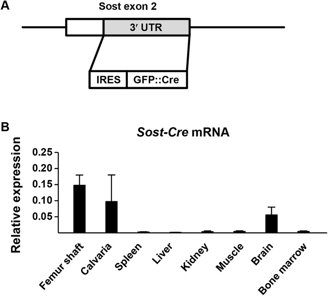
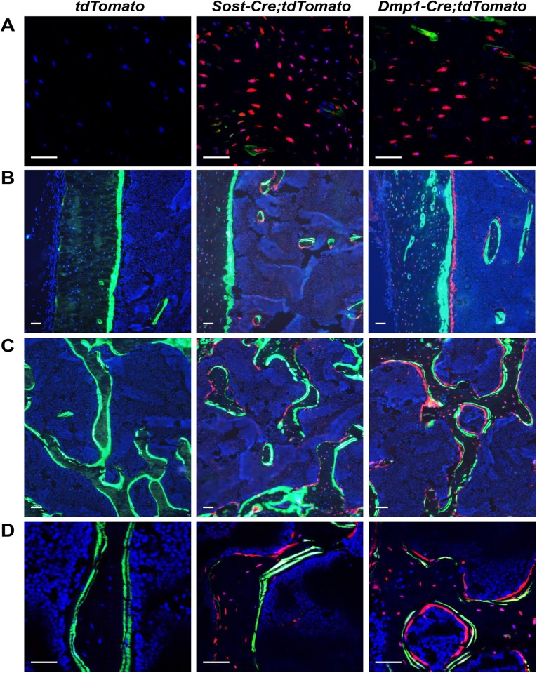
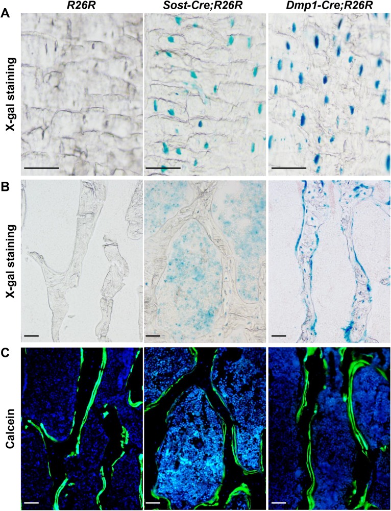
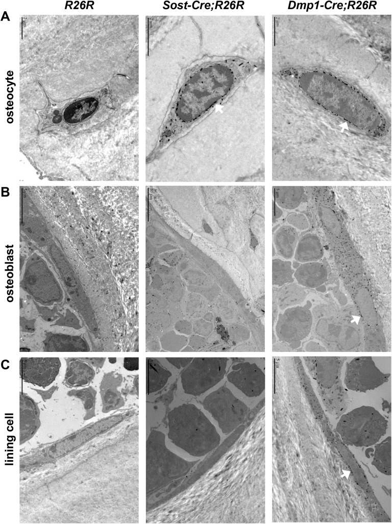
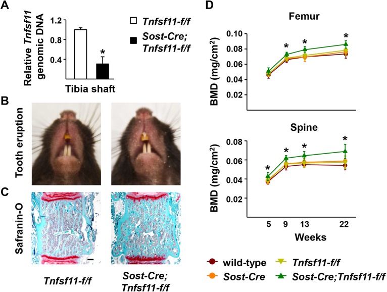
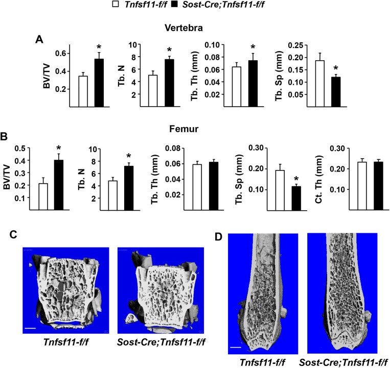
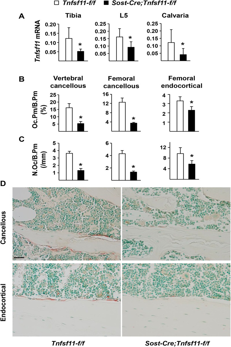
The cytokine receptor activator of nuclear factor kappa B ligand (RANKL), encoded by the Tnfsf11 gene, is essential for osteoclastogenesis and previous studies have shown that deletion of the Tnfsf11 gene using a Dmp1-Cre transgene reduces osteoclast formation in cancellous bone by more than 70%. However, the Dmp1-Cre transgene used in those studies leads to recombination in osteocytes, osteoblasts, and lining cells making it unclear whether one or more of these cell types produce the RANKL required for osteoclast formation in cancellous bone. Because osteoblasts, osteocytes, and lining cells have distinct locations and functions, distinguishing which of these cell types are sources of RANKL is essential for understanding the orchestration of bone remodeling. To distinguish between these possibilities, we have now created transgenic mice expressing the Cre recombinase under the control of regulatory elements of the Sost gene, which is expressed in osteocytes but not osteoblasts or lining cells in murine bone. Activity of the Sost-Cre transgene in osteocytes, but not osteoblast or lining cells, was confirmed by crossing Sost-Cre transgenic mice with tdTomato and R26R Cre-reporter mice, which express tdTomato fluorescent protein or LacZ, respectively, only in cells expressing the Cre recombinase or their descendants. Deletion of the Tnfsf11 gene in Sost-Cre mice led to a threefold decrease in osteoclast number in cancellous bone and increased cancellous bone mass, mimicking the skeletal phenotype of mice in which the Tnfsf11 gene was deleted using the Dmp1-Cre transgene. These results demonstrate that osteocytes, not osteoblasts or lining cells, are the main source of the RANKL required for osteoclast formation in remodeling cancellous bone.
Overview
The claims and statistics of this paper haven’t been checked yet.
Run the check — free, no account needed. It reads the open-access text and checks the sources it cites.
Integrity signals
Registry
Cites
Statistics
Figures
Verification
Claims the paper makes
The claims and statistics of this paper haven’t been checked yet.
Run the check — free, no account needed. It reads the open-access text and checks the sources it cites.
Claims other papers make about this one
Each is checked against this paper’s own text: does it say what the citing paper says it does?
In the citing paper
“As the activation of Dmp1-Cre promoters is not exclusive to osteocytes,53 the conditional deletion of RANKL expression was performed under the control of the more specific SOST promoter. This conditional deletion generated a phenotype resembling the previous one, with a significant decrease in osteoclasts number in cancellous bone.54”
From Bone remodeling: an operational process ensuring survival and bone mechanical competence (2022) · cites this paper as [54]
Self-check
Percentages, sums and reported differences re-computed from the paper’s own numbers.
The self-check needs the paper’s full text.
Citations
Cites
Analysis of Relative Gene Expression Data Using Real-Time Quantitative PCR and the 2−ΔΔCT Method
Livak, K. J., & Schmittgen, T. D. (2001) · Methods · doi:10.1006/meth.2001.1262 · Cited by 180,860
A robust and high-throughput Cre reporting and characterization system for the whole mouse brain
Madisen, L., Zwingman, T. A., Sunkin, S. M., Oh, S. W., Zariwala, H. A., Gu, H., Ng, L. L., Palmiter, R. D., Hawrylycz, M. J., Jones, A. R., et al. (2010) · Nature Neuroscience · doi:10.1038/nn.2467 · Cited by 6,907
Osteoprotegerin ligand is a cytokine that regulates osteoclast differentiation and activation.
Lacey, D. L., Timms, E., Tan, H.-L., Kelley, M. J., Dunstan, C. R., Burgess, T., Elliott, R., Colombero, A., Elliott, G., Scully, S., et al. (1998) · Cell · doi:10.1016/s0092-8674(00)81569-x · Cited by 5,439
Generalized lacZ expression with the ROSA26 Cre reporter strain
Soriano, P. (1999) · Nature Genetics · doi:10.1038/5007 · Cited by 5,237
OPGL is a key regulator of osteoclastogenesis, lymphocyte development and lymph node organogenesis.
Kong, Y.-Y., Yoshida, H., Sarosi, I., Tan, H.-L., Timms, E., Capparelli, C., Morony, S., Oliveira-dos-Santos, A. J., Van, G., Itie, A., et al. (1999) · Nature · doi:10.1038/16852 · Cited by 3,351
Manolagas, S. C. (2000) · Endocrine Reviews · doi:10.1210/edrv.21.2.0395 · Cited by 2,639
Self-citationKong, Y.-Y., Feige, U., Sarosi, I., Bolon, B., Tafuri, A., Morony, S., Capparelli, C., Li, J., Elliott, R., McCabe, S., et al. (1999) · Nature · doi:10.1038/46303 · Cited by 1,856
Evidence for osteocyte regulation of bone homeostasis through RANKL expression
Nakashima, T., Hayashi, M., Fukunaga, T., Kurata, K., Oh-hora, M., Feng, J. Q., Bonewald, L. F., Kodama, T., Wutz, A., Wagner, E. F., et al. (2011) · Nature Medicine · doi:10.1038/nm.2452 · Cited by 1,778
Self-citationManolagas, S. C. (2000) · Endocrine Reviews · doi:10.1210/er.21.2.115 · Cited by 1,318
Self-citationMatrix-embedded cells control osteoclast formation
Xiong, J., Onal, M., Jilka, R. L., Weinstein, R. S., Manolagas, S. C., & O'Brien, C. A. (2011) · Nature Medicine · doi:10.1038/nm.2448 · Cited by 1,304
Self-citation
Cited by
Sorted by citations
Mechanisms of bone development and repair
Salhotra, A., Shah, H. N., Lévi, B., & Longaker, M. T. (2020) · Nature Reviews Molecular Cell Biology · doi:10.1038/s41580-020-00279-w · Cited by 1,302
Kenkre, J. E., & Bassett, J. H. D. (2018) · Annals of Clinical Biochemistry International Journal of Laboratory Medicine · doi:10.1177/0004563218759371 · Cited by 775
RANKL biology: bone metabolism, the immune system, and beyond
Ono, T., Hayashi, M., Sasaki, F., & Nakashima, T. (2020) · Inflammation and Regeneration · doi:10.1186/s41232-019-0111-3 · Cited by 597
Robling, A. G., & Bonewald, L. F. (2020) · Annual Review of Physiology · doi:10.1146/annurev-physiol-021119-034332 · Cited by 539
Understanding the Bone in Cancer Metastasis
Fornetti, J., Welm, A. L., & Stewart, S. A. (2018) · Journal of Bone and Mineral Research · doi:10.1002/jbmr.3618 · Cited by 540
Metadata
- Authors
- Xiong, J., Piemontese, M., Onal, M., Campbell, J., Goellner, J. J., Dusevich, V., Bonewald, L., Manolagas, S. C., & O’Brien, C. A.
- Affiliations
- Central Arkansas Veterans Healthcare System (US)
- University of Missouri–Kansas City (US)
- Corresponding author
- O’Brien, Charles A.
- ORCID
- Venue
- PLOS ONE
- Publisher
- Public Library of Science (PLoS)
- Published
- 2015-09-22
- Volume
- 10
- Issue
- 9
- Pages
- e0138189
- ISSN
- 1932-6203 (ISSN-L)
- PMC
- PMC4578942
- OpenAlex
- W2195625763
- Cited by
- 323
- References
- 37
- Citations by year
- 2026: 20 · 2025: 19 · 2024: 28 · 2023: 44 · 2022: 29 · 2021: 28 · 2020: 40 · 2019: 35 · 2018: 26 · 2017: 31 · 2016: 21 · 2015: 1
- FWCI
- 11.42
- Type
- journal article
- Language
- en
- Open access
- gold
- doi.org
- Licence
- public-domain
- creativecommons.org/publicdomain/zero/1.0/ (vor)
- Locations
- PLoS ONE (publishedVersion, public-domain)
- Europe PMC (PubMed Central) (publishedVersion, public-domain)
- PubMed Central (publishedVersion, public-domain)
- PubMed (publishedVersion)
- DOAJ (DOAJ: Directory of Open Access Journals) (submittedVersion, cc-by-sa)
- Figshare (submittedVersion, cc-by)
- Funders
- U.S. Department of Veterans Affairs (I01 BX000294)
- National Institutes of Health (RR029884, AG13918)
- Translational Research Institute, University of Arkansas for Medical Sciences (RR029884, UL1 RR029884)
- National Institute on Aging (AG13918, P01AG013918)
- National Institute of Arthritis and Musculoskeletal and Skin Diseases (AR049794, R01AR049794)
- National Center for Research Resources (UL1RR029884)
- Topics
- Life Sciences › Biochemistry, Genetics and Molecular Biology › Molecular Biology › Bone Metabolism and Diseases
- Health Sciences › Medicine › Orthopedics and Sports Medicine › Bone health and osteoporosis research
- Health Sciences › Medicine › Rheumatology › Osteomyelitis and Bone Disorders Research
- Keywords
- RANKL, osteocytes, osteoclastogenesis, bone remodeling, osteoblasts, bone lining cells, trabecular bone, bone mass
- MeSH terms
- Animals, Cell Division, Mice, Transgenic, Osteoblasts, Osteoclasts, Osteocytes, Recombination, Genetic, Bone Density, Bone Remodeling, Mice, RANK Ligand
About these checks
Registry, retraction, Cites and statistics results are computed, not judged, with one exception: a printed reference that no index lists is read once by a model before it counts as a citation error. Claims are extracted by a model and kept only when their quote appears verbatim in the text; each cited source is then checked, with a model’s help, for whether it says what the claim relies on. Those verdicts can be wrong — read the claim’s quote and the cited source before relying on one.
Statistics and arithmetic are recomputed, not judged.
Confidence bands are shown only for fields calibrated on our annotated test set. None is calibrated yet, so no band is shown.
What counts as a claim ›How we measure confidence ›How we check this ›
Are you an author, or do you see a problem with this page? Request a review
