

The DOI matches its Crossref record; 35 of the 36 references it cites were found in a registry or an index.
Enhanced Performance of Brain Tumor Classification via Tumor Region Augmentation and Partition
Open accessAbstract
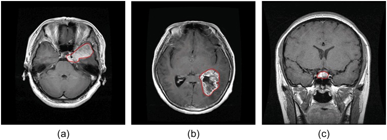
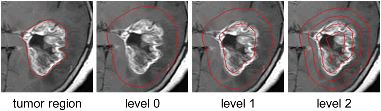
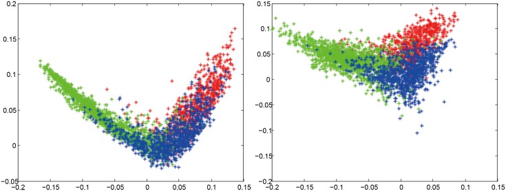
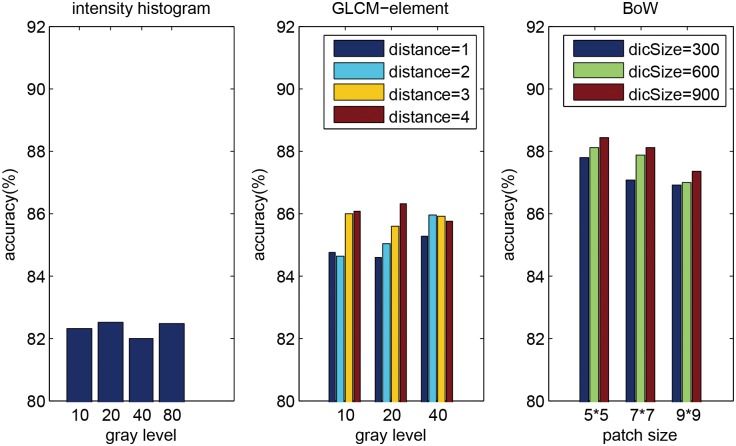
Automatic classification of tissue types of region of interest (ROI) plays an important role in computer-aided diagnosis. In the current study, we focus on the classification of three types of brain tumors (i.e., meningioma, glioma, and pituitary tumor) in T1-weighted contrast-enhanced MRI (CE-MRI) images. Spatial pyramid matching (SPM), which splits the image into increasingly fine rectangular subregions and computes histograms of local features from each subregion, exhibits excellent results for natural scene classification. However, this approach is not applicable for brain tumors, because of the great variations in tumor shape and size. In this paper, we propose a method to enhance the classification performance. First, the augmented tumor region via image dilation is used as the ROI instead of the original tumor region because tumor surrounding tissues can also offer important clues for tumor types. Second, the augmented tumor region is split into increasingly fine ring-form subregions. We evaluate the efficacy of the proposed method on a large dataset with three feature extraction methods, namely, intensity histogram, gray level co-occurrence matrix (GLCM), and bag-of-words (BoW) model. Compared with using tumor region as ROI, using augmented tumor region as ROI improves the accuracies to 82.31% from 71.39%, 84.75% from 78.18%, and 88.19% from 83.54% for intensity histogram, GLCM, and BoW model, respectively. In addition to region augmentation, ring-form partition can further improve the accuracies up to 87.54%, 89.72%, and 91.28%. These experimental results demonstrate that the proposed method is feasible and effective for the classification of brain tumors in T1-weighted CE-MRI.
Overview
The claims and statistics of this paper haven’t been checked yet.
Run the check — free, no account needed. It reads the open-access text and checks the sources it cites.
Integrity signals
Registry
Cites
Statistics
Figures
Verification
Claims the paper makes
The claims and statistics of this paper haven’t been checked yet.
Run the check — free, no account needed. It reads the open-access text and checks the sources it cites.
Claims other papers make about this one
Each is checked against this paper’s own text: does it say what the citing paper says it does?
In the citing paper
“Data collected from three different kinds of brain tumors consisted of 708 meningiomas, 1426 gliomas, and 930 pituitaries images from the General Hospital and Nanfang Hospital, Tianjin Medical University, China [72].”
From A Comprehensive Analysis of Recent Deep and Federated-Learning-Based Methodologies for Brain Tumor Diagnosis (2022) · cites this paper as [72]
In the citing paper
“This contains 3064 T1-weighted contrast-enhanced images collected from 233 patients with a resolution of 512 × 512 pixels.”
From A Comprehensive Analysis of Recent Deep and Federated-Learning-Based Methodologies for Brain Tumor Diagnosis (2022) · cites this paper as [72]
Self-check
Percentages, sums and reported differences re-computed from the paper’s own numbers.
The self-check needs the paper’s full text.
Citations
Cites
Distinctive Image Features from Scale-Invariant Keypoints
Lowe, D. G. (2004) · International Journal of Computer Vision · doi:10.1023/b:visi.0000029664.99615.94 · Cited by 55,589 · cited 1×
“(cid:129) Local features: The next step is to extract local features within the ROI, such as scale-invariant feature transform (SIFT) descriptor [20] and raw patch [21].”
Background$rm K$-SVD: An Algorithm for Designing Overcomplete Dictionaries for Sparse Representation
Aharon, M., Elad, M., & Bruckstein, A. M. (2006) · IEEE Transactions on Signal Processing · doi:10.1109/tsp.2006.881199 · Cited by 9,464
Robust Face Recognition via Sparse Representation
Wright, J. N., Yang, A., Ganesh, A., Sastry, S., & Ma, Y. (2009) · IEEE Transactions on Pattern Analysis and Machine Intelligence · doi:10.1109/tpami.2008.79 · Cited by 9,398 · cited 1×
“We also compared three classification methods (i.e. SVM, sparse representation-based classification (SRC) [31], and k nearest neighbors (kNN) classifier) using 2-region BoW representations.”
ResultBeyond Bags of Features: Spatial Pyramid Matching for Recognizing Natural Scene Categories
Lazebnik, S., Schmid, C., & Ponce, J. (2006) · 2006 IEEE Computer Society Conference on Computer Vision and Pattern Recognition - Volume 2 (CVPR'06) · doi:10.1109/cvpr.2006.68 · Cited by 7,916 · cited 4×
“Among those methods, the most notable work is SPM [15], which splits the image into hierarchical cells, computes BoW representation for each cell, and finally weights and concatenates the results.”
BackgroundMethodInfluentialVideo Google: a text retrieval approach to object matching in videos
Sivic, & Zisserman (2003) · Proceedings Ninth IEEE International Conference on Computer Vision · doi:10.1109/iccv.2003.1238663 · Cited by 6,367 · cited 1×
“The BoW method was originally used in the text retrieval domain, and it has been successfully adapted to the visual analysis domain [13,14].”
MethodRobust wide-baseline stereo from maximally stable extremal regions
Matas, J., Chum, O., Urban, M., & Pajdla, T. (2004) · Image and Vision Computing · doi:10.1016/j.imavis.2004.02.006 · Cited by 3,760 · cited 1×
“As pointed out in [29,30], it is beneficial to capture a certain amount of context.”
BackgroundA Bayesian Hierarchical Model for Learning Natural Scene Categories
Fei-Fei, L., & Perona, P. (2005) · IEEE Computer Society Conference on Computer Vision and Pattern Recognition/Proceedings - IEEE Computer Society Conference on Computer Vision and Pattern Recognition/Proceedings · doi:10.1109/cvpr.2005.16 · Cited by 3,665 · cited 1×
“The BoW method was originally used in the text retrieval domain, and it has been successfully adapted to the visual analysis domain [13,14].”
MethodLocality-constrained Linear Coding for image classification
Wang, J., Yang, J., Yu, K., Lv, F., Huang, T., & Gong, Y. (2010) · 2010 IEEE Computer Society Conference on Computer Vision and Pattern Recognition · doi:10.1109/cvpr.2010.5540018 · Cited by 3,100 · cited 2×
“Other more sophisticated methods may also be used, such as SC, soft assignment (SA) [24,25], orthogonal matching pursuit (OMP), locality-constrained linear coding [26], and local anchor embedding [27].”
MethodA comparison of affine region detectors
Mikolajczyk, K., Tuytelaars, T., Schmid, C., Zisserman, A., Matas, J., Schaffalitzky, F., Kadir, T., & Gool, L. V. (2005) · International Journal of Computer Vision · doi:10.1007/s11263-005-3848-x · Cited by 2,939
Linear spatial pyramid matching using sparse coding for image classification
Yang, J., Yu, K., Gong, Y., & Huang, T. S. (2009) · IEEE Computer Society Conference on Computer Vision and Pattern Recognition/Proceedings - IEEE Computer Society Conference on Computer Vision and Pattern Recognition/Proceedings · doi:10.1109/cvpr.2009.5206757 · Cited by 2,895
Research works printed but not linked: 1
- S Patil, 2013, A Computer Aided Diagnostic System For Classification Of Brain Tumors Using Texture Features And Probabilistic Neural Network, Int J Comput Sci Eng Inf Technol Res, 3, 61
Cited by
Sorted by citations
Items 601–700 of 879
Image Classification of Brain Tumors Using Improved CNN Framework with Data Augmentation
Ning, X., Li, Z., & Pang, H. (2020) · Lecture notes of the Institute for Computer Sciences, Social Informatics and Telecommunications Engineering · doi:10.1007/978-3-030-64214-3_6 · Cited by 2
Ramasamy, J., Doshi, R., & Hiran, K. K. (2023) · International Journal on Recent and Innovation Trends in Computing and Communication · doi:10.17762/ijritcc.v11i3s.6155 · Cited by 2
Spatial pyramid match kernels for brain image classification
Young, J. (2016) · International Workshop on Pattern Recognition in NeuroImaging · doi:10.1109/prni.2016.7552330 · Cited by 2
“However in [3] SPMK was used to provide a fast and accurate nearest neighbour search for MRI image retrieval, and [4] applied it to segment brain tumours.”
MethodRevealing Brain Tumor Using Cross-Validated NGBoost Classifier: NG Boost Classifier
Dutta, S., & Bandyopadhyay, S. K. (2020) · International Journal of Machine Learning and Networked Collaborative Engineering · Cited by 2
Brain Tumor Classification using Transfer Learning from MRI Images
Akter, N., Junjun, J. A., Nahar, N., Hossain, M. S., Andersson, K., & Hoassain, M. S. (2022) · Lecture notes in networks and systems · doi:10.1007/978-981-19-2445-3_40 · Cited by 2
A Review of Recent Advances in Classification and Prediction of Brain Tumor using Deep Learning
Reddy, D. L., Soumya, D. R., Nagar, A., & Rajpoot, A. K. (2022) · International Conference on Computing, Communication, and Intelligent Systems (ICCCIS) · doi:10.1109/icccis56430.2022.10037668 · Cited by 2
“Figshare[14] As three of the most popular varieties of brain cancer, 708 slices of Meningioma, 1426 slices of glioma, and 930 slices of pituitary tumors - are represented in this BraTS dataset of 3064 T1-weighted contrast-enhanced images from 233 people.”
BackgroundSegmentation and classification of specific pattern of Brain tumor using CNN
Das, S., Ghosh, P., & Chaudhuri, A. K. (2023) · International Journal of Engineering Technology and Management Sciences · doi:10.46647/ijetms.2023.v07i02.004 · Cited by 2
Brain Tumor Classification Using Deep CNN-Based Transfer Learning Approach
Arya, M. K., & Agrawal, R. K. (2023) · International Journal of Biology and Biomedical Engineering · doi:10.46300/91011.2023.17.1 · Cited by 2
Retracted: Brain Tumor Classification with Inception V3 Network Model Using Transfer Learning
Kumar, T. S., Lohanandd, R., Kaviyanandh, K., & Gunabharathi, J. (2023) · International Conference on Advanced Computing and Communication Systems (ICACCS) · doi:10.1109/icaccs57279.2023.10112951 · Cited by 2
RetractedBrain Tumor Classification Based on Multi-Level Attention
Shi, H., Wu, X., & Li, X. (2023) · IEEE International Conference on Electronic Information and Communication Technology (ICEICT) · doi:10.1109/iceict57916.2023.10245579 · Cited by 2
“This paper makes use of a brain tumor digital shared dataset that is openly accessible and was released by Cheng et al [8].”
MethodInfluentialLocal Style Transfer via Latent Space Manipulation for Cross-Disease Lesion Segmentation
Lyu, F., Ye, M., Yip, T. C., Wong, G. L., & Yuen, P. C. (2023) · IEEE Journal of Biomedical and Health Informatics · doi:10.1109/jbhi.2023.3327726 · Cited by 2
“For brain tumor segmentation, CE-MRI [47] is used.”
MethodNijaguna, G. S., Kumar, D. P., Manjunath, B. N., Jain, T. J. S., & Lal, N. D. (2023) · Internet Technology Letters · doi:10.1002/itl2.484 · Cited by 2
“31 The data composition of every slice in a dataset includes the patient’s tumor kinds, including meningioma, glioma, and pituitary tumor.”
Classification of Brain Tumors via Deep Learning Models
DAGLI, K., & Eroğul, O. (2020) · Medical Technologies Congress (TIPTEKNO) · doi:10.1109/tiptekno50054.2020.9299231 · Cited by 2
“Çalışmada evrişimli sinir ağlarını eğitmek için kullanılan veri seti uzmanlar tarafından işaretlenmiş ve etiketlenmiş olup, 3064 adet T-1 ağırlıklı manyetik rezonans beyin görüntüsü içermektedir [9].”
Jamshidi, B., & Rostamy‐Malkhalifeh, M. (2023) · SSRN Electronic Journal · doi:10.2139/ssrn.4624936 · Cited by 3
Paluh, J. L., Sunil, S., Rajeev, R., Chatterjee, A., Pilitsis, J. G., & Mukherjee, A. (2024) · Research Square · doi:10.21203/rs.3.rs-4087784/v1 · Cited by 3
Brain Tumor Classification and Detection Using Machine Learning by Analyzing MRI Images
Roy, C., Parvathavarthini, K., Gomathi, M., Fatangare, M. P., Kishore, D., & Suthar, A. C. (2025) · doi:10.1002/9781394272464.ch13 · Cited by 2
Qi, L. J., & Alias, N. B. (2020) · International Annals of Science · doi:10.21467/ias.9.1.160-173 · Cited by 1
“[20] SVM BoW model with region partitioning 91.”
MethodClassification of Brain Tumor Using CoAtNet Model
Mattar, E. A. (2023) · American Journal of Biomedical Science & Research · doi:10.34297/ajbsr.2023.20.002687 · Cited by 1
Alzheimer disease (AD) medical image analysis with convolutional neural networks
Sohail, A., Fiaz, M., Nutini, A., & Iqbal, M. S. (2024) · Elsevier eBooks · doi:10.1016/b978-0-44-318468-0.00017-9 · Cited by 1
Swamy, P. K., Yamsani, Y. N., Kumar, V. M., Kumar, B. V., & Marrikukkala, R. K. (2024) · AIP conference proceedings · doi:10.1063/5.0196697 · Cited by 1
Adaptive Spatially-Weighted Region-Based Active Contour
Kumar, B., Panda, R., & Agrawal, S. (2024) · International Journal of Image and Graphics · doi:10.1142/s0219467826500208 · Cited by 1
Implementation of Vision Transformer Method in Detecting Brain Tumor Based on Medical Image
Arief, R. M., & Kurniawan, I. (2024) · International Conference on Information Technology, Information Systems and Electrical Engineering (ICITISEE) · doi:10.1109/icitisee63424.2024.10730634 · Cited by 1
“Nowadays, the use of digital images in the medical field is increasingly common for diagnosis [13].”
BackgroundMethodInfluentialAsthana, P., & Kamboj, D. (2024) · IEEE International Conference for Women in Computing (InCoWoCo) · doi:10.1109/incowoco64194.2024.10863200 · Cited by 1
“The proposed model is verified on the two-class and three-class datasets [18-20] using MATLAB 2022a.”
Dheepak, G., & Vaishali, D. (2024) · Global Conference on Communications and Information Technologies (GCCIT) · doi:10.1109/gccit63234.2024.10862572 · Cited by 1
Aggrey, E. S. E. B., Zhen, Q., Kodjiku, S. L., Fiasam, L. D., Sey, C., Ejiyi, C. J., & Ntia, N. W. (2024) · International Computer Conference on Wavelet Active Media Technology and Information Processing (ICCWAMTIP) · doi:10.1109/iccwamtip64812.2024.10873647 · Cited by 1
Analysis and prediction of different stages of brain tumor from mri using ai models
Navruzov, R., Ismoilov, J., Bahadirkhanov, M., Almatova, U., Djuraev, J. A., Mikhiddinov, A., & Mirametova, N. (2024) · Health Leadership and Quality of Life · doi:10.56294/hl2024.188 · Cited by 1
Self-supervised 3D Vision Transformer Pre-training for Robust Brain Tumor Classification
Nunes, D. W., Rauber, D., & Palm, C. (2025) · Informatik aktuell · doi:10.1007/978-3-658-47422-5_69 · Cited by 1
A High-Accuracy Brain Tumor Classification Method Based onVIT in the Context of Small Sample Size
Ding, J., Fu, B., & Xie, X. (2024) · International Conference on Health Big Data and Intelligent Healthcare (ICHIH) · doi:10.1109/ichih63459.2024.11064889 · Cited by 1
“Cheng et al. [2] proposed an image extension technique to expand the tumor region and divide it into sub-regions, aiming to improve brain tumor classification performance.”
Tummala, S., Chauhdary, S. H., Singh, V., Kumar, R., Kadry, S., & Kim, J. (2025) · Computer Modeling in Engineering & Sciences · doi:10.32604/cmes.2025.069731 · Cited by 1
“Further details regarding this dataset can be found in [24,25].”
Chakraborty, S., Miah, M. S., Habib, A., Rohman, M. M., Ahmed, K. M., & Islam, M. K. (2025) · IEEE International Conference on Quantum Photonics, Artificial Intelligence & Networking (QPAIN) · doi:10.1109/qpain66474.2025.11171912 · Cited by 1
“Gliomas, meningiomas, and pituitary tumors stand out among the different kinds of brain tumors [3].”
Hark, B. D., & Yildizli, M. G. (2025) · IET Image Processing · doi:10.1049/ipr2.70251 · Cited by 1
EfficientNet-based deep learning approach for early detection of brain tumors
Verma, N., & Bohat, V. K. (2025) · Quality & Quantity · doi:10.1007/s11135-025-02499-8 · Cited by 1
Comparative Analysis of Deep Learning Architectures for Brain Tumor Classification using MRI Images
Shahriar, M. M., Hafiz, M., Rabbi, R. I., Liew, T. H., Tarin, F. M., & Tusher, E. H. (2025) · International Conference on New Media Studies (CONMEDIA) · doi:10.1109/conmedia67140.2025.11290384 · Cited by 1
“Afshar et al. [16] applied capsule networks to retain relational information between tumor areas, thus alleviatinge the restriction of CNN to pose and orientation variations.”
Deep Learning Advances for Brain Tumor Diagnosis and Multimodal MRI Registration
Kothapalle, M. S. R., & Nelakuditi, U. R. (2025) · International Conference on Electronic Circuits and Signaling Technologies (ICECST) · doi:10.1109/icecst66106.2025.11307326 · Cited by 1
Karaca, K. Ö., Arslan, B., Atay, Y., & Sagiroglu, S. (2025) · Gazi University Journal of Science Part A Engineering and Innovation · doi:10.54287/gujsa.1822726 · Cited by 1
Desai, K., Patel, P. K., & Barve, A. (2025) · International Conference on Applied Artificial Intelligence and Computing (ICAAIC) · doi:10.1109/icaaic64647.2025.11330252 · Cited by 2
“By offering methods and tools for peering into the black box, XAI helps humans comprehend AI judgments [14][19][23].”
DeepWave: A Hybrid CNN-Wavelet Approach for Brain Tumor MRI Classification
Panda, N. R., Muduli, D., Parija, S., Sharma, S. K., & Kumar, R. R. (2025) · International Conference on Cognitive, Green and Ubiquitous Computing (IC-CGU) · doi:10.1109/ic-cgu67042.2025.11338053 · Cited by 1
“Cheng [4] showed that CNNs are very good at capturing high-level abstractions from medical images.”
Toward the Detection of Brain Tumor Using MRI Images: A Deep Learning-Based Approach
Soni, J., & Upadhyay, H. (2026) · Signals and communication technology · doi:10.1007/978-3-031-89700-9_9 · Cited by 1
Annadurai, A., Joseph, B., Prusty, M. R., Ananthakrishnan, B., & Shaik, A. (2026) · In Silico Research in Biomedicine · doi:10.1016/j.insi.2026.100239 · Cited by 1
Machine learning and digital images of porous materials: from rock to the human brain
Sahimi, A., & Sahimi, M. (2026) · Frontiers in Physics · doi:10.3389/fphy.2025.1742403 · Cited by 2
Digital Fiqh and Maqāṣid al-Sharīʿah in the Onlife Condition
Ja’far, S., Yaqin, H., Iffah, I., Hodri, H., & Sanuri, S. (2026) · SAMARAH Jurnal Hukum Keluarga dan Hukum Islam · doi:10.22373/sjhk.v10.i1.31868 · Cited by 1
Brain Tumor Detection Using Optimization with Genetic and Heuristic Algorithms
Indurekaa, S. R., Ranjana, R., Pavitradevi, S., Rajarajeswari, R., Srinathi, K., Harini, M., & Madhumitha, V. (2026) · International Conference on Innovative Computing, Intelligent Communication and Smart Electrical Systems (ICSES) · doi:10.1109/icses66558.2026.11479025 · Cited by 1
Basha, C. Z., & Yalla, P. (2026) · Journal of Innovative Image Processing · doi:10.36548/jiip.2026.2.014 · Cited by 1
“Although these methods offered a baseline, they were heavily sensitive to noise and did not generalize well across different MRI scanners and protocols [12].”
Lu, D., Zhang, Y., & Chaudhary, D. (2026) · Journal of Medical Systems · doi:10.1007/s10916-026-02418-w · Cited by 4
“To further assess cross-dataset generalization, we additionally evaluated Swin-T and MTA-Swin on the independent Figshare brain tumor MRI dataset (3,064 T1CE images from 233 patients, collected from clinical cases and labeled into three tumor classes (meningioma/glioma/pituitary) [ 58].”
Saifullah, S., Dreżewski, R., Yudhana, A., Caesarendra, W., & Sela, E. I. (2026) · Journal of Artificial Intelligence and Soft Computing Research · doi:10.2478/jaiscr-2026-0025 · Cited by 1
“This study utilizes three benchmark datasets for brain tumor segmentation: the Figshare Brain Tumor Segmentation (FBTS) dataset [63] and the BraTS (Brain Tumor Segmentation) 2021 [64] and 2018 [65, 66, 67] datasets.”
A class-wise quantum relational calibration network for brain tumor diagnosis
Sheu, J.-R., Xie, J., Tsai, C.-N., Hsieh, C.-Y., & Lin, C.-S. (2026) · Scientific Reports · doi:10.1038/s41598-026-61891-y · Cited by 1
“In the context of brain tumor diagnosis, CNN-based models have demonstrated strong performance in tumor detection, segmentation, and multi-class classification tasks, benefiting from their ability to capture hierarchical spatial features and complex visual patterns 14,15 .”
Quantum-Based Deep Learning Approach for Brain Tumour MRI Detection
Diviya, M., Subramanian, M., Divya, P., & Utthirakumari, R. (2026) · Journal of The Institution of Engineers (India) Series B · doi:10.1007/s40031-026-01368-8 · Cited by 1
“1 Different types of brain tumour (representative MRI slices from the publicly available figshare brain tumour dataset [46, 47]) neural network utilizes sparse quantum embedding that significantly reduces the computational costs [10].”
Chhetri, L., Datta, A., & Ghosal, P. (2026) · IEEE Guwahati Subsection Conference (GCON) · doi:10.1109/gcon69192.2026.11648823 · Cited by 1
“The size of medical image datasets is much smaller compared to natural image datasets, with only 3,064 images of 233 patients in the popular Figshare brain tumor dataset [5], in contrast to ImageNet’s 14 million images [6].”
Khan, M. A., Miah, A. S. M., Rahim, M. A., Shin, J., & Husen, M. N. (2026) · Computers · doi:10.3390/computers15090586 · Cited by 1
FRI Modelling of Fourier Descriptors
Kamath, A. J., Rudresh, S., & Seelamantula, C. S. (2019) · IEEE International Conference on Acoustics Speech and Signal Processing · doi:10.1109/icassp.2019.8682685 · Cited by 1
“1: [Colour online] Outlining (curves in red) of tumours in T1 contrast enhanced brain MRI [9, 10] and images of melanoma [11] using the proposed method.”
BackgroundInfluentialLocal Ensemble Approach for Meningioma Tumor Firmness Detection in MRI Images
Ismail, M. M. B. (2020) · International Conference on Image, Video and Signal Processing (IVSP) · doi:10.1145/3388818.3389158 · Cited by 1
“Despite researchers’ efforts to propose accurate CAD systems in terms of tumor classification as malignant or benign [5-9], supervised learning techniques have not been used yet to investigate the Meningioma firmness.”
BackgroundWeakly-Supervised Brain Tumor Classification with Global Diagnosis Label
Zhou, Y., Li, Z., Ma, C., Chen, C., Gao, M., Zhu, H., & Xu, J. (2020) · IEEE International Symposium on Biomedical Imaging (ISBI) · doi:10.1109/isbi45749.2020.9098640 · Cited by 1
“Different from existing public datasets with detailed slicelevel labels [6], our dataset has only global labels in patient level.”
BackgroundMethodInfluentialA Brief Survey on Brain Tumor Identification Models Using Deep Neural Networks
Revathi, V., & Muddana, A. L. (2021) · International Conference on Smart Electronics and Communication (ICOSEC) · doi:10.1109/icosec51865.2021.9591832 · Cited by 1
“[19] separates the three stages of glioma and differentiates between the many meningioma and pituitary tumour types, all of which originate in the brain (Grade II, Grade III and Grade IV).”
BackgroundDetection and Classification of Brain Tumor Using MRI Images
Koli, R., Lotya, S., Govekar, P., Sachdev, K., & Bhatia, G. S. (2022) · Lecture notes in networks and systems · doi:10.1007/978-981-16-5655-2_37 · Cited by 1
Brain Tumor Detection and Classification Using Transfer Learning Technique
ram, A. V., Kuchulakanti, H., & Raj, T. S. (2022) · Advances in intelligent systems and computing · doi:10.1007/978-981-16-9573-5_36 · Cited by 1
Brain Tumor Classification using Deep Learning Methods
Rehman, H. U., Masood, S., & Khan, M. A. (2022) · International Journal of Information Systems and Computer Technologies · doi:10.58325/ijisct.001.01.0016 · Cited by 1
Experimental Analysis of Deep Learning Algorithms Used in Brain Tumor Classification
Mundada, K., Rahatwilkar, T., & Kulkarni, J. V. (2023) · Lecture notes in networks and systems · doi:10.1007/978-981-19-9225-4_8 · Cited by 1
Yousif, A. S., Omar, Z., Hamoodat, H., & Morad, N. Y. A. (2022) · IEEE-EMBS Conference on Biomedical Engineering and Sciences · doi:10.1109/iecbes54088.2022.10079290 · Cited by 1
“2 shown the instances of tumor diseases [21].”
BackgroundMISM: A Medical Image Segmentation Metric for Evaluation of Weak Labeled Data
Hartmann, D., Schmid, V., Meyer, P., Auer, F., Soto‐Rey, I., Müller, D., & Krämer, F. (2023) · Diagnostics · doi:10.3390/diagnostics13162618 · Cited by 1
RETRACTED: An efficient hybrid bert model for brain tumor classification
Kumar, S. S. P., Kumar, C. A., Venugopal, A., & Sharma, A. (2024) · Journal of Intelligent & Fuzzy Systems · doi:10.3233/jifs-237653 · Cited by 1
RetractedMulticlass Brain Tumor Detection using Deep Transfer Learning
Wagh, K. S., Dhotre, A., Lagad, K., Yadav, A., & Kamble, A. (2023) · International Conference on Cognitive Computing and Engineering Education (ICCCEE) · doi:10.1109/icccee55951.2023.10424539 · Cited by 1
“They used dataset presented by Cheng [26] that contains 3054 T1 weighted magnetic resonanse pictures images of brain.”
MethodEnhanced Brain Tumor Classification from MRI Images Using Deep Learning Model
Rahman, A. B., Islam, M. T., Islam, M. R., Sohrawordi, M., & Sultan, M. N. (2023) · International Conference on Computer and Information Technology (ICCIT) · doi:10.1109/iccit60459.2023.10441064 · Cited by 1
PrinciResNet Brain Tumor Classification Technique for Multimodal Input-level Fusion Network
Usha, M. P., Kannan, G., Sai Akshay., S., Giri, S., & Huzaifa, S. M. (2024) · International Journal of Computing and Digital Systems · doi:10.12785/ijcds/150176 · Cited by 1
Distance Analysis and Dimensionality Reduction using PCA on Brain Tumour MRI Scans
Jhariya, A., Parekh, D., Lobo, J., Bongale, A. M., Jayaswal, R., Kadam, P., Patil, S., & Choudhury, T. (2024) · EAI Endorsed Transactions on Pervasive Health and Technology · doi:10.4108/eetpht.10.5632 · Cited by 1
“Cheng and collaborators [16] revisited their work on enhanced brain tumor classification through tumor region augmentation and partition, reinforcing the significance of their proposed techniques in the broader landscape of brain tumor research.”
BackgroundPOD: PCM-Based Computing Platform for Object Detection in Biomedical Imaging Application
Chen, D., Amirsoleimani, A., Azghadi, M. R., Genov, R., & Ahmadi, M. A. (2024) · IEEE Latin American Symposium on Circuits and Systems (LASCAS) · doi:10.1109/lascas60203.2024.10506128 · Cited by 1
“Thus, we choose the a dataset with T1-weighted contrast-inhanced images from 233 patients with three kinds of brain tumor: meningioma, glioma, and pituitary tumor [24].”
MethodA Review of Capsule Network Limitations, Modifications, and Applications in Object Recognition
Haq, M. U., Sethi, M. A. J., & Rehman, A. U. (2024) · Advances in geospatial technologies book series · doi:10.4018/979-8-3693-2913-9.ch005 · Cited by 1
Brain Tumor Classification Using CNNs
Saveetha, D., Prathaban, B. P., Rashmi, A., & Purushothaman, K. E. (2024) · International Conference on Power, Energy, Control and Transmission Systems (ICPECTS) · doi:10.1109/icpects62210.2024.10780208 · Cited by 1
Meta-constrained extreme learning machines: a novel approach for classification problems
Serin, Z., Karakuzu, C., & Yüzgeç, U. (2026) · The Computer Journal · doi:10.1093/comjnl/bxag071 · Cited by 1
Brain Tumor Detection Using Deep Learning: ResNet+LSTM
Balaji, S., Krithika, T., & Joshi, S. (2024) · International Conference on IoT Based Control Networks and Intelligent Systems (ICICNIS) · doi:10.1109/icicnis64247.2024.10823218 · Cited by 1
“While existing studies have explored either ResNet or LSTM in isolation, few have combined these architectures to fully capture the spatial-temporal complexities inherent in MRI data [17] [18].”
Brain Tumor Detection via Transfer Learning and Ensemble Learning
Hun, W.-J., Lim, J.-Y., Lim, K. M., Lee, C. P., Tan, Y.-X., & Pa-Pa-Min (2024) · IEEE Conference on Systems, Process and Control · doi:10.1109/icspc63060.2024.10862279 · Cited by 1
“…Dataset-I: The brain tumor dataset [28] used in this study, referred to as Figshare Dataset-I, includes 3,064 T1-weighted contrast-enhanced images from 233 patients, classified into three types of brain tumors: Meningioma (708 slices), Glioma (1,426 slices), and Pituitary (930 slices) [29], [30].”
Fuzzy-Based Atrous Convolution for Brain Tumor Detection Using MRI
Irfan, M., Subaşı, A., MEHDI, H. A., Westerlund, T., & Chen, W. (2024) · IEEE International Conference on Progress in Informatics and Computing · doi:10.1109/pic62406.2024.10892686 · Cited by 1
“Chang et al. [16] applied K-Nearest Neighbors (KNN) and support vector machine (SVM) clas-sifiers to obtain 91.28% accuracy.”
InfluentialYamcharoen, N., Piboonthummasak, T., Nobnop, N., & Charoenpong, T. (2024) · Biomedical Engineering International Conference · doi:10.1109/bmeicon64021.2024.10896305 · Cited by 1
“Our dataset sourced from Cheng Jun et al. [8] which proposed a method to enhance performance of tumor classification in three typical brain tumors: Meningioma, Glioma and Pituitary tumor, We selected of 474 MRI images and ground truth of the Glioblastoma type in the transverse plane only.”
Mask Augmentation For Tumor Classification In Medical Images
Weng, C.-C., Chen, H., Tzeng, J.-T., Lin, C., Kuo, P., & Lee, C.-C. (2025) · IEEE International Conference on Acoustics Speech and Signal Processing · doi:10.1109/icassp49660.2025.10890833 · Cited by 1
“• CE-MRI [28] is a public 2D slice brain MRI dataset with 233 patients and a total of 3064 images with tumor segmentation labels.”
Fathima, M. N., Singh, P., & Singh, S. (2024) · International Conference on Advances in Computation, Communication and Information Technology (ICAICCIT) · doi:10.1109/icaiccit64383.2024.10912286 · Cited by 1
Transfer Learning with Mobile Net for Brain Tumor Detection in Neuroimaging
Verma, N., & Bohat, V. K. (2025) · Lecture notes in networks and systems · doi:10.1007/978-981-97-3859-5_10 · Cited by 1
EnSLDe: an enhanced short-range and long-range dependent system for brain tumor classification
Chen, W., Liu, J., Tan, X., Zhang, J., Du, G., Fu, Q., & Jiang, H. (2025) · Frontiers in Oncology · doi:10.3389/fonc.2025.1512739 · Cited by 1
Detection of Brain Tumor Using a Deep Learning Approach
Arun Kumar, S., Kumar, M. A., Vincent, R., Anusha, M. U., Hegde, U. U., & Pasha, N. (2025) · International Conference on Artificial Intelligence and Data Engineering (AIDE) · doi:10.1109/aide64228.2025.10987329 · Cited by 1
Optimized Neural Network Architectures for Brain Tumor Classification and Precision Medicine
Papadimitriou, O., Kanavos, A., Μaragoudakis, M., & Karamitsos, I. (2025) · International Journal of Artificial Intelligence Tools · doi:10.1142/s0218213025400044 · Cited by 1
Deep Learning-Driven Brain Tumor Sectionalisation: Leveraging YOLO11 for MRI Analysis
Khan, M. A., Sutradhar, U., Sarkar, P., Imran, M. I. A., & Dipa, S. A. (2025) · IEEE International Conference on Quantum Photonics, Artificial Intelligence & Networking (QPAIN) · doi:10.1109/qpain66474.2025.11172256 · Cited by 1
RAY-BASED SEGMENTATION ALGORITHM FOR MEDICAL IMAGING
Данилов, В., Skirnevskiy, I. P., Manakov, R. A., Kolpashchikov, D. Y., Gerget, O. M., & Frangi, A. F. (2019) · The international archives of the photogrammetry, remote sensing and spatial information sciences/International archives of the photogrammetry, remote sensing and spatial information sciences · doi:10.5194/isprs-archives-xlii-2-w12-37-2019 · Cited by 1
Segmentation Algorithm Based on Square Blocks Propagation
Данилов, В., Данилов, В., Петрович, С. И., Skirnevskiy, I. P., Manakov, R. A., Kolpashchikov, D. Y., & Gerget, O. M. (2019) · International Conference on Computer Graphics and Vision (GraphiCon) · doi:10.30987/graphicon-2019-2-148-154 · Cited by 1
Bayesian Deep Active Learning for Medical Image Analysis
Ghoshal, B., Swift, S., & Tucker, A. (2021) · Lecture notes in computer science · doi:10.1007/978-3-030-77211-6_4 · Cited by 1
Deep Learning Based Classification of Brain Tumor Types from MRI Scans
Das, J., Ghosh, S., Chakraborty, R., & Pramanik, A. (2021) · Intelligent systems reference library · doi:10.1007/978-3-030-71975-3_16 · Cited by 1
CNN Parameter Adjustment for Brain Tumor Classification
Ho, T. P., & Hoang, V. T. (2021) · Advances in intelligent systems and computing · doi:10.1007/978-981-16-2008-9_1 · Cited by 1
AUTOMATIC SEGMENTATION AND CLASSIFICATION OF BRAIN TUMOR USING DEEP LEARNING
Prasad, K. G. D., Murthy, A. J. N., Narasimha, G., & Sinha, N. (2021) · International Journal of Engineering Applied Sciences and Technology · doi:10.33564/ijeast.2021.v06i01.057 · Cited by 1
Active Contour with Adaptive Timestep for Brain Tumor Segmentation
Kumar, B., Panda, R., & Agrawal, S. (2022) · IEEE International Conference for Convergence in Technology (I2CT) · doi:10.1109/i2ct54291.2022.9825091 · Cited by 1
Robust Brain Diseases Classification Using CNN and Soft Computing Techniques
Gore, D. V., Sinha, A. K., & Deshpande, V. (2022) · Cognitive science and technology · doi:10.1007/978-981-19-2350-0_25 · Cited by 1
DETEKSI TUMOR OTAK PADA MAGNETIC RESONANCE IMAGING MENGGUNAKAN YOLOv7
Passa, R. S., Nurmaini, S., & Rini, D. P. (2023) · Jurnal Ilmiah Matrik · doi:10.33557/jurnalmatrik.v25i2.2404 · Cited by 1
Brain Tumor Classification Using Hit-or-Miss Capsule Layers
Chang, S. J. (2019) · doi:10.15368/theses.2019.26 · Cited by 1
Supervised Classification of Normal and Tumorous Brain MR Images Using Machine Learning SchemesPreprint
Qi, L. J., Alias, N. B., & Johar, F. (2020) · Preprints.org · doi:10.20944/preprints202007.0688.v1 · Cited by 1
“The slice thickness is 6 mm and the slice gap is 1 mm [9, 10].”
BackgroundMAG-Net: Mutli-task attention guided network for brain tumor segmentation and classification.Preprint
Gupta, S., Punn, N. S., Sonbhadra, S. K., & Agarwal, S. (2021) · arXiv (Cornell University) · Cited by 1
Saha, P., Das, R., & Das, S. K. (2021) · Research Square · doi:10.21203/rs.3.rs-1100868/v1 · Cited by 1
Spiking convolutional neural network for brain tumor classificationPreprint
Niepceron, B., Moh, A. N. S., & Grassia, F. (2022) · Research Square · doi:10.21203/rs.3.rs-1129282/v1 · Cited by 1
Singh, K., Kaur, A., & Kaur, P. (2023) · Research Square · doi:10.21203/rs.3.rs-3423149/v1 · Cited by 1
Zia, R., Akhtar, P., Aziz, A., & Kundi, D. (2018) · International Journal of Wavelets Multiresolution and Information Processing · doi:10.1142/s0219691318500480
Qi, L. J., Alias, N. B., & Johar, F. (2020) · AIP conference proceedings · doi:10.1063/5.0018056
Deep learning architectures for the analysis and classification of brain tumors in MR images
Osorio-Barone, A., & Contreras-Ortiz, S. H. (2020) · International Symposium on Medical Information Processing and Analysis (SIPAIM) · doi:10.1117/12.2579618
Tetik, B., UCUZAL, H., Yaşar, Ş., & Çolak, C. (2021) · Konuralp Tıp Dergisi · doi:10.18521/ktd.889777
Convolutional Neural Networks for brain tumor images classification
Ho, T. P. (2021) · รายงานการประชุมวิชาการเสนอผลงานวิจัย ระดับชาติและนานาชาติ
Risk Stratification of Brain lesions in MRI Images using Cascaded Machine Learning Paradigm
Agrawal, U. K. (2021) · Turkish Journal of Computer and Mathematics Education (TURCOMAT)
Metadata
- Authors
- Cheng, J., Huang, W., Cao, S., Yang, R., Yang, W., Yun, Z., Wang, Z., & Feng, Q.
- Affiliations
- Southern Medical University (CN)
- Nanfang Hospital (CN)
- Corresponding author
- Feng, Qianjin
- Venue
- PLOS ONE
- Publisher
- Public Library of Science (PLoS)
- Published
- 2015-10-08
- Volume
- 10
- Issue
- 10
- Pages
- e0140381
- ISSN
- 1932-6203 (ISSN-L)
- PMC
- PMC4598126
- OpenAlex
- W2182098131
- Cited by
- 879
- References
- 36
- Citations by year
- 2026: 110 · 2025: 134 · 2024: 142 · 2023: 152 · 2022: 128 · 2021: 85 · 2020: 63 · 2019: 44 · 2018: 14 · 2017: 4 · 2016: 2 · 2015: 1
- FWCI
- 3.74
- Type
- journal article
- Language
- en
- Update notices
- correction (2015-12-02)
- Open access
- gold
- doi.org
- Licence
- Locations
- PLoS ONE (publishedVersion, cc-by)
- Europe PMC (PubMed Central) (publishedVersion, cc-by)
- PubMed Central (publishedVersion, cc-by)
- PubMed (publishedVersion)
- DOAJ (DOAJ: Directory of Open Access Journals) (submittedVersion)
- Funders
- National Natural Science Foundation of China (81101109, 31371009, 2012BAI14B02, 863 Program)
- National High-tech Research and Development Program
- Topics
- Physical Sciences › Computer Science › Computer Vision and Pattern Recognition › Brain Tumor Detection and Classification
- Health Sciences › Medicine › Radiology, Nuclear Medicine and Imaging › Radiomics and Machine Learning in Medical Imaging
- Physical Sciences › Computer Science › Artificial Intelligence › AI in cancer detection
- Keywords
- brain tumor classification, meningioma, glioma, pituitary tumors, region of interest, image dilation, spatial pyramid matching, contrast-enhanced T1-weighted imaging, computer-aided diagnosis, gray level co-occurrence matrix, bag-of-words model, intensity histogram
- MeSH terms
- Brain Neoplasms, Glioma, Humans, Image Interpretation, Computer-Assisted, Magnetic Resonance Imaging, Meningioma, Sensitivity and Specificity
About these checks
Registry, retraction, Cites and statistics results are computed, not judged, with one exception: a printed reference that no index lists is read once by a model before it counts as a citation error. Claims are extracted by a model and kept only when their quote appears verbatim in the text; each cited source is then checked, with a model’s help, for whether it says what the claim relies on. Those verdicts can be wrong — read the claim’s quote and the cited source before relying on one.
Statistics and arithmetic are recomputed, not judged.
Confidence bands are shown only for fields calibrated on our annotated test set. None is calibrated yet, so no band is shown.
What counts as a claim ›How we measure confidence ›How we check this ›
Are you an author, or do you see a problem with this page? Request a review
