

The DOI matches its Crossref record; 35 of the 36 references it cites were found in a registry or an index.
Enhanced Performance of Brain Tumor Classification via Tumor Region Augmentation and Partition
Open accessAbstract
Automatic classification of tissue types of region of interest (ROI) plays an important role in computer-aided diagnosis. In the current study, we focus on the classification of three types of brain tumors (i.e., meningioma, glioma, and pituitary tumor) in T1-weighted contrast-enhanced MRI (CE-MRI) images. Spatial pyramid matching (SPM), which splits the image into increasingly fine rectangular subregions and computes histograms of local features from each subregion, exhibits excellent results for natural scene classification. However, this approach is not applicable for brain tumors, because of the great variations in tumor shape and size. In this paper, we propose a method to enhance the classification performance. First, the augmented tumor region via image dilation is used as the ROI instead of the original tumor region because tumor surrounding tissues can also offer important clues for tumor types. Second, the augmented tumor region is split into increasingly fine ring-form subregions. We evaluate the efficacy of the proposed method on a large dataset with three feature extraction methods, namely, intensity histogram, gray level co-occurrence matrix (GLCM), and bag-of-words (BoW) model. Compared with using tumor region as ROI, using augmented tumor region as ROI improves the accuracies to 82.31% from 71.39%, 84.75% from 78.18%, and 88.19% from 83.54% for intensity histogram, GLCM, and BoW model, respectively. In addition to region augmentation, ring-form partition can further improve the accuracies up to 87.54%, 89.72%, and 91.28%. These experimental results demonstrate that the proposed method is feasible and effective for the classification of brain tumors in T1-weighted CE-MRI.
Overview
The claims and statistics of this paper haven’t been checked yet.
Run the check — free, no account needed. It reads the open-access text and checks the sources it cites.
Integrity signals
Registry
Cites
Statistics
Figures
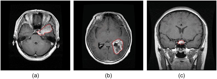
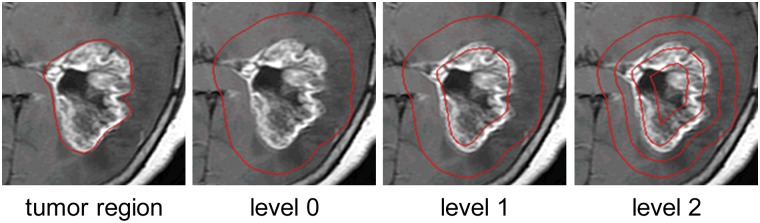
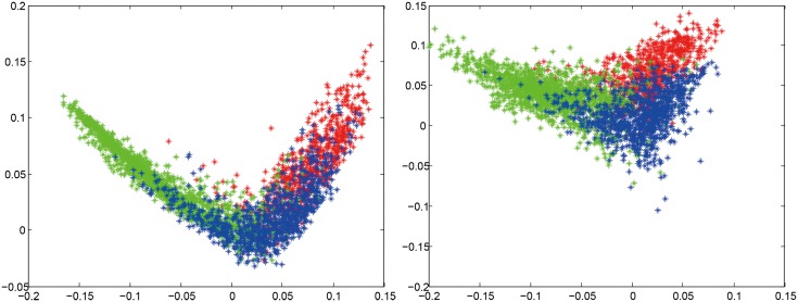
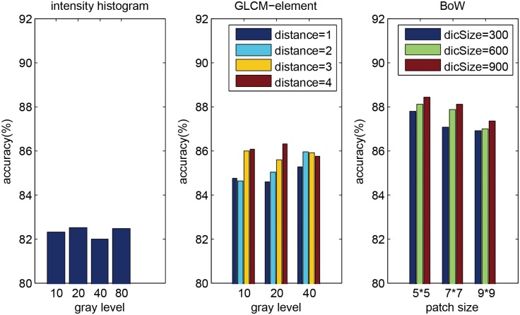
Verification
Claims the paper makes
The claims and statistics of this paper haven’t been checked yet.
Run the check — free, no account needed. It reads the open-access text and checks the sources it cites.
Claims other papers make about this one
Each is checked against this paper’s own text: does it say what the citing paper says it does?
In the citing paper
“Data collected from three different kinds of brain tumors consisted of 708 meningiomas, 1426 gliomas, and 930 pituitaries images from the General Hospital and Nanfang Hospital, Tianjin Medical University, China [72].”
From A Comprehensive Analysis of Recent Deep and Federated-Learning-Based Methodologies for Brain Tumor Diagnosis (2022) · cites this paper as [72]
In the citing paper
“This contains 3064 T1-weighted contrast-enhanced images collected from 233 patients with a resolution of 512 × 512 pixels.”
From A Comprehensive Analysis of Recent Deep and Federated-Learning-Based Methodologies for Brain Tumor Diagnosis (2022) · cites this paper as [72]
Self-check
Percentages, sums and reported differences re-computed from the paper’s own numbers.
The self-check needs the paper’s full text.
Citations
Cites
Distinctive Image Features from Scale-Invariant Keypoints
Lowe, D. G. (2004) · International Journal of Computer Vision · doi:10.1023/b:visi.0000029664.99615.94 · Cited by 55,589 · cited 1×
“(cid:129) Local features: The next step is to extract local features within the ROI, such as scale-invariant feature transform (SIFT) descriptor [20] and raw patch [21].”
Background$rm K$-SVD: An Algorithm for Designing Overcomplete Dictionaries for Sparse Representation
Aharon, M., Elad, M., & Bruckstein, A. M. (2006) · IEEE Transactions on Signal Processing · doi:10.1109/tsp.2006.881199 · Cited by 9,464
Robust Face Recognition via Sparse Representation
Wright, J. N., Yang, A., Ganesh, A., Sastry, S., & Ma, Y. (2009) · IEEE Transactions on Pattern Analysis and Machine Intelligence · doi:10.1109/tpami.2008.79 · Cited by 9,398 · cited 1×
“We also compared three classification methods (i.e. SVM, sparse representation-based classification (SRC) [31], and k nearest neighbors (kNN) classifier) using 2-region BoW representations.”
ResultBeyond Bags of Features: Spatial Pyramid Matching for Recognizing Natural Scene Categories
Lazebnik, S., Schmid, C., & Ponce, J. (2006) · 2006 IEEE Computer Society Conference on Computer Vision and Pattern Recognition - Volume 2 (CVPR'06) · doi:10.1109/cvpr.2006.68 · Cited by 7,916 · cited 4×
“Among those methods, the most notable work is SPM [15], which splits the image into hierarchical cells, computes BoW representation for each cell, and finally weights and concatenates the results.”
BackgroundMethodInfluentialVideo Google: a text retrieval approach to object matching in videos
Sivic, & Zisserman (2003) · Proceedings Ninth IEEE International Conference on Computer Vision · doi:10.1109/iccv.2003.1238663 · Cited by 6,367 · cited 1×
“The BoW method was originally used in the text retrieval domain, and it has been successfully adapted to the visual analysis domain [13,14].”
MethodRobust wide-baseline stereo from maximally stable extremal regions
Matas, J., Chum, O., Urban, M., & Pajdla, T. (2004) · Image and Vision Computing · doi:10.1016/j.imavis.2004.02.006 · Cited by 3,760 · cited 1×
“As pointed out in [29,30], it is beneficial to capture a certain amount of context.”
BackgroundA Bayesian Hierarchical Model for Learning Natural Scene Categories
Fei-Fei, L., & Perona, P. (2005) · IEEE Computer Society Conference on Computer Vision and Pattern Recognition/Proceedings - IEEE Computer Society Conference on Computer Vision and Pattern Recognition/Proceedings · doi:10.1109/cvpr.2005.16 · Cited by 3,665 · cited 1×
“The BoW method was originally used in the text retrieval domain, and it has been successfully adapted to the visual analysis domain [13,14].”
MethodLocality-constrained Linear Coding for image classification
Wang, J., Yang, J., Yu, K., Lv, F., Huang, T., & Gong, Y. (2010) · 2010 IEEE Computer Society Conference on Computer Vision and Pattern Recognition · doi:10.1109/cvpr.2010.5540018 · Cited by 3,100 · cited 2×
“Other more sophisticated methods may also be used, such as SC, soft assignment (SA) [24,25], orthogonal matching pursuit (OMP), locality-constrained linear coding [26], and local anchor embedding [27].”
MethodA comparison of affine region detectors
Mikolajczyk, K., Tuytelaars, T., Schmid, C., Zisserman, A., Matas, J., Schaffalitzky, F., Kadir, T., & Gool, L. V. (2005) · International Journal of Computer Vision · doi:10.1007/s11263-005-3848-x · Cited by 2,939
Linear spatial pyramid matching using sparse coding for image classification
Yang, J., Yu, K., Gong, Y., & Huang, T. S. (2009) · IEEE Computer Society Conference on Computer Vision and Pattern Recognition/Proceedings - IEEE Computer Society Conference on Computer Vision and Pattern Recognition/Proceedings · doi:10.1109/cvpr.2009.5206757 · Cited by 2,895
Research works printed but not linked: 1
- S Patil, 2013, A Computer Aided Diagnostic System For Classification Of Brain Tumors Using Texture Features And Probabilistic Neural Network, Int J Comput Sci Eng Inf Technol Res, 3, 61
Cited by
Sorted by citations
Items 801–879 of 879
Santhosh, A. J. S., Lingaswamy, S., & Ramamurthy, K. (2026) · Elsevier eBooks · doi:10.1016/b978-0-443-41459-6.00006-7
Binish, M. C., & Thomas, V. (2026) · Egyptian Informatics Journal · doi:10.1016/j.eij.2026.100951
A study of machine learning and deep learning methods and their application to medical imaging
Wallis, D. I. (2021) · theses.fr (ABES) · doi:10.70675/136c540fz5d49z41f4zb634zbd9663332582
Brain Tumor Classification Using a Fine-Tuned Enhanced CNN
Alaeddine, H., & Kouka, N. (2025) · IEEE Afro-Mediterranean Conference on Artificial Intelligence (AMCAI) · doi:10.1109/amcai66110.2025.11474320
Subasree, S., Sakthivel, N. K., Gayathri, M., & Manikandan, M. (2026) · International Journal of Pattern Recognition and Artificial Intelligence · doi:10.1142/s0218001426570168
Hakata, Y., Oikawa, M., & Fujisawa, S. (2026) · medRxiv · doi:10.64898/2026.04.15.26351000
Visumathi, J., VijayaBaskaran, S., & Pratheesh, E. R. (2026) · International Conference on Innovative Computing, Intelligent Communication and Smart Electrical Systems (ICSES) · doi:10.1109/icses66558.2026.11479022
“The images are obtained from three publicly available datasets including the Figshare brain tumor dataset [7], the SARTAJ brain MRI dataset [8], and the Br35H brain MRI dataset [9].”
Nirob, A. A., Apon, T. S., Tahsin, A., Alam, M. G. R., Costanzo, S., Fortino, G., Aliverti, A., & Hassan, M. M. (2026) · Computer Methods and Programs in Biomedicine · doi:10.1016/j.cmpb.2026.109409
Hybrid Deep Learning for Enhanced Brain Tumor Detection and Classification in MRI Images
Kumar, M. V., & Vedantham, R. (2026) · International Conference on Emerging Systems and Intelligent Computing (ESIC) · doi:10.1109/esic68176.2026.11495667
Optimizing Brain Tumor Classification for Edge Devices with Knowledge Distillation
Islam, M. S., Nath, S. D., Azhar, T., Hossen, M. M., & Rafi, M. S. M. (2025) · International Conference on Computer and Information Technology (ICCIT) · doi:10.1109/iccit68739.2025.11490190
“Glioma, meningioma, and pituitary tumors are among the notable types [2].”
Ravindran, M., & Kalyane, S. (2026) · Lecture notes in networks and systems · doi:10.1007/978-3-032-18346-0_40
Yang, C., & Wang, M. (2026) · Applied Sciences · doi:10.3390/app16104708
A Deep Learning-Based Model for the Detection and Classification of Brain Tumors
Abdelhamied, M. M., Madbouly, A. M. M., Mostafa, S. I., & Reda, N. M. (2026) · International Conference on Emerging Technologies and Engineering Systems (ICETES) · doi:10.1109/icetes68504.2026.11518665
Pande, T. (2026) · International Conference on Augmented Intelligence and Sustainable Systems (ICAISS) · doi:10.1109/icaiss68683.2026.11526530
“In particular, the application of deep learning to brain imaging has resulted in the implementation of such technologies to tumor segmentation, classification [6], and survival prediction tasks.”
InfluentialFan, L., Wang, C., Ye, M., Yang, Q., Wu, Z., Wang, L., Ding, X., Huang, Y., Bao, J., Cai, S., et al. (2026) · Pattern Recognition · doi:10.1016/j.patcog.2026.114135
Ali, T., Sheihan, M., Ahmad, F., & Ahmad, M. (2026) · International Conference on Device Intelligence, Computing and Communication Technologies (DICCT) · doi:10.1109/dicct69099.2026.11535989
“The Figshare Brain Tumour Dataset [26] contains 3,064 axial slices of brain MRI scans.”
Tumor-Swin Transformer model for brain Tumor classification using MRI
Shahzad, T., Saqib, S. M., Mazhar, T., Jiang, W., & Ouahada, K. (2026) · Array · doi:10.1016/j.array.2026.100954
Swain, K., Nayak, S. R., Swain, S. K., & Singh, P. (2026) · Biomedical & Pharmacology Journal · doi:10.13005/bpj/3414
Yılmaz, C., SUİÇMEZ, Ç., SUİÇMEZ, A., & Işık, M. F. (2026) · Aksaray University Journal of Science and Engineering · doi:10.29002/asujse.1950042
An attention-based hybrid deep learning technique for brain tumor classification
Han, Q., & Ren, C. (2026) · Signal Image and Video Processing · doi:10.1007/s11760-026-05455-7
MRI Brain Tumor Segmentation Using Segmentation-Guided Deep Ensembles with Self-Supervised Learning
Babu, S. R., Chowdary, G. H., Reddy, G. G., Rachana, J., & Malavika, K. S. (2026) · International Conference Big Data and Machine Learning · doi:10.1109/icbdml68582.2026.11544756
Uncertainty-Aware Fuzzy Weighted Ensemble Learning for Biomedical Image Classification
Raut, D. K., Rao, K. S. N., Shasidhar, M. B. S., Venkatesh, J., Sathwik, D. S. N., & Sowkuntla, P. (2026) · International Conference on Signal, Systems, and Computing for Next-Gen Automation (ICSSCNA) · doi:10.1109/icsscna68616.2026.11546722
“Equally, CNN-based methods have also shown encouraging sources of brain MRI classification in tumor detection and neurological disorders analysis [4], [5].”
Grad-CAM Integrated CNN for Explainable Brain Tumor Classification and Localization
Manikandan, R., Bindu, V., Bhavyasree, M., Reddy, K. D., Pavan, M. G., & Dinesh, K. (2026) · International Conference on Signal, Systems, and Computing for Next-Gen Automation (ICSSCNA) · doi:10.1109/icsscna68616.2026.11547147
“In order to ensure consistency between classification and explanation, the Grad-CAM stage was applied to the final convolutional representation of the trained model rather than on an external localization network [16].”
VGG19 encoder-ISCFSO: An enhanced evolutionary framework for brain tumor segmentation
Muthulakshmi, K., & Mohan, J. (2026) · Cluster Computing · doi:10.1007/s10586-026-06161-2
“This dataset includes 708 meningioma, 1426 glioma, and 930 pituitary tumor slices, stored in MATLAB format [38 ], where each file cor - responds to an individual MRI slice along with its associated annotations.”
Saifullah, S., Dreżewski, R., & Yudhana, A. (2026) · Lecture notes in computer science · doi:10.1007/978-3-032-29912-3_38
“FBTS consists of contrast-enhanced T1-weighted MRI slices for meningioma, glioma, and pituitary tumors with binary masks [7], while BraTS 2021 provides multimodal MRI (FLAIR, T1, T1ce, T2) with annotations for whole tumor (WT), tumor core (TC), and enhancing tumor (ET) [4].”
Lamba, K., & Rani, S. (2026) · ICT Express · doi:10.1016/j.icte.2026.06.007
DBAF-Net: Dual-Branch Attention Fusion Network for brain tumor classification in MRI
Kumar, M., & Vedantham, R. (2026) · Biomedical Signal Processing and Control · doi:10.1016/j.bspc.2026.110878
Explainability-Centric Benchmarking of CNN and Transformer Models for Brain Tumor MRI Classification
Padmaja, R., Khanduja, R. S., Dudekula, W. R., Nelatur, A. V., Das, S., & Nunna, S. K. (2026) · International Conference on Connected Intelligence for Industrial Applications (CI2A) · doi:10.1109/ci2a69097.2026.11576827
“These data augmentation strategies simulate real-world variations typically observed in clinical MRI imaging, as shown in previous works by Cheng et al. [5] and Rehman et al. [7].”
An Enhanced Approach in Brain Tumor Classification with Generative Adversarial Networks
Remya, R., Singh, O. J., Sharma, R. R., Dhivyasri, & Manikandan (2026) · Lecture notes in networks and systems · doi:10.1007/978-3-032-22289-3_13
Using Explainability Techniques for Alzheimer Disease and Cancer Detection
Moreira, A., Ferreira, A. J., & Leite, N. (2026) · Studies in computational intelligence · doi:10.1007/978-3-032-22633-4_8
Improved Brain Tumor Classification Using Capsule Networks with SAMME.R Boosting
Sandhya, G., Babu, B. J., Prameela, K. S., Vani, M., Sree, T. N., & Tejaswini, P. (2026) · Smart innovation, systems and technologies · doi:10.1007/978-3-032-22118-6_12
Bhagavatula, V. S. N., & Panda, M. R. (2026) · Smart innovation, systems and technologies · doi:10.1007/978-3-032-18849-6_21
Robust MRI Brain Tumor Classification Using Resnet-Based Transfer Learning
Babu, S. R., Chowdary, G. H., Reddy, G. G., Rachana, J., & Malavika, K. S. (2026) · International Conference on Computing Theory and Wireless Communications (ICCTWC) · doi:10.1109/icctwc68241.2026.11583701
When to Adapt? Adapting the Model or Data in Federated Medical Imaging
Shiranthika, C., & Saeedi, P. (2026) · Lecture notes in computer science · doi:10.1007/978-3-032-30710-1_18
Enhanced Brain Tumor Classification: Integrating ResNet And PCA With GAT For Superior Accuracy
Ashok, A., Yaswanth, S., Durgalakshmi, P. C., & Nair, M. A. (2026) · Procedia Computer Science · doi:10.1016/j.procs.2026.06.169
Evaluating the detection of small brain lesions in magnetic resonance using deep learning
Nogales, A., de la Pinta, C., García-Tejedor, Á. J., García‐Barriocanal, E., Guadalupe, D., & Sicilia, M.-A. (2026) · Frontiers in Neuroscience · doi:10.3389/fnins.2026.1833713 · Cited by 1
“This data was compiled and obtained by Nanfang Hospital and General Hospital, Tianjing Medical University of China (Cheng et al., 2015).”
Computationally Efficient Lightweight Model for Multi-Class Brain Tumor Diagnosis
Devi, Y. D., & Kumar, G. S. (2026) · International Conference on Next Generation Electronics (NELEX) · doi:10.1109/nelex69209.2026.11590253
“…DenseNet, ResNet, EfficientNet, and MobileNet families are commonly adopted due to their strong representational power and pretrained feature extractors, and several studies report promising classification and segmentation accuracies on public MRI benchmarks when fine-tuned for BT tasks [10]–[12].”
Fusion Direction Matters: Alignment-Adaptive Cross-Modal Fusion for Medical Image Segmentation
Guo, Z., Yang, H., Yin, H., & Xu, J. (2026) · Lecture notes in computer science · doi:10.1007/978-981-92-3495-0_21
Brain Tumor Segmentation and Classification using NVIDIA Jetson Nano
Chennamsetti, T., Raj, N. P., V, Raja, A. R., Chintapalli, M. V. R., & Gaigulla, T. R. (2026) · International Conference on Innovations in Communication, Geoscience and Robotics (ICICGR) · doi:10.1109/icicgr68236.2026.11600109
“This makes the dataset useful for evaluating how well deep learning models work and how well they can be generalized [24].”
MRI-Based Deep Learning for Brain Tumor Prediction
Tashkandi, A., & Wally, S. F. (2026) · Communications in computer and information science · doi:10.1007/978-3-032-22199-5_31
Deep Learning Approach for Automated Brain Tumor Detection Using MRI Image
Hossen, B., Manjar, M. T. A., Sultana, S., Shihab, M. A., Zabed, M. A. A., & Akash, K. M. K. (2026) · International Conference on Innovations in Cybersecurity and Data Science (ICICDS) · doi:10.1109/icicds70526.2026.11604682
“Because it offers precise soft-tissue contrast and makes it easier for medical professionals to see tumor location, size, and structural abnormalities than many other imaging modalities, MRI is frequently utilized for the detection of brain tumors [2].”
Xu, Q. (2026) · Lecture notes in computer science · doi:10.1007/978-981-92-3513-1_2
Towards Development of an Optimized Brain Tumor Classification Model
Talukdar, G., Dutta, P., & Roy, M. (2026) · International Conference on Cognitive Computing and Networking Systems (ICC-CNS) · doi:10.1109/icc-cns70518.2026.11606042
“Cheng et al. introduced one of the early CNN-based approaches for brain tumor classification using the Figshare MRI dataset [10].”
Qausain, S., Pramodh, B., Dar, A. A., Basheeruddin, M., Fayaz, M., & Ingole, R. P. (2026) · IEEE International Conference on Intelligent Technologies for a Sustainable and Inclusive Future (ICITSIF) · doi:10.1109/icitsif69060.2026.11608354
“A comprehensive collection of brain MRI scans aggregated from multiple publicly available repositories, including the Figshare dataset [18] and the BraTS challenge datasets [14].”
Alnageeb, M. H. O., & Supriya, M. H. (2026) · Engineering Applications of Artificial Intelligence · doi:10.1016/j.engappai.2026.115727
Yurdakul, M., Zeynalov, J., Ersoy, M. T., Özger, F., & Paçal, İ. (2026) · Diagnostics · doi:10.3390/diagnostics16152407
“All experiments were conducted on the public Figshare brain tumour dataset originally described by Cheng et al. [22].”
Deep Learning-Based Brain Tumor Identification: A Lightweight CNN Approach for MRI Analysis
Raj, U., Choubey, D. K., & Bharodiya, R. B. (2026) · Lecture notes in electrical engineering · doi:10.1007/978-3-032-25176-3_22
Verma, K., Mishra, R. K., & Bhandari, A. K. (2026) · Multimedia Tools and Applications · doi:10.1007/s11042-026-21826-1
A Modular Framework for Brain Tumor Segmentation and Explainable-by-Design Classification from MRI
Miglionico, G. C., Ducange, P., Fazzolari, M., & Marcelloni, F. (2026) · IEEE International Conference on Fuzzy Systems (FUZZ-IEEE) · doi:10.1109/fuzz69877.2026.11626386
“In the context of Brain Tumor Classification (BTC), restricting the analysis to tumor-centered ROIs, obtained through manual annotation or automated procedures, has been shown to improve classification accuracy by reducing background variability and emphasizing clinically relevant structures [10].”
MoBRiX: An Attention-Enhanced MobileNetV3 Framework for Brain Tumor Classification
Wu, Y., Wang, R., Lin, J., & Li, B. (2026) · IEEE International Conference on Computer Vision and Deep Learning (DLCV) · doi:10.1109/dlcv69906.2026.11635231
“These difficulties have motivated extensive research on data-driven brain tumor recognition, including tumor-region augmentation [1], transfer-learning CNN features [2], large-scale data augmentation [3], lightweight convolutional architectures [4], and multi-class deep learning pipelines [5].”
Brain tumor detection in MRI using YOLO architectures
Hammad, M. S., & Makram, A. W. (2026) · Biomedical Signal Processing and Control · doi:10.1016/j.bspc.2026.111242
Eassa, M., HEGAZY, N. Y., & Elbehiery, H. (2026) · Computers · doi:10.3390/computers15080519
Kumar, S., Chakraborty, M., Hazarika, D., & Nath, V. K. (2026) · IEEE Guwahati Subsection Conference (GCON) · doi:10.1109/gcon69192.2026.11648797
“To evaluate the performance of proposed framework, we conducted experiments on two publicly available datasets i.e. Figshare Brain MRI [13, 14] and Sartaj brain MRI [15] as summarized in Table-I”
Nawathey, N., Bachir, M. A., Gill, S., Reddy, A. J., Desai, A., Vyas, A., & Patel, R. (2026) · Cureus · doi:10.7759/cureus.114682
“The results are consistent with previous research demonstrating the effectiveness of convolutional neural networks and other deep-learning methods for brain tumor classification [12-21,25, 26] .”
Optimized approach to image-based automatic disease classification using improved capsule networks
Yadav, S., & Dhage, S. (2026) · Multimedia Tools and Applications · doi:10.1007/s11042-026-21868-5
“● MRI Dataset : The brain tumor dataset [50], as shown in The overall details pertaining to the three datasets, including the total number of sam - ples, the number of samples used for training, and the number of samples used for testing, are presented in Table 5 .”
Advanced Faster R-CNN Framework for Accurate Real-Time Brain Tumor Diagnosis
BENABOUD, O., Mhasni, K., & Alaoui, E. M. I. (2026) · International Conference on Circuit, Systems and Communication (ICCSC) · doi:10.1109/iccsc71566.2026.11650455
Hybrid Deep Learning Approach for Brain Tumor Detection Using Swin Transformer
Srivastava, A., Singh, D., Verma, A., Pandey, D., & Singh, K. K. (2026) · International Conference on Emerging VLSI and Semiconductor Technology for AI and Computing Applications (EVST) · doi:10.1109/evst69093.2026.11660600
“In response to this, lighter CNNs with reduced layers have been suggested that are competitive in combination with preprocessing and augmentation [2][15].”
Cancer Imaging Datasets: A Review
Nuseir, A. (2026) · Lecture notes in networks and systems · doi:10.1007/978-3-032-21200-9_16
Nguyen, N., Huynh, T. V., & Dang, L. V. (2026) · IEEE Access · doi:10.1109/access.2026.3729887
Kadiri, C., Saheed, Y. K., Chukwuere, J. E., Jimoh, K., Omole, A. I., Usman, T. M., & Mustapha, A. (2026) · Intelligence-Based Medicine · doi:10.1016/j.ibmed.2026.100473
Popescu, A., Ionescu, M., Stan, E., & Radu, C. (2026) · Journal of Medical Sciences and Interdisciplinary Research · doi:10.51847/onimjzlq0c
TopoAgent: An Agentic Framework for Automated Topology Learning in Medical Imaging
Meng, G., Gu, P., Li, X., Shi, Y., Chambers, E. W., & Chen, D. Z. (2026) · Lecture notes in computer science · doi:10.1007/978-3-032-37032-7_17
Large Language Model Benchmarks in Medical Tasks
Yan, L. K. Q., Niu, Q., Li, M., Zhang, Y., Yin, C. H., Fei, C., Peng, B., Bi, Z., Feng, P., Chen, K., et al. (2026) · Medicine Advances · doi:10.1002/med4.70085
The impact of region of interest size on brain tumor classification
Guassu, R. A. C., & de Pina, D. R. (2026) · Imaging · doi:10.1556/1647.2026.00420
Zhang, J., Cai, C., Tan, X., Chen, W., Zhao, X., 杜敢琴, & Jiang, H. (2026) · PLoS ONE · doi:10.1371/journal.pone.0343457
“The dataset, sourced from Cheng et al.[30], comprises 3064 grayscale brain MR images categorized into three groups, 708 meningioma samples, 1426 glioma samples, and 930 pituitary tumor samples.”
Brain Tumor Classification Using Computer Vision and Deep Learning Techniques
Alshahrani, A. (2026) · Engineering Technology & Applied Science Research · doi:10.48084/etasr.21079
“It was first introduced for brain tumor classification using tumor region augmentation and subsequently utilized for brain tumor retrieval based on adaptive spatial pooling and Fisher vector representation [18, 19].”
Jeyshri, J., Jessica, A., & MonishLingam, T. (2026) · International Conference on Soft Computing for Security Applications (ICSCSA) · doi:10.1109/icscsa69367.2026.11707944
The Explanatory Deep Learning Model for Brain Tumor Detection Based on MRI Images
Kumar, T. R., Kumar, G. V. S., Jayalakshmi, D., & Mahaveerakannan, R. (2026) · International Conference on Ambient Intelligence, Knowledge Informatics and Industrial Electronics (AIKIIE) · doi:10.1109/aikiie69997.2026.11707878
“But the manual extraction of the images taken from MRI is time consuming, subjective, subjective and the diagnosis and treatment time may vary depending on the expertise of different radiologists [3].”
TP-Seg: Task-Prototype Framework for Unified Medical Lesion Segmentation
Xu, J., Zhou, Q., Zhu, D., Chen, Y., Yi, Y., & Zhao, X. (2026) · doi:10.1109/cvprf71909.2026.00524
“MLS encompasses a wide variety of lesion types, such as wet age-related macular degeneration [23], brain tumors [12], adenocarcinoma [33], thyroid nodules [55], colorectal polyps [35], lung infection [17], breast lesions [1], and skin lesions [14].”
Zhang, S., Arshad, M. R., Ibrahim, H., & Zou, J. (2026) · Physical and Engineering Sciences in Medicine · doi:10.1007/s13246-026-01816-1
Cai, C., Zhang, J., Zhang, D., Chen, W., Tan, X., Du, G., & Jiang, H. (2026) · Frontiers in Neuroscience · doi:10.3389/fnins.2026.1986322
Kumar, A., Agarwal, M., & Srivastava, A. K. (2026) · International Journal of Computational Intelligence and Applications · doi:10.1142/s1469026826500628
Smart Brain Tumor Classification Using Deep Learning with Channel Attention Networks
Das, V., Mohapatra, A., & Rout, T. (2026) · International Conference on Intelligent Computing and Sustainable Innovations in Technology (IC-SIT) · doi:10.1109/ic-sit69723.2026.11735814
“Because incorrect classifications can significantly affect the choice of treatment and prognosis, reliable multiclass identification of brain tumors is crucial [1].”
Ali, M., Hashmi, U., Azam, M., & Sarwar, R. (2026) · Vector The Journal of Engineering · doi:10.1049/tje2.70241
An Explainable XAIFusion Deep Learning Framework for Brain Tumour MRI Classification
Vedanarayanan, V., Sakthi Prabha, R., Rajinikanth, E., Gomathi, T., S.Manodhiya, & Aranganathan, A. (2026) · International Conference on Intelligent Cyber Physical Systems and Internet of Things (ICoICI) · doi:10.1109/icoici69824.2026.11724036
“ResNet18 is fine-tuned on the publicly available Cheng et al. dataset [6].”
Duong, L. T., Maswita, P. W., Sumaryada, T. I., Yani, S., Tài, D. T., Omer, H. B., Tamam, N. M., & Sulieman, A. (2026) · Diagnostics · doi:10.3390/diagnostics16213485
Saifullah, S., Dreżewski, R., & Yudhana, A. (2026) · International Journal of Machine Learning and Cybernetics · doi:10.1007/s13042-026-03348-2
“Three benchmark datasets were employed: (i) FBTS, consisting of T1-CE MRI slices with meningioma, glioma, and pituitary tumor classes [70 ]; (ii) BraTS 2021, containing multimodal scans comprising T1, T1CE, T2, and FLAIR sequences with tumor-subregion annotations subsequently converted into binary…”
Gayathridevi, R., & Venkatesan, R. (2026) · National Academy Science Letters · doi:10.1007/s40009-026-02464-5
Metadata
- Authors
- Cheng, J., Huang, W., Cao, S., Yang, R., Yang, W., Yun, Z., Wang, Z., & Feng, Q.
- Affiliations
- Southern Medical University (CN)
- Nanfang Hospital (CN)
- Corresponding author
- Feng, Qianjin
- Venue
- PLOS ONE
- Publisher
- Public Library of Science (PLoS)
- Published
- 2015-10-08
- Volume
- 10
- Issue
- 10
- Pages
- e0140381
- ISSN
- 1932-6203 (ISSN-L)
- PMC
- PMC4598126
- OpenAlex
- W2182098131
- Cited by
- 879
- References
- 36
- Citations by year
- 2026: 110 · 2025: 134 · 2024: 142 · 2023: 152 · 2022: 128 · 2021: 85 · 2020: 63 · 2019: 44 · 2018: 14 · 2017: 4 · 2016: 2 · 2015: 1
- FWCI
- 3.74
- Type
- journal article
- Language
- en
- Update notices
- correction (2015-12-02)
- Open access
- gold
- doi.org
- Licence
- Locations
- PLoS ONE (publishedVersion, cc-by)
- Europe PMC (PubMed Central) (publishedVersion, cc-by)
- PubMed Central (publishedVersion, cc-by)
- PubMed (publishedVersion)
- DOAJ (DOAJ: Directory of Open Access Journals) (submittedVersion)
- Funders
- National Natural Science Foundation of China (81101109, 31371009, 2012BAI14B02, 863 Program)
- National High-tech Research and Development Program
- Topics
- Physical Sciences › Computer Science › Computer Vision and Pattern Recognition › Brain Tumor Detection and Classification
- Health Sciences › Medicine › Radiology, Nuclear Medicine and Imaging › Radiomics and Machine Learning in Medical Imaging
- Physical Sciences › Computer Science › Artificial Intelligence › AI in cancer detection
- Keywords
- brain tumor classification, meningioma, glioma, pituitary tumors, region of interest, image dilation, spatial pyramid matching, contrast-enhanced T1-weighted imaging, computer-aided diagnosis, gray level co-occurrence matrix, bag-of-words model, intensity histogram
- MeSH terms
- Brain Neoplasms, Glioma, Humans, Image Interpretation, Computer-Assisted, Magnetic Resonance Imaging, Meningioma, Sensitivity and Specificity
About these checks
Registry, retraction, Cites and statistics results are computed, not judged, with one exception: a printed reference that no index lists is read once by a model before it counts as a citation error. Claims are extracted by a model and kept only when their quote appears verbatim in the text; each cited source is then checked, with a model’s help, for whether it says what the claim relies on. Those verdicts can be wrong — read the claim’s quote and the cited source before relying on one.
Statistics and arithmetic are recomputed, not judged.
Confidence bands are shown only for fields calibrated on our annotated test set. None is calibrated yet, so no band is shown.
What counts as a claim ›How we measure confidence ›How we check this ›
Are you an author, or do you see a problem with this page? Request a review
