

The DOI matches its Crossref record; 35 of the 36 references it cites were found in a registry or an index.
Enhanced Performance of Brain Tumor Classification via Tumor Region Augmentation and Partition
Open accessAbstract
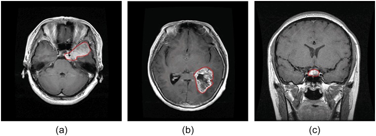
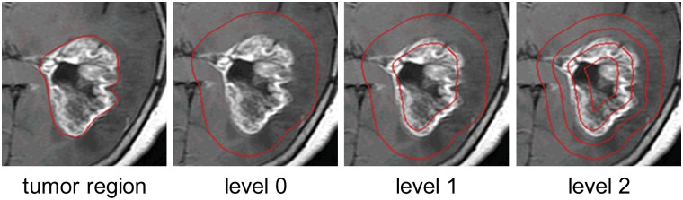
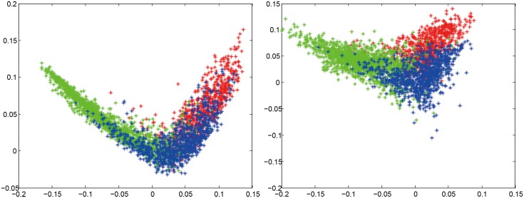
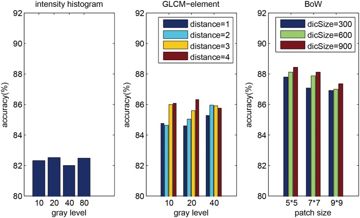
Automatic classification of tissue types of region of interest (ROI) plays an important role in computer-aided diagnosis. In the current study, we focus on the classification of three types of brain tumors (i.e., meningioma, glioma, and pituitary tumor) in T1-weighted contrast-enhanced MRI (CE-MRI) images. Spatial pyramid matching (SPM), which splits the image into increasingly fine rectangular subregions and computes histograms of local features from each subregion, exhibits excellent results for natural scene classification. However, this approach is not applicable for brain tumors, because of the great variations in tumor shape and size. In this paper, we propose a method to enhance the classification performance. First, the augmented tumor region via image dilation is used as the ROI instead of the original tumor region because tumor surrounding tissues can also offer important clues for tumor types. Second, the augmented tumor region is split into increasingly fine ring-form subregions. We evaluate the efficacy of the proposed method on a large dataset with three feature extraction methods, namely, intensity histogram, gray level co-occurrence matrix (GLCM), and bag-of-words (BoW) model. Compared with using tumor region as ROI, using augmented tumor region as ROI improves the accuracies to 82.31% from 71.39%, 84.75% from 78.18%, and 88.19% from 83.54% for intensity histogram, GLCM, and BoW model, respectively. In addition to region augmentation, ring-form partition can further improve the accuracies up to 87.54%, 89.72%, and 91.28%. These experimental results demonstrate that the proposed method is feasible and effective for the classification of brain tumors in T1-weighted CE-MRI.
Overview
The claims and statistics of this paper haven’t been checked yet.
Run the check — free, no account needed. It reads the open-access text and checks the sources it cites.
Integrity signals
Registry
Cites
Statistics
Figures
Verification
Claims the paper makes
The claims and statistics of this paper haven’t been checked yet.
Run the check — free, no account needed. It reads the open-access text and checks the sources it cites.
Claims other papers make about this one
Each is checked against this paper’s own text: does it say what the citing paper says it does?
In the citing paper
“Data collected from three different kinds of brain tumors consisted of 708 meningiomas, 1426 gliomas, and 930 pituitaries images from the General Hospital and Nanfang Hospital, Tianjin Medical University, China [72].”
From A Comprehensive Analysis of Recent Deep and Federated-Learning-Based Methodologies for Brain Tumor Diagnosis (2022) · cites this paper as [72]
In the citing paper
“This contains 3064 T1-weighted contrast-enhanced images collected from 233 patients with a resolution of 512 × 512 pixels.”
From A Comprehensive Analysis of Recent Deep and Federated-Learning-Based Methodologies for Brain Tumor Diagnosis (2022) · cites this paper as [72]
Self-check
Percentages, sums and reported differences re-computed from the paper’s own numbers.
The self-check needs the paper’s full text.
Citations
Cites
Distinctive Image Features from Scale-Invariant Keypoints
Lowe, D. G. (2004) · International Journal of Computer Vision · doi:10.1023/b:visi.0000029664.99615.94 · Cited by 55,589 · cited 1×
“(cid:129) Local features: The next step is to extract local features within the ROI, such as scale-invariant feature transform (SIFT) descriptor [20] and raw patch [21].”
Background$rm K$-SVD: An Algorithm for Designing Overcomplete Dictionaries for Sparse Representation
Aharon, M., Elad, M., & Bruckstein, A. M. (2006) · IEEE Transactions on Signal Processing · doi:10.1109/tsp.2006.881199 · Cited by 9,464
Robust Face Recognition via Sparse Representation
Wright, J. N., Yang, A., Ganesh, A., Sastry, S., & Ma, Y. (2009) · IEEE Transactions on Pattern Analysis and Machine Intelligence · doi:10.1109/tpami.2008.79 · Cited by 9,398 · cited 1×
“We also compared three classification methods (i.e. SVM, sparse representation-based classification (SRC) [31], and k nearest neighbors (kNN) classifier) using 2-region BoW representations.”
ResultBeyond Bags of Features: Spatial Pyramid Matching for Recognizing Natural Scene Categories
Lazebnik, S., Schmid, C., & Ponce, J. (2006) · 2006 IEEE Computer Society Conference on Computer Vision and Pattern Recognition - Volume 2 (CVPR'06) · doi:10.1109/cvpr.2006.68 · Cited by 7,916 · cited 4×
“Among those methods, the most notable work is SPM [15], which splits the image into hierarchical cells, computes BoW representation for each cell, and finally weights and concatenates the results.”
BackgroundMethodInfluentialVideo Google: a text retrieval approach to object matching in videos
Sivic, & Zisserman (2003) · Proceedings Ninth IEEE International Conference on Computer Vision · doi:10.1109/iccv.2003.1238663 · Cited by 6,367 · cited 1×
“The BoW method was originally used in the text retrieval domain, and it has been successfully adapted to the visual analysis domain [13,14].”
MethodRobust wide-baseline stereo from maximally stable extremal regions
Matas, J., Chum, O., Urban, M., & Pajdla, T. (2004) · Image and Vision Computing · doi:10.1016/j.imavis.2004.02.006 · Cited by 3,760 · cited 1×
“As pointed out in [29,30], it is beneficial to capture a certain amount of context.”
BackgroundA Bayesian Hierarchical Model for Learning Natural Scene Categories
Fei-Fei, L., & Perona, P. (2005) · IEEE Computer Society Conference on Computer Vision and Pattern Recognition/Proceedings - IEEE Computer Society Conference on Computer Vision and Pattern Recognition/Proceedings · doi:10.1109/cvpr.2005.16 · Cited by 3,665 · cited 1×
“The BoW method was originally used in the text retrieval domain, and it has been successfully adapted to the visual analysis domain [13,14].”
MethodLocality-constrained Linear Coding for image classification
Wang, J., Yang, J., Yu, K., Lv, F., Huang, T., & Gong, Y. (2010) · 2010 IEEE Computer Society Conference on Computer Vision and Pattern Recognition · doi:10.1109/cvpr.2010.5540018 · Cited by 3,100 · cited 2×
“Other more sophisticated methods may also be used, such as SC, soft assignment (SA) [24,25], orthogonal matching pursuit (OMP), locality-constrained linear coding [26], and local anchor embedding [27].”
MethodA comparison of affine region detectors
Mikolajczyk, K., Tuytelaars, T., Schmid, C., Zisserman, A., Matas, J., Schaffalitzky, F., Kadir, T., & Gool, L. V. (2005) · International Journal of Computer Vision · doi:10.1007/s11263-005-3848-x · Cited by 2,939
Linear spatial pyramid matching using sparse coding for image classification
Yang, J., Yu, K., Gong, Y., & Huang, T. S. (2009) · IEEE Computer Society Conference on Computer Vision and Pattern Recognition/Proceedings - IEEE Computer Society Conference on Computer Vision and Pattern Recognition/Proceedings · doi:10.1109/cvpr.2009.5206757 · Cited by 2,895
Research works printed but not linked: 1
- S Patil, 2013, A Computer Aided Diagnostic System For Classification Of Brain Tumors Using Texture Features And Probabilistic Neural Network, Int J Comput Sci Eng Inf Technol Res, 3, 61
Cited by
Sorted by citations
Items 301–400 of 879
An Improved Deep Structure for Accurately Brain Tumor Recognition
Ata, M. M., Yousef, R. N., Karim, F. K., & Khafaga, D. S. (2023) · Computer Systems Science and Engineering · doi:10.32604/csse.2023.034375 · Cited by 20
Recurrent Attention Mechanism Networks for Enhanced Classification of Biomedical Images
Shaikh, M., Kollerathu, V. A., & Krishnamurthi, G. (2019) · IEEE International Symposium on Biomedical Imaging (ISBI) · doi:10.1109/isbi.2019.8759214 · Cited by 19
“We explore two datasets in our work viz; a dataset[17] comprising of 3064 brain MRI and a dataset[18] consisting of 516 images of the retina.”
MethodRagupathy, B., & Karunakaran, M. (2020) · International Journal of Imaging Systems and Technology · doi:10.1002/ima.22464 · Cited by 20
Mengash, H. A., & Mahmoud, H. A. H. (2021) · Computers, materials & continua/Computers, materials & continua (Print) · doi:10.32604/cmc.2021.016907 · Cited by 20
“The authors in [13] performed tumor classification by enhancing the region of interest and the image ring segmentation.”
BackgroundA successive framework for brain tumor interpretation using Yolo variants
Priyadharshini, S., Bhoopalan, R., Manikandan, D., & Ramaswamy, K. (2025) · Scientific Reports · doi:10.1038/s41598-025-13155-4 · Cited by 19
“Several studies have explored YOLO-based models for brain tumour diagnosis 50–53 , mostly using older versions like YOLOv5 and YOLOv7.”
Kazihise, N. G., Zhao, J., Sabuni, F. M., & Wang, J. (2020) · Information Communication Technologies Conference (ICTC) · doi:10.1109/ictc49638.2020.9123249 · Cited by 19
“However, Cheng, Jun et al [12] used the augmented tumor region via image dilation as the ROI instead of the original tumor region because tumor surrounding tissues can also offer important clues for tumor types, which highlighted the added advantage of the information provided by the tissue…”
MethodAlmalki, Y. E., Ali, M. U., Ahmed, W., Kallu, K. D., Zafar, A., Alduraibi, S. K., Irfan, M., Basha, M. A. A., Alshamrani, H. A., & Alduraibi, A. K. (2022) · Life · doi:10.3390/life12071084 · Cited by 19
“In one study [24], the authors hybridized the gray-level co-occurrence matrix, histogram intensity, and bag of words to classify brain MRI images.”
BackgroundBrain tumor classification in magnetic resonance imaging images using convolutional neural network
Remzan, N., Tahiry, K., & Farchi, A. (2022) · International Journal of Power Electronics and Drive Systems/International Journal of Electrical and Computer Engineering · doi:10.11591/ijece.v12i6.pp6664-6674 · Cited by 19
“Cheng et al. [12] have developed a tumor classification method, that consists of two stages.”
MethodA Reversible Medical Image Watermarking for ROI Tamper Detection and Recovery
Bhalerao, S., Ansari, I. A., & Kumar, A. (2023) · Circuits Systems and Signal Processing · doi:10.1007/s00034-023-02416-0 · Cited by 19
“The MRI and CT images have been obtained from [6, 15], and other databases [2, 30] present at “The Cancer Imaging Archive” [7].”
MethodPreetha, R., Priyadarsini, M. J. P., & Nisha, J. S. (2023) · IEEE Access · doi:10.1109/access.2023.3340443 · Cited by 24
“Different types of tumor types with publicly available dataset is given in Table 5 [33], [37], [38], [39], [40], [41], [42], [43], [44], [45], [46], [47], [48], [49], [50], [51], [52], [53].”
BackgroundBrain Tumor Semi-automatic Segmentation on MRI T1-weighted Images using Active Contour Models
Thias, A. H., Mubarok, A. F. A., Handayani, A., Danudirdjo, D., & Rajab, T. L. E. (2019) · International Conference on Mechatronics, Robotics and Systems Engineering (MoRSE) · doi:10.1109/morse48060.2019.8998651 · Cited by 19
“This research utilizes MRI T1-weighted images collected from 233 brain tumor patients in the Nanfang Hospital and General Hospital of Tianjin University in China between 2005-2010 [6].”
MethodZahoor, M. M., Qureshi, S. A., Khan, A., Rehman, A. U., & Rafique, M. (2022) · Waves in Random and Complex Media · doi:10.1080/17455030.2022.2070683 · Cited by 20
“Cheng et al. [53] proposed a method with three different feature extraction techniques, namely bag-of-words (BoW) model, intensity histogram, and gray-level co-occurrence matrix (GLCM).”
RAMS: Remote and automatic mammogram screening
Cogan, T., Cogan, M., & Tamil, L. (2019) · Computers in Biology and Medicine · doi:10.1016/j.compbiomed.2019.01.024 · Cited by 18
A Hybrid Learnable Fusion of ConvNeXt and Swin Transformer for Optimized Image Classification
Qezelbash-Chamak, J., & Hicklin, K. (2025) · IoT · doi:10.3390/iot6020030 · Cited by 20
“We used a 7023 image dataset comprising various tumor types (e.g., glioma, meningioma, pituitary tumor, healthy) at mixed resolutions [51].”
Brain Tumour Classification Based on Deep Convolutional Neural Networks
El‐Feshawy, S. A., Saad, W., Shokair, M., & Dessouky, M. I. (2021) · International Conference on Electronic Engineering (ICEEM) · doi:10.1109/iceem52022.2021.9480637 · Cited by 19
EFF_D_SVM: a robust multi-type brain tumor classification system
Zhang, J., Tan, X., Chen, W., Du, G., Fu, Q., Zhang, H., & Jiang, H. (2023) · Frontiers in Neuroscience · doi:10.3389/fnins.2023.1269100 · Cited by 18
“The Chen dataset is the CE-MRI dataset shared by Cheng et al. (2015), which consists of 3,064 brain MRI images from 233 patients, including three types of brain tumors, namely glioma, meningioma, and pituitary tumors.”
MethodContourTL-Net: Contour-Based Transfer Learning Algorithm for Early-Stage Brain Tumor Detection
Ashafuddula, N. I. M., & Islam, R. (2024) · International Journal of Biomedical Imaging · doi:10.1155/2024/6347920 · Cited by 18
Saifullah, S., & Dreżewski, R. (2024) · Genetic and Evolutionary Computation Conference Companion (GECCO Companion) · doi:10.1145/3638530.3654339 · Cited by 18
“Our study utilized a Kaggle.com dataset comprising 3064 Brain Images from the T1-CE MRI dataset [4], with consistent 512 × 512 pixels and 24-bit depth, crucial for training our segmentation model [23].”
Saifullah, S., & Dreżewski, R. (2024) · Lecture notes in computer science · doi:10.1007/978-3-031-70071-2_21 · Cited by 18
Generating 3D brain tumor regions in MRI using vector-quantization Generative Adversarial Networks
Zhou, M., Wagner, M. W., Tabori, U. Y., Hawkins, C. E., Ertl-Wagner, B. B., & Khalvati, F. (2024) · Computers in Biology and Medicine · doi:10.1016/j.compbiomed.2024.109502 · Cited by 27
Detection and Classification of Brain Tumor Using Machine Learning Algorithms
Refaat, F. M., Gouda, M. M., & Omar, M. A. (2022) · Biomedical & Pharmacology Journal · doi:10.13005/bpj/2576 · Cited by 18
Khan, M. A., Hussain, M. Z., Mehmood, S., Khan, M. F., Ahmad, M., Mazhar, T., Shahzad, T., & Saeed, M. M. A. (2025) · Discover Oncology · doi:10.1007/s12672-025-02671-4 · Cited by 17
“Using a brain tumor dataset created by Cheng in 2015 called Figshare [23], Cheng et al. [24] attempted to address the three-class challenge of brain tumor identification using brain T1 MR images.”
Vamsidhar, D., Desai, P., Joshi, S., Kolhar, S., Deshpande, N. M., & Gite, S. (2025) · Scientific Reports · doi:10.1038/s41598-025-06455-2 · Cited by 26
Deep AutoEncoder-Decoder Framework for Semantic Segmentation of Brain Tumor.
Rehman, A., Naz, S., Naseem, U., Razzak, I., & Hameed, I. A. (2019) · Cited by 17
MRI-Image based Brain Tumor Detection and Classification using CNN-KNN
Shanjida, S., Islam, M. S., & Mohiuddin, M. (2022) · IEEE IAS Global Conference on Emerging Technologies (GlobConET) · doi:10.1109/globconet53749.2022.9872168 · Cited by 17
“Dissimilar forms of transforms from the Gabor filter and Demographic features from the wavelet presented with an accuracy of 91.9% [5].”
BackgroundMethodInfluentialLee, W., Yang, H., Lee, C., Lu, C., Wu, C., Chung, W., Wu, H., Guo, W., & Wu, Y. (2022) · Computer Methods and Programs in Biomedicine · doi:10.1016/j.cmpb.2022.107311 · Cited by 17
A Convolutional Neural Network for Automatic Brain Tumor Detection
Mohsen, S., Abdel-Rehim, W. M. F., Emam, A. Z., & Kasem, H. M. (2023) · Proceedings of Engineering and Technology Innovation · doi:10.46604/peti.2023.10307 · Cited by 17
Singh, Y. P., & Lobiyal, D. K. (2023) · Multimedia Tools and Applications · doi:10.1007/s11042-023-16637-7 · Cited by 19
“Researchers of [12]suggested in 2015 that brain tumor classification could be made more accurate by using image augmentation to make the tumor region bigger and then divide it into smaller parts.”
TE-CapsNet: time efficient capsule network for automatic disease classification from medical images
Yadav, S., & Dhage, S. (2023) · Multimedia Tools and Applications · doi:10.1007/s11042-023-17458-4 · Cited by 17
“As shown in Table 2, another dataset is the brain tumor dataset [50, 51], which contains 3064 T1-weighted contrast-enhancing MRI images from 233 individuals with three types of brain tumors: pituitary tumor (930 samples), glioma (1426 samples), and meningioma (708 samples).”
Saifullah, S., & Dreżewski, R. (2024) · Lecture notes in computer science · doi:10.1007/978-3-031-63772-8_30 · Cited by 17
“In this study, a comprehensive dataset comprising 3064 TI-CE MRIs obtained from 233 patients forms the foundation of our research [6].”
Li, G., Sun, J., Song, Y., Qu, J., Zhu, Z., & Khosravi, M. R. (2021) · Journal of Real-Time Image Processing · doi:10.1007/s11554-021-01072-4 · Cited by 17
Deep Learning of Forced Convection Heat Transfer
Kang, M., & Kwon, B. (2021) · ASME Journal of Heat and Mass Transfer · doi:10.1115/1.4052893 · Cited by 17
Hussain, L., Malibari, A., Alzahrani, J. S., Alamgeer, M., Obayya, M. I., Al‐Wesabi, F. N., Mohsen, H., & Hamza, M. A. (2022) · Scientific Reports · doi:10.1038/s41598-022-19563-0 · Cited by 18
“A publicly dataset was taken available at (https:// github. com/ cheng jun583/ brain Tumor Retri eval) which is described in 43,44 .”
MethodCDANet: Channel Split Dual Attention Based CNN for Brain Tumor Classification In Mr Images
Dutta, T. K., & Nayak, D. R. (2022) · Proceedings - International Conference on Image Processing · doi:10.1109/icip46576.2022.9897799 · Cited by 17
“To evaluate the proposed approach, a publicly available brain tumor dataset [14, 15] is used, which includes 3064 T1weighted MRI images of three tumor types: glioma (1426 images), meningioma (708 images), and pituitary (930 images).”
MethodAutomatic detection of the meningioma tumor firmness in MRI images
Alkubeyyer, A., Ismail, M. M. B., Bchir, O., & Alkubeyyer, M. (2020) · Journal of X-Ray Science and Technology · doi:10.3233/xst-200644 · Cited by 17
A Deep learning framework for brain tumor detection using CNNs and transfer learning on MRI scans
Kandagatla, S. P., Basem, A., Lakshmi, L., Talha, A., Mohammed, S. H., Khan, M. I., & Khedher, N. B. (2025) · Systems and Soft Computing · doi:10.1016/j.sasc.2025.200389 · Cited by 25
Bayoumi, E. S., Abd-Ellah, M. K., Khalaf, A. A. M., & Gharieb, R. R. (2021) · Journal of Advanced Engineering Trends · doi:10.21608/jaet.2020.42896.1051 · Cited by 16
Saifullah, S., Dreżewski, R., & Yudhana, A. (2025) · Multimedia Tools and Applications · doi:10.1007/s11042-025-20702-8 · Cited by 16
“The dataset used in this study comprises 3064 MRI images collected [58] and curated by Cheng et al. [22] It features high-resolution magnetic resonance imaging (MRI) scans segmented into three distinct tumor classes: Meningioma, Glioma, and Pituitary.”
ConvCaps: Multi-input Capsule Network for Brain Tumor Classification
Cheng, Y., Qin, G., Zhao, R., Liang, Y., & Sun, M. (2019) · Lecture notes in computer science · doi:10.1007/978-3-030-36708-4_43 · Cited by 18
Stacked Sparse Autoencoder and Softmax Classifier Framework to Classify MRI of Brain Tumor Images
Aldhahab, A., Ibrahim, S. K., & Mikhael, W. B. (2020) · International journal of intelligent engineering and systems · doi:10.22266/ijies2020.0630.25 · Cited by 16
“Furthermore, the classification technique implemented by stacking SSA and SMC shows better performance compared with BPA classifier and those classification techniques discussed in [23-26].”
BackgroundMethodInfluentialQuickTumorNet: Fast Automatic Multi-Class Segmentation of Brain Tumors
Maas, B., Zabeh, E., & Arabshahi, S. (2021) · International IEEE/EMBS Conference on Neural Engineering · doi:10.1109/ner49283.2021.9441286 · Cited by 16
“The modified network has an encoder/decoder like 2D FCNN architecture with 4 encoders and 4 decoders separated by a bottleneck layer shown in Fig.”
InfluentialDeep Transfer Learning for Brain Magnetic Resonance Image Multi-class Classification
Brima, Y., Tushar, M. H. K., Kabir, U., & Islam, T. (2022) · Dhaka University Journal of Applied Science and Engineering · doi:10.3329/dujase.v6i2.59215 · Cited by 18
Brain Magnetic Resonance Image Tumor Detection and Segmentation Using Edgeless Active Contour
Kumar, S. B., Panda, R., & Agrawal, S. (2020) · International Conference on Computing, Communication and Networking Technologies (ICCCNT) · doi:10.1109/icccnt49239.2020.9225296 · Cited by 16
“This dataset has been previously used for the brain tumor classification [16] and in another case for the content based image retrieval system to retrieve the brain tumors of similar pathological type, as the query tumor image [17].”
MethodLiu, X., Liang, J., Zhang, J., Qian, Z., Xing, P., Chen, T., Yang, S. J., Chukwudi, C. C., Qiu, L., Liu, D., et al. (2025) · Computer Methods and Programs in Biomedicine · doi:10.1016/j.cmpb.2025.108705 · Cited by 17
Bilal, O., & Asif, S. (2025) · Multiscale and Multidisciplinary Modeling Experiments and Design · doi:10.1007/s41939-025-00889-x · Cited by 15
SVM Based Brain Tumor Detection and Classification System
Kiran, Parameshachari, B. D., & Kumar, D. S. S. (2022) · IEEE Mysore Sub Section International Conference (MysuruCon) · doi:10.1109/mysurucon55714.2022.9972652 · Cited by 15
“The brain tumour dataset from fig share was used for the investigation [3, 4].”
MethodSowparnika, B., Yamini, K., Walid, M. A. A., Prasad, J., Aparna, N., & Chauhan, A. (2023) · International Conference on Applied Artificial Intelligence and Computing (ICAAIC) · doi:10.1109/icaaic56838.2023.10140207 · Cited by 15
“Meningioma, glioma, and pituitary tumor are the three forms of brain tumors that [11] primarily categorized.”
BackgroundDeep-Net: Fine-Tuned Deep Neural Network Multi-Features Fusion for Brain Tumor Recognition
Khan, M. A., Mostafa, R. R., Zhang, Y., Baili, J., Alhaisoni, M. M., Tariq, U., Khan, J. A., Kim, Y. J., & Cha, J. (2023) · Computers, materials & continua/Computers, materials & continua (Print) · doi:10.32604/cmc.2023.038838 · Cited by 15
Prediction of microRNAs involved in immune system diseases through network based features
Prabahar, A., & Natarajan, J. (2016) · Journal of Biomedical Informatics · doi:10.1016/j.jbi.2016.11.003 · Cited by 15
Ahmmed, S., Bhuia, M. S., Chowdhury, R., Munshi, M. H., Eity, T. A., Kamli, H. M., & Islam, M. T. (2025) · International Journal of Food Science · doi:10.1155/ijfo/2256871 · Cited by 14
Divya, S., Suresh, L. P., & John, A. (2022) · Pattern Analysis and Applications · doi:10.1007/s10044-022-01064-5 · Cited by 23
A Novel Brain Tumor Classification Model Using Machine Learning Techniques
Pal, S. S., Raymahapatra, P., Paul, S., Dolui, S., Chaudhuri, A. K., & Das, S. (2023) · International Journal of Engineering Technology and Management Sciences · doi:10.46647/ijetms.2023.v07i02.011 · Cited by 14
An Efficient Ensemble Approach for Brain Tumors Classification Using Magnetic Resonance Imaging
Saeed, Z., Torfeh, T., Aouadi, S., Ji, X., & Bouhali, O. (2024) · Information · doi:10.3390/info15100641 · Cited by 14
A new deep CNN for brain tumor classification
Ayadi, W., Elhamzi, W., & Atri, M. (2020) · International Conference on Sciences and Techniques of Automatic Control and Computer Engineering · doi:10.1109/sta50679.2020.9329328 · Cited by 14
Chaieb, M., Hfidhi, Y., Sassi, D. B., & Ltaief, M. A. B. (2026) · Neural Computing and Applications · doi:10.1007/s00521-026-12110-2 · Cited by 13
Ouyang, Z.-Q., He, S.-N., Zeng, Y.-Z., Zhu, Y., Ling, B.-B., Sun, X.-J., Gu, H.-Y., He, B., Han, D., & Lu, Y. (2023) · Quantitative Imaging in Medicine and Surgery · doi:10.21037/qims-22-689 · Cited by 13
Billingsley, G., Dietlmeier, J., Narayanaswamy, V., Spanias, A., & O'Connor, N. E. (2023) · Proceedings - International Conference on Image Processing · doi:10.1109/icip49359.2023.10222887 · Cited by 13
“The 2D T1-weighted CE-MRI dataset 1 used in this work, first mentioned in a 2015 study by Cheng et al, is publicly available [4].”
MethodInfluentialA Unified Approach Addressing Class Imbalance in Magnetic Resonance Image for Deep Learning Models
Cui, L., Li, D., Yang, X., Liu, C., & Yan, X. (2024) · IEEE Access · doi:10.1109/access.2024.3365544 · Cited by 15
“In recent years, deep learning models have shown remarkable achievement in analyzing medical images, including Magnetic Resonance (MR) images [2], [3], [4], [5], [6].”
BackgroundTransfer Learning for Cancer Detection based on Images Analysis
Bechar, A., Elmir, Y., Medjoudj, R., Himeur, Y., & Amira, A. (2024) · Procedia Computer Science · doi:10.1016/j.procs.2024.06.373 · Cited by 13
Accurate MRI-Based Brain Tumor Diagnosis: Integrating Segmentation and Deep Learning Approaches
Ashimgaliyev, M., Matkarimov, B. T., Barlybayev, A., Li, R. Y. M., & Zhumadillayeva, А. (2024) · Applied Sciences · doi:10.3390/app14167281 · Cited by 13
Combined Oriented Data Augmentation Method for Brain MRI Images
Farhan, A. S., Khalid, M., & Manzoor, U. (2025) · IEEE Access · doi:10.1109/access.2025.3526684 · Cited by 13
“It includes 1426 MRI images of glioma tumors, 708 MRI images of meningioma tumors, and 930 MRI images of pituitary tumors [59], [61].”
A novel generative model for brain tumor detection using magnetic resonance imaging
Nascimento, J. J. D. C., Marques, A. G., Souza, L. D. N., Dourado, C. M. J. D. M., Barros, A. C. D. S., de Albuquerque, V. H. C., & Sousa, L. F. D. F. (2025) · Computerized Medical Imaging and Graphics · doi:10.1016/j.compmedimag.2025.102498 · Cited by 14
Adaptive image encryption approach using an enhanced swarm intelligence algorithm
Minocha, S., Sharma, S. R., Singh, B., & Gandomi, A. H. (2025) · Scientific Reports · doi:10.1038/s41598-025-86569-9 · Cited by 13
Veri çoğaltma kullanılarak derin öğrenme ile beyin tümörlerinin sınıflandırılması
Gürkahraman, K., & Karakış, R. (2021) · Gazi Üniversitesi Mühendislik-Mimarlık Fakültesi Dergisi · doi:10.17341/gazimmfd.762056 · Cited by 13
Brain Tumor Detection using Convolution Neural Network with Data Augmentation
Kumar, E. V., & Kollem, S. (2022) · International Conference on Smart Electronics and Communication (ICOSEC) · doi:10.1109/icosec54921.2022.9951958 · Cited by 12
“…is taken, and a new version of the input image is created, which is 2*2, resulting in a smaller feature map after Pooling (Fig.4) Data Augmentation [7] is a method of increasing the quantity of data available by adding slightly changed copies to current data in order to generate new copies. it…”
MethodEvaluating the Potential of Wavelet Pooling on Improving the Data Efficiency of Light-Weight CNNs
El-Bana, S., Al-Kabbany, A., Elragal, H. M., & El‐Khamy, S. E. (2023) · IEEE Access · doi:10.1109/access.2023.3280191 · Cited by 12
“1, namely CIFAR-10 [25], CINIC-10 [26], STL-10 [27], INTEL [28], land scene remote sensing [29], Colon Disease [30], Malaria Cell [31], and Brain Tumor datasets [32], [33].”
BackgroundTiwari, N., Omar, M., & Ghadi, Y. Y. (2023) · Advances in business strategy and competitive advantage book series · doi:10.4018/979-8-3693-1634-4.ch023 · Cited by 12
Medtransnet: advanced gating transformer network for medical image classification
Shaik, N. S., Cherukuri, T. K., Veeranjaneyulu, N., & Bodapati, J. D. (2024) · Machine Vision and Applications · doi:10.1007/s00138-024-01542-2 · Cited by 13
Kadhim, Y. A., Güzel, M. S., & Mishra, A. (2024) · Diagnostics · doi:10.3390/diagnostics14141469 · Cited by 12
“This approach to identifying the features of medical images is ineffective, and in [30,33,34] the authors employed deep learning with the SVM classifier as their sole feature selector in order to choose the most useful features and produce high-quality results.”
BackgroundMethodEARLY DETECTION OF BRAIN TUMOR USING MRI AND TRANSFER LEARNING
Korani, W., Domakonda, S. S., & Kumar, P. M. (2024) · Biomedical Engineering Applications Basis and Communications · doi:10.4015/s1016237224300062 · Cited by 12
A Hybrid Deep Learning Model with Data Augmentation to Improve Tumor Classification Using MRI Images
Younis, E. M. G., Mahmoud, M. N., Albarrak, A. M., & Ibrahim, I. A. (2024) · Diagnostics · doi:10.3390/diagnostics14232710 · Cited by 17
Navigating prevalence shifts in image analysis algorithm deployment
Godau, P., Kalinowski, P., Christodoulou, E., Reinke, A., Tizabi, M. D., Ferrer, L., Jäger, P. F., & Maier-Hein, L. (2025) · Medical Image Analysis · doi:10.1016/j.media.2025.103504 · Cited by 13
CARISI: Convolutional Autoencoder-Based Inter-Slice Interpolation of Brain Tumor Volumetric Images
Afshar, P., Shahroudnejad, A., Mohammadi, A., & Plataniotis, K. N. (2018) · Proceedings - International Conference on Image Processing · doi:10.1109/icip.2018.8451759 · Cited by 12
“More specifically, to evaluate the proposed CARISI framework, we use the brain tumor data-set described in [19], which consists of 3064 segmented brain tumor images.”
MethodAli, M. B., Gu, I. Y., Lidemar, A., Berger, M. S., Widhalm, G., & Jakola, A. S. (2022) · BMC Biomedical Engineering · doi:10.1186/s42490-022-00061-3 · Cited by 12
“As mentioned in [34–36], we believe that this strategy helps to capture certain amount of information not only in tumor region but also information from the surrounding tissue that may not cause a major problem in recognition of glioma-subtypes.”
BackgroundBrain Tumor Classification using Fine-Tuning based Deep Transfer Learning and Support Vector Machine
Karim, P. J., Mahmood, S. R., & Şah, M. (2023) · International Journal of Computing and Digital Systems · doi:10.12785/ijcds/130108 · Cited by 12
Tumor-Net: convolutional neural network modeling for classifying brain tumors from MRI images
Bitto, A. K., Bijoy, M. H. I., Yesmin, S., Mahmud, I., Mia, M. J., & Biplob, K. B. M. B. (2023) · International Journal of Advances in Intelligent Informatics · doi:10.26555/ijain.v9i2.872 · Cited by 12
“Jun Cheng [34] Brain tumor prediction via tumor region augmentation BoW-SVM 91.28% Ismael [35] Brain tumor classification via statistical features DWT-Gabor-NN 91.90% Pashaei [36] Brain tumor classification CNN-ELM 93.68% Abiwinanda [37] Brain tumor classification CNN 84.19% Afshar [38] Brain”
MethodInfluentialAiCNNs (Artificially-integrated Convolutional Neural Networks) for Brain Tumor Prediction
Mittal, A. P., & Kumar, D. (2019) · EAI Endorsed Transactions on Pervasive Health and Technology · doi:10.4108/eai.12-2-2019.161976 · Cited by 15
“Later, Jun Cheng et al [41] performed brain tumor classification using ROI (as described in [36]) on 3 classes (i.e. meningioma, glioma, and pituitary brain tumors).”
MethodAsthana, P., Hanmandlu, M., & Vashisth, S. (2021) · International Journal of Imaging Systems and Technology · doi:10.1002/ima.22619 · Cited by 12
“Cheng et al. 20 have extracted features from intensity histograms, gray-level co-occurrence matrix (GLCM), and bags-of-words for the classification of the brain tumor using support-vector machine (SVM).”
MethodA Novel Approach to Predict Brain Cancerous Tumor Using Transfer Learning
Khan, M. M., Omee, A. S., Tazin, T., Almalki, F. A., Aljohani, M., & Algethami, H. (2022) · Computational and Mathematical Methods in Medicine · doi:10.1155/2022/2702328 · Cited by 11
“But using the deep learning techniques described [23, 24], they achieved lower accuracy than our proposed model.”
MethodRetractedBrain Tumor Synthetic Data Generation with Adaptive StyleGANs
Tariq, U., Qureshi, R., Zafar, A., Aftab, D., Wu, J., Alam, T., Shah, Z., & Ali, H. (2023) · Communications in computer and information science · doi:10.1007/978-3-031-26438-2_12 · Cited by 11
MRI Brain Tumor Classification based on Federated Deep Learning
Viet, K. L. D., Ha, K. L., Quoc, T. N., & Hoang, V. T. (2023) · Zooming Innovation in Consumer Technologies Conference (ZINC) · doi:10.1109/zinc58345.2023.10174015 · Cited by 11
“In this study, the dataset [10], which consists of 3064 MRI T1-weight brain tumor images, is used to demonstrate the effectiveness of the federated learning algorithm for classifying medical images.”
MethodBrain tumour classification using BoF-SURF with filter-based feature selection methods
Mohammed, Z. F., & Mussa, D. J. (2024) · Multimedia Tools and Applications · doi:10.1007/s11042-024-18171-6 · Cited by 11
“The proposed system was applied to a public dataset [27, 28] containing 3064 512 × 512 MRI scans of malignant brain tumours that are labelled with three different classes.”
Saman, S., & Narayanan, S. J. (2024) · Evolutionary Intelligence · doi:10.1007/s12065-024-00925-8 · Cited by 11
Bhattacharjee, S., Prakash, D., Kim, C., Kim, H., & Choi, H. (2022) · Healthcare Informatics Research · doi:10.4258/hir.2022.28.1.46 · Cited by 11
“[10] Intensity histogram Multiclass SVM 87.”
BackgroundMethodInfluentialA deep learning based system for accurate diagnosis of brain tumors using T1-w MRI
Ahmed, M., Khalifa, F., Moustafa, H. E.-D., Saleh, G. A., & AbdElhalim, E. (2022) · Indonesian Journal of Electrical Engineering and Computer Science · doi:10.11591/ijeecs.v28.i2.pp1192-1202 · Cited by 11
“Method Model Ac F1-score Pr Rr Cheng et al.[13] SVM, KNN 91.”
MethodInfluentialEnhancing Brain Tumor Detection on MRI Images Using an Innovative VGG-19 Model-Based Approach
Şener, A., & Ergen, B. (2023) · Sakarya University Journal of Science · doi:10.16984/saufenbilder.1302803 · Cited by 11
MEHW‐SVM multi‐kernel approach for improved brain tumour classification
Dheepak, G., Christaline, J. A., & Vaishali, D. (2023) · IET Image Processing · doi:10.1049/ipr2.12990 · Cited by 11
Identification of Astrocytoma Grade Using Intensity, Texture, and Shape Based Features
Mitra, A., Tripathi, P. C., & Bag, S. (2019) · Advances in intelligent systems and computing · doi:10.1007/978-981-15-0035-0_36 · Cited by 11
Brain Tumor Classification in MRI Scans Using Edge Computing and a Shallow Attention-Guided CNN
Babar, N. A., Lateef, J., Syed, S., Dietlmeier, J., O'Connor, N. E., Raupp, G. B., & Spanias, A. (2025) · Biomedicines · doi:10.3390/biomedicines13102571 · Cited by 10
“[10] used in this study showing images containing three types of brain tumors (meningioma, glioma, and pituitary) in axial, coronal, and sagittal views with ground truth segmentation masks overlaid.”
InfluentialEvaluating the Impact of Filtering Techniques on Deep Learning-Based Brain Tumour Segmentation
Rosa, S., Vasconcelos, V., & Caridade, P. J. S. B. (2024) · Computers · doi:10.3390/computers13090237 · Cited by 10
Adaptive Fusion Attention for enhanced classification and interpretability in medical imaging
Shaik, N. S., Veeranjaneyulu, N., & Bodapati, J. D. (2025) · Machine Vision and Applications · doi:10.1007/s00138-025-01665-0 · Cited by 10
“For brain tumor classification, the Figshare dataset [ 23] is employed as it supports multi-class classification across different tumor types, offering a robust basis for assessing model performance in identifying varied tumor types.”
T3SSLNet: Triple-Method Self-Supervised Learning for Enhanced Brain Tumor Classification in MRI
Safwan, M. N., Rahman, S., Mahadi, M. H., Mobin, I., Jabir, T. M., Aung, Z., & Mridha, M. F. (2025) · IEEE Access · doi:10.1109/access.2025.3589619 · Cited by 10
A novel pruning-enhanced hybrid approach for efficient and accurate brain tumor diagnosis
Doğan, Y. (2025) · Biomedical Signal Processing and Control · doi:10.1016/j.bspc.2025.108466 · Cited by 10
A two-stage classification method for borehole-wall images with support vector machine
Deng, Z., Cao, M., Rai, L., & Wei, G. (2018) · PLoS ONE · doi:10.1371/journal.pone.0199749 · Cited by 10
ASystematic Mapping Study on Brain Tumors Recognition Based on Machine Learning Algorithms
Flayyih, H. Q., Waleed, J., & Albawi, S. (2020) · International Conference on Engineering Technology and its Applications (IICETA) · doi:10.1109/iiceta50496.2020.9318886 · Cited by 10
“Early detection of brain tumors is critical for the successful treatment of tumors [6].”
BackgroundTürkoğlu, M. (2021) · European Journal of Science and Technology · doi:10.31590/ejosat.963609 · Cited by 11
“(Cheng et al., 2015; Cheng et al., 2016; Abir et al., 2018; Ayadi et al., 2020) studies for classification of brain tumors applied feature extraction methods such as Fisher Vector, GLCM, Bag of words, and DSURF/HoG, respectively.”
MethodInfluentialDeep residual architectures and ensemble learning for efficient brain tumour classification
Ali, H. S., Ismail, A. I., El‐Rabaie, E. M., & El‐Samie, F. E. A. (2022) · Expert Systems · doi:10.1111/exsy.13226 · Cited by 10
“The complex nature of brain tumours also limits the discriminative capability of handcrafted features, which is noticed with the low classification accuracies obtained in (Cheng et al., 2015; Cheng et al., 2016; Ismael & Abdel-Qader, 2018).”
BackgroundMethodInfluentialAl-Dabagh, M. Z. N. (2021) · IAES International Journal of Artificial Intelligence · doi:10.11591/ijai.v10.i2.pp284-290 · Cited by 10
“Results of some successful tumor segmentation, (a) original image [26], [27], (b) Original image after skull removal[26], [27], (c) Segmentation result of FCM[26], [27], (d) Segmentation result of suggested method [26], [27].”
MethodResultBlockchain-based Knapsack System for Security and Privacy Preserving to Medical Data
Ranjith, J., & Mahantesh, K. (2021) · SN Computer Science · doi:10.1007/s42979-021-00568-x · Cited by 10
“Whenever terminal data were sent on gateways, IoT protocol dealt with blockchain technology, then routed these data stream using smart contracts by private blockchain network Cheng [22].”
MethodWang, Z., He, C., Hu, Y., Luo, H., Li, C., Wu, X., Zhang, Y., Li, J., & Cai, J. (2024) · Frontiers in Oncology · doi:10.3389/fonc.2024.1363756 · Cited by 10
Self-citation
Metadata
- Authors
- Cheng, J., Huang, W., Cao, S., Yang, R., Yang, W., Yun, Z., Wang, Z., & Feng, Q.
- Affiliations
- Southern Medical University (CN)
- Nanfang Hospital (CN)
- Corresponding author
- Feng, Qianjin
- Venue
- PLOS ONE
- Publisher
- Public Library of Science (PLoS)
- Published
- 2015-10-08
- Volume
- 10
- Issue
- 10
- Pages
- e0140381
- ISSN
- 1932-6203 (ISSN-L)
- PMC
- PMC4598126
- OpenAlex
- W2182098131
- Cited by
- 879
- References
- 36
- Citations by year
- 2026: 110 · 2025: 134 · 2024: 142 · 2023: 152 · 2022: 128 · 2021: 85 · 2020: 63 · 2019: 44 · 2018: 14 · 2017: 4 · 2016: 2 · 2015: 1
- FWCI
- 3.74
- Type
- journal article
- Language
- en
- Update notices
- correction (2015-12-02)
- Open access
- gold
- doi.org
- Licence
- Locations
- PLoS ONE (publishedVersion, cc-by)
- Europe PMC (PubMed Central) (publishedVersion, cc-by)
- PubMed Central (publishedVersion, cc-by)
- PubMed (publishedVersion)
- DOAJ (DOAJ: Directory of Open Access Journals) (submittedVersion)
- Funders
- National Natural Science Foundation of China (81101109, 31371009, 2012BAI14B02, 863 Program)
- National High-tech Research and Development Program
- Topics
- Physical Sciences › Computer Science › Computer Vision and Pattern Recognition › Brain Tumor Detection and Classification
- Health Sciences › Medicine › Radiology, Nuclear Medicine and Imaging › Radiomics and Machine Learning in Medical Imaging
- Physical Sciences › Computer Science › Artificial Intelligence › AI in cancer detection
- Keywords
- brain tumor classification, meningioma, glioma, pituitary tumors, region of interest, image dilation, spatial pyramid matching, contrast-enhanced T1-weighted imaging, computer-aided diagnosis, gray level co-occurrence matrix, bag-of-words model, intensity histogram
- MeSH terms
- Brain Neoplasms, Glioma, Humans, Image Interpretation, Computer-Assisted, Magnetic Resonance Imaging, Meningioma, Sensitivity and Specificity
About these checks
Registry, retraction, Cites and statistics results are computed, not judged, with one exception: a printed reference that no index lists is read once by a model before it counts as a citation error. Claims are extracted by a model and kept only when their quote appears verbatim in the text; each cited source is then checked, with a model’s help, for whether it says what the claim relies on. Those verdicts can be wrong — read the claim’s quote and the cited source before relying on one.
Statistics and arithmetic are recomputed, not judged.
Confidence bands are shown only for fields calibrated on our annotated test set. None is calibrated yet, so no band is shown.
What counts as a claim ›How we measure confidence ›How we check this ›
Are you an author, or do you see a problem with this page? Request a review
