

The DOI matches its Crossref record; 35 of the 36 references it cites were found in a registry or an index.
Enhanced Performance of Brain Tumor Classification via Tumor Region Augmentation and Partition
Open accessAbstract
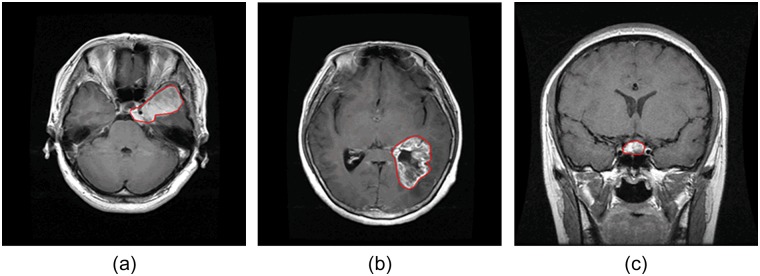
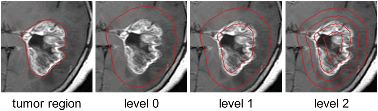
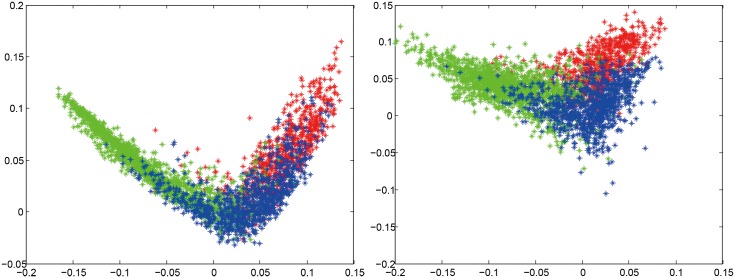
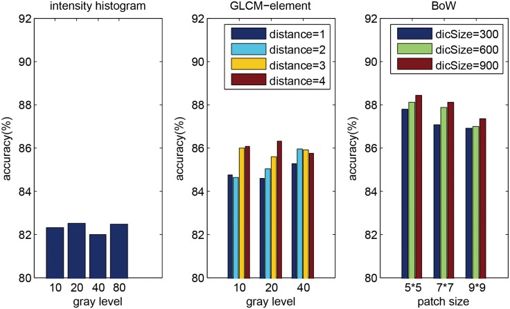
Automatic classification of tissue types of region of interest (ROI) plays an important role in computer-aided diagnosis. In the current study, we focus on the classification of three types of brain tumors (i.e., meningioma, glioma, and pituitary tumor) in T1-weighted contrast-enhanced MRI (CE-MRI) images. Spatial pyramid matching (SPM), which splits the image into increasingly fine rectangular subregions and computes histograms of local features from each subregion, exhibits excellent results for natural scene classification. However, this approach is not applicable for brain tumors, because of the great variations in tumor shape and size. In this paper, we propose a method to enhance the classification performance. First, the augmented tumor region via image dilation is used as the ROI instead of the original tumor region because tumor surrounding tissues can also offer important clues for tumor types. Second, the augmented tumor region is split into increasingly fine ring-form subregions. We evaluate the efficacy of the proposed method on a large dataset with three feature extraction methods, namely, intensity histogram, gray level co-occurrence matrix (GLCM), and bag-of-words (BoW) model. Compared with using tumor region as ROI, using augmented tumor region as ROI improves the accuracies to 82.31% from 71.39%, 84.75% from 78.18%, and 88.19% from 83.54% for intensity histogram, GLCM, and BoW model, respectively. In addition to region augmentation, ring-form partition can further improve the accuracies up to 87.54%, 89.72%, and 91.28%. These experimental results demonstrate that the proposed method is feasible and effective for the classification of brain tumors in T1-weighted CE-MRI.
Overview
The claims and statistics of this paper haven’t been checked yet.
Run the check — free, no account needed. It reads the open-access text and checks the sources it cites.
Integrity signals
Registry
Cites
Statistics
Figures
Verification
Claims the paper makes
The claims and statistics of this paper haven’t been checked yet.
Run the check — free, no account needed. It reads the open-access text and checks the sources it cites.
Claims other papers make about this one
Each is checked against this paper’s own text: does it say what the citing paper says it does?
In the citing paper
“Data collected from three different kinds of brain tumors consisted of 708 meningiomas, 1426 gliomas, and 930 pituitaries images from the General Hospital and Nanfang Hospital, Tianjin Medical University, China [72].”
From A Comprehensive Analysis of Recent Deep and Federated-Learning-Based Methodologies for Brain Tumor Diagnosis (2022) · cites this paper as [72]
In the citing paper
“This contains 3064 T1-weighted contrast-enhanced images collected from 233 patients with a resolution of 512 × 512 pixels.”
From A Comprehensive Analysis of Recent Deep and Federated-Learning-Based Methodologies for Brain Tumor Diagnosis (2022) · cites this paper as [72]
Self-check
Percentages, sums and reported differences re-computed from the paper’s own numbers.
The self-check needs the paper’s full text.
Citations
Cites
Distinctive Image Features from Scale-Invariant Keypoints
Lowe, D. G. (2004) · International Journal of Computer Vision · doi:10.1023/b:visi.0000029664.99615.94 · Cited by 55,589 · cited 1×
“(cid:129) Local features: The next step is to extract local features within the ROI, such as scale-invariant feature transform (SIFT) descriptor [20] and raw patch [21].”
Background$rm K$-SVD: An Algorithm for Designing Overcomplete Dictionaries for Sparse Representation
Aharon, M., Elad, M., & Bruckstein, A. M. (2006) · IEEE Transactions on Signal Processing · doi:10.1109/tsp.2006.881199 · Cited by 9,464
Robust Face Recognition via Sparse Representation
Wright, J. N., Yang, A., Ganesh, A., Sastry, S., & Ma, Y. (2009) · IEEE Transactions on Pattern Analysis and Machine Intelligence · doi:10.1109/tpami.2008.79 · Cited by 9,398 · cited 1×
“We also compared three classification methods (i.e. SVM, sparse representation-based classification (SRC) [31], and k nearest neighbors (kNN) classifier) using 2-region BoW representations.”
ResultBeyond Bags of Features: Spatial Pyramid Matching for Recognizing Natural Scene Categories
Lazebnik, S., Schmid, C., & Ponce, J. (2006) · 2006 IEEE Computer Society Conference on Computer Vision and Pattern Recognition - Volume 2 (CVPR'06) · doi:10.1109/cvpr.2006.68 · Cited by 7,916 · cited 4×
“Among those methods, the most notable work is SPM [15], which splits the image into hierarchical cells, computes BoW representation for each cell, and finally weights and concatenates the results.”
BackgroundMethodInfluentialVideo Google: a text retrieval approach to object matching in videos
Sivic, & Zisserman (2003) · Proceedings Ninth IEEE International Conference on Computer Vision · doi:10.1109/iccv.2003.1238663 · Cited by 6,367 · cited 1×
“The BoW method was originally used in the text retrieval domain, and it has been successfully adapted to the visual analysis domain [13,14].”
MethodRobust wide-baseline stereo from maximally stable extremal regions
Matas, J., Chum, O., Urban, M., & Pajdla, T. (2004) · Image and Vision Computing · doi:10.1016/j.imavis.2004.02.006 · Cited by 3,760 · cited 1×
“As pointed out in [29,30], it is beneficial to capture a certain amount of context.”
BackgroundA Bayesian Hierarchical Model for Learning Natural Scene Categories
Fei-Fei, L., & Perona, P. (2005) · IEEE Computer Society Conference on Computer Vision and Pattern Recognition/Proceedings - IEEE Computer Society Conference on Computer Vision and Pattern Recognition/Proceedings · doi:10.1109/cvpr.2005.16 · Cited by 3,665 · cited 1×
“The BoW method was originally used in the text retrieval domain, and it has been successfully adapted to the visual analysis domain [13,14].”
MethodLocality-constrained Linear Coding for image classification
Wang, J., Yang, J., Yu, K., Lv, F., Huang, T., & Gong, Y. (2010) · 2010 IEEE Computer Society Conference on Computer Vision and Pattern Recognition · doi:10.1109/cvpr.2010.5540018 · Cited by 3,100 · cited 2×
“Other more sophisticated methods may also be used, such as SC, soft assignment (SA) [24,25], orthogonal matching pursuit (OMP), locality-constrained linear coding [26], and local anchor embedding [27].”
MethodA comparison of affine region detectors
Mikolajczyk, K., Tuytelaars, T., Schmid, C., Zisserman, A., Matas, J., Schaffalitzky, F., Kadir, T., & Gool, L. V. (2005) · International Journal of Computer Vision · doi:10.1007/s11263-005-3848-x · Cited by 2,939
Linear spatial pyramid matching using sparse coding for image classification
Yang, J., Yu, K., Gong, Y., & Huang, T. S. (2009) · IEEE Computer Society Conference on Computer Vision and Pattern Recognition/Proceedings - IEEE Computer Society Conference on Computer Vision and Pattern Recognition/Proceedings · doi:10.1109/cvpr.2009.5206757 · Cited by 2,895
Research works printed but not linked: 1
- S Patil, 2013, A Computer Aided Diagnostic System For Classification Of Brain Tumors Using Texture Features And Probabilistic Neural Network, Int J Comput Sci Eng Inf Technol Res, 3, 61
Cited by
Sorted by citations
Items 201–300 of 879
Brain Tumor Classification With Inception Network Based Deep Learning Model Using Transfer Learning
Soumik, M. F. I., & Hossain, M. A. (2020) · IEEE Region 10 Symposium (TENSYMP) · doi:10.1109/tensymp50017.2020.9230618 · Cited by 39
“Cheng et al.[2] proposed an approach which relied upon manually delineated tumor borders to extract feature from the region of interests of T-1 MRI images where SVM model on bag of words(BOW) features produced best result and this is also regarded as one of the first works on figshare brain MRI image…”
BackgroundMethodSingh, P. (2021) · Applied Soft Computing · doi:10.1016/j.asoc.2021.107119 · Cited by 39
Identification of Tumor-Specific MRI Biomarkers Using Machine Learning (ML)
Hajjo, R., Sabbah, D. A., Bardaweel, S. K., & Tropsha, A. (2021) · Diagnostics · doi:10.3390/diagnostics11050742 · Cited by 38
Multiclass convolutional neural network based classification for the diagnosis of brain MRI images
Jaspin, K., & Selvan, S. (2022) · Biomedical Signal Processing and Control · doi:10.1016/j.bspc.2022.104542 · Cited by 40
Sailunaz, K., Beştepe, D., Alhajj, S., Özyer, T., Rokne, J. G., & Alhajj, R. (2023) · PLoS ONE · doi:10.1371/journal.pone.0284418 · Cited by 38
Asiri, A. A. M., Shaf, A., Ali, T. M., Aamir, M., Usman, A., Irfan, M., Alshamrani, H. A., Mehdar, K. M., Alshehri, O. M., & Alqhtani, S. M. (2022) · Intelligent Automation & Soft Computing · doi:10.32604/iasc.2023.032391 · Cited by 37
Çınar, N., Kaya, M., & Kaya, B. (2022) · International Journal of Imaging Systems and Technology · doi:10.1002/ima.22839 · Cited by 37
A Hybrid Deep CNN-SVM Approach for Brain Tumor Classification
Biswas, A., & Islam, M. S. (2023) · Journal of Information Systems Engineering and Business Intelligence · doi:10.20473/jisebi.9.1.1-15 · Cited by 37
“62 % [21] 2015 Augmentation+ Intensity histogram+ BoW+ SVM 91.”
BackgroundMethodBathla, G., Durjoy, D., Soni, N., Liu, Y., Larson, N. B., Kassmeyer, B. A., Mohan, S., Roberts-Wolfe, D., Rathore, S., Le, N. H., et al. (2023) · Journal of Neuroradiology · doi:10.1016/j.neurad.2023.08.007 · Cited by 39
Surdo, S., & Barillaro, G. (2024) · Small · doi:10.1002/smll.202400499 · Cited by 37
Zhu, M., Li, S., Kuang, Y., Hill, V. B., Heimberger, A. B., Zhai, L., & Zhai, S. (2022) · Frontiers in Oncology · doi:10.3389/fonc.2022.924245 · Cited by 45
Türk, Ö., Özhan, D., Acar, E., Akıncı, T. Ç., & Yılmaz, M. (2022) · Zeitschrift für Medizinische Physik · doi:10.1016/j.zemedi.2022.11.010 · Cited by 37
Onakpojeruo, E. P., Mustapha, M. T., Ozsahin, D. U., & Özşahin, İ. (2024) · Brain Communications · doi:10.1093/braincomms/fcae372 · Cited by 36
“Recent models frequently employ the Figshare benchmark brain tumour dataset 36 or the Brain Tumor Classification dataset 61 from the Kaggle open-source data repository for performance evaluation.”
MethodOnaizah, A. N., Xia, Y., & Hussain, K. (2024) · Alexandria Engineering Journal · doi:10.1016/j.aej.2024.11.063 · Cited by 40
Almalki, Y. E., Ali, M. U., Kallu, K. D., Masud, M., Zafar, A., Alduraibi, S. K., Irfan, M., Basha, M. A. A., Alshamrani, H. A., Alduraibi, A. K., et al. (2022) · Diagnostics · doi:10.3390/diagnostics12081793 · Cited by 41
A comparative study of brain tumor classification on unbalanced dataset using deep neural networks
Agrawal, T., Choudhary, P., Kumar, V., Singh, P., Diwakar, M., & Kumar, S. S. (2024) · Biomedical Signal Processing and Control · doi:10.1016/j.bspc.2024.106256 · Cited by 38
Comprehensive Review on MRI-Based Brain Tumor Segmentation: A Comparative Study from 2017 Onwards
Verma, A., Shivhare, S. N., Singh, S. P., Kumar, N., & Nayyar, A. (2024) · Archives of Computational Methods in Engineering · doi:10.1007/s11831-024-10128-0 · Cited by 35
Capsule Networks’ Interpretability for Brain Tumor Classification Via Radiomics Analyses
Afshar, P., Plataniotis, K. N., & Mohammadi, A. (2019) · Proceedings - International Conference on Image Processing · doi:10.1109/icip.2019.8803615 · Cited by 35
“To test the explainability properties of the Capsule network for brain tumor classification, we have adopted a data set [24], consisting of MRI images of 233 patients diagnosed with brain cancer.”
MethodAdvanced image generation for cancer using diffusion models
Kidder, B. L. (2024) · Biology Methods and Protocols · doi:10.1093/biomethods/bpae062 · Cited by 40
“We trained separate fine-tuned models on datasets of clinically curated medical images for various brain cancers—specifically gliomas, meningiomas, and pituitary tumors [45, 46], as cata-loged in the “Brain Tumor Image Dataset” from Kaggle (see Materials and methods section).”
MethodHybrid 3B Net and EfficientNetB2 Model for Multi-Class Brain Tumor Classification
Preetha, R., Priyadarsini, M. J. P., & Nisha, J. S. (2025) · IEEE Access · doi:10.1109/access.2025.3558411 · Cited by 34
“The methods evaluated include traditional machine learning approaches like SVM and KNN, as used by Cheng et al. [25], and more advanced techniques such as Genetic Algorithm-CNN by Anaraki et al. [47] and CNN-based models by Afshar et al. [48].”
Chandra, S. K., & Bajpai, M. K. (2020) · Biomedical Signal Processing and Control · doi:10.1016/j.bspc.2019.101841 · Cited by 34
Segmentation of Brain Tumors from MRI Images Using Convolutional Autoencoder
Atanasijević, M. B., & Barjaktarović, M. Č. (2021) · Applied Sciences · doi:10.3390/app11094317 · Cited by 34
“Automatic segmentation and classification of multiple changes in images, such as magnetic resonance imaging (MRI) and computer tomogra‐ phy, by computer vision methods, can offer valuable support to physicians as a second opinion [1,2].”
BackgroundMethodBashir-Gonbadi, F., & Khotanlou, H. (2021) · Multimedia Tools and Applications · doi:10.1007/s11042-021-10637-1 · Cited by 33
Badrigilan, S., Nabavi, S., Abin, A. A., Rostampour, N., Abedi, I., Shirvani, A., & Moghaddam, M. E. (2021) · International Journal of Computer Assisted Radiology and Surgery · doi:10.1007/s11548-021-02326-z · Cited by 33
A variate brain tumor segmentation, optimization, and recognition framework
Balaha, H. M., & Hassan, A. E.-S. (2022) · Artificial Intelligence Review · doi:10.1007/s10462-022-10337-8 · Cited by 35
Chaudhary, Q.-U.-A., Qureshi, S. A., Sadiq, T., Usman, A., Khawar, A., Shah, S. T. H., & Rehman, A. U. (2025) · Results in Engineering · doi:10.1016/j.rineng.2025.104025 · Cited by 36
Sobhaninia, Z., Karimi, N., Khadivi, P., & Samavi, S. (2023) · Biomedical Signal Processing and Control · doi:10.1016/j.bspc.2023.104834 · Cited by 40
MEEDNets: Medical Image Classification via Ensemble Bio-inspired Evolutionary DenseNets
Zhu, H., Wang, W., Ulidowski, I., Zhou, Q., Wang, S., Chen, H., & Zhang, Y. (2023) · Knowledge-Based Systems · doi:10.1016/j.knosys.2023.111035 · Cited by 32
Zia, R., Akhtar, P., & Aziz, A. (2017) · International Journal of Imaging Systems and Technology · doi:10.1002/ima.22266 · Cited by 31
“17,27 There are 2 types of studies that deals with the classification of brain neoplasms.”
BackgroundDeepak, S., & Ameer, P. M. (2021) · International Journal of Imaging Systems and Technology · doi:10.1002/ima.22543 · Cited by 34
“30 Recent works used deep transfer learning to address the same research problem and the results improved significantly.”
BackgroundDeep Transfer Learning Based Multi-Class Brain Tumors Classification Using MRI Images
Mondal, M., Faruk, M. F., Raihan, N., & Ahammed, P. (2021) · International Conference on Electrical & Electronic Engineering (ICEEE) · doi:10.1109/iceee54059.2021.9719003 · Cited by 31
Block-Wise Neural Network for Brain Tumor Identification in Magnetic Resonance Images
Asiri, A. A. M., Aamir, M., Shaf, A., Ali, T. M., Zeeshan, M., Irfan, M., Alshamrani, K. A., Alshamrani, H. A., Alqahtani, F. F., & Alshehri, A. H. D. (2022) · Computers, materials & continua/Computers, materials & continua (Print) · doi:10.32604/cmc.2022.031747 · Cited by 30
Karacı, A., & Akyol, K. (2023) · Neural Computing and Applications · doi:10.1007/s00521-023-08395-2 · Cited by 30
“These images were taken in three different planes such as sagittal (1025 images), axial (994 images), and coronal plane (1045 images) [22, 40].”
BackgroundMethodDeep Learning-based MRI Analysis for Brain Tumor Detection: Insights from ResNet and DenseNet Models
Padmakala, S., & Maheswari, S. U. (2024) · International Conference on Electronics, Communication and Aerospace Technology (ICECA) · doi:10.1109/iceca63461.2024.10800894 · Cited by 29
Koong, K., Preda, V. A., Jian, A., Liquet, B., & Ieva, A. D. (2021) · Neuroradiology · doi:10.1007/s00234-021-02845-1 · Cited by 27
“The most important factor affecting the progression of this field is the need for multiple public datasets with a large cohort reflective of the general population like Figshare dataset, but larger and more international [30].”
Multilayer Stacked Probabilistic Belief Network-Based Brain Tumor Segmentation and Classification
Raghavendra, S., Harshavardhan, A., Neelakandan, S., Partheepan, R., Walia, R., & Rao, V. C. S. (2022) · International Journal of Foundations of Computer Science · doi:10.1142/s0129054122420047 · Cited by 29
Brain Tumor Detection and Classification Using PSO and Convolutional Neural Network
Ali, M. J., Shah, J. H., Khan, M. A., Alhaisoni, M. M., Tariq, U., Akram, T., Kim, Y. J., & Chang, B. (2022) · Computers, materials & continua/Computers, materials & continua (Print) · doi:10.32604/cmc.2022.030392 · Cited by 29
Comparative evaluation for detection of brain tumor using machine learning algorithms
Kareem, S. W., Abdulrahman, B. F., Hawezi, R. S., Khoshaba, F. S., Askar, S. K., Muheden, K., & Abdulkhaleq, I. S. (2022) · IAES International Journal of Artificial Intelligence · doi:10.11591/ijai.v12.i1.pp469-477 · Cited by 29
Sánchez-Moreno, L., Pérez-Peña, A. M., Durán-López, L., & Dominguez-Morales, J. P. (2025) · Computers in Biology and Medicine · doi:10.1016/j.compbiomed.2025.110555 · Cited by 33
Rui, W., Wu, Y., Ma, Z., Wang, Y., Wang, Y., Xu, X., Zhang, J., & Yao, Z. (2018) · European Journal of Radiology · doi:10.1016/j.ejrad.2018.12.002 · Cited by 28
IoT framework for brain tumor detection based on optimized modified ResNet 18 (OMRES)
El‐Feshawy, S. A., Saad, W., Shokair, M., & Dessouky, M. I. (2022) · The Journal of Supercomputing · doi:10.1007/s11227-022-04678-y · Cited by 28
Verma, A., & Singh, V. P. (2022) · Multimedia Tools and Applications · doi:10.1007/s11042-022-13545-0 · Cited by 28
“[6] authors extracted BoW features to attain 91.”
MethodA CNN-Based Strategy to Classify MRI-Based Brain Tumors Using Deep Convolutional Network
Reza, A. W., Hossain, M. S., Wardiful, M. A., Farzana, M., Ahmad, S., Alam, F., Nandi, R. N., & Siddique, N. H. (2022) · Applied Sciences · doi:10.3390/app13010312 · Cited by 30
A robust approach for multi-type classification of brain tumor using deep feature fusion
Chen, W., Tan, X., Zhang, J., Du, G., Fu, Q., & Jiang, H. (2024) · Frontiers in Neuroscience · doi:10.3389/fnins.2024.1288274 · Cited by 29
Özdemir, C., & Doğan, Y. (2024) · Medical & Biological Engineering & Computing · doi:10.1007/s11517-024-03064-5 · Cited by 27
Deep Neural Networks with Transfer Learning Model for Brain Tumors Classification
Bulla, P., Anantha, N. L., & Peram, S. R. (2020) · Traitement du signal · doi:10.18280/ts.370407 · Cited by 27
“Cheng et al. [12] implemented tumor classification using augmented tumor region of interest.”
BackgroundMethodBrain tumor magnetic resonance images classification based machine learning paradigms
Pattnaik, B. B., Anitha, K., Rathore, S., Biswas, P., Sethy, P. K., & Behera, S. K. (2022) · Współczesna Onkologia · doi:10.5114/wo.2023.124612 · Cited by 27
GT-Net: global transformer network for multiclass brain tumor classification using MR images
Dutta, T. K., Nayak, D. R., & Pachori, R. B. (2024) · Biomedical Engineering Letters · doi:10.1007/s13534-024-00393-0 · Cited by 26
“We validate our approach on an openly available brain tumor dataset comprises of 3064 T1-weighted contrast-enhanced MR images from three classes [4, 25].”
Improved brain tumor classification through DenseNet121 based transfer learning
Rasheed, M., Jaffar, M. A., Akram, A., Rashid, J., Alshalali, T. A. N., Irshad, A., & Sarwar, N. (2025) · Discover Oncology · doi:10.1007/s12672-025-03501-3 · Cited by 26
“Using a small dataset, Sarah Zuhair Kurdi et al. achieved 98% accuracy in binary brain tumor classification using CNN. Prasun Chandra Tripathi et al. developed a model based on deep residual networks that accurately detected two kinds of gliomas 95.67% of the time [32].”
InfluentialDilated Capsule Network for Brain Tumor Type Classification Via MRI Segmented Tumor Region
Adu, K., Yu, Y., Cai, J., & Tashi, N. (2019) · IEEE International Conference on Robotics and Biomimetics (ROBIO) · doi:10.1109/robio49542.2019.8961610 · Cited by 26
A survey on machine learning based brain retrieval algorithms in medical image analysis
Sharma, A. K., Nandal, A., Dhaka, A., & Dixit, R. (2020) · Health and Technology · doi:10.1007/s12553-020-00471-0 · Cited by 27
“…overall precision of 97% and 98% respectively Complex Structure Bag of Words (BOW), Gray Level Co-occurrence Matrix (GLCM) and intensity histogram [24] It enhances the performance of brain tumor classification by using image dilation and afterwards by separating into sub regions Low accuracy of…”
BackgroundMethodA survey on artificial intelligence techniques for chronic diseases: open issues and challenges
Patel, K., Mistry, C., Mehta, D., Thakker, U., Tanwar, S., Gupta, R., & Kumar, N. (2021) · Artificial Intelligence Review · doi:10.1007/s10462-021-10084-2 · Cited by 30
Qin, C., Li, B., & Han, B. (2023) · Engineering Applications of Artificial Intelligence · doi:10.1016/j.engappai.2022.105816 · Cited by 26
Ensemble Deep Learning Technique for Detecting MRI Brain Tumor
Jader, R. F., Kareem, S. W., & Awla, H. Q. (2024) · Applied Computational Intelligence and Soft Computing · doi:10.1155/2024/6615468 · Cited by 25
BrainNet: A Deep Learning Approach for Brain Tumor Classification
Singh, T., Nair, R. R., Babu, T., Wagh, A., Bhosalea, A., & Duraisamy, P. (2024) · Procedia Computer Science · doi:10.1016/j.procs.2024.04.310 · Cited by 24
Khaniki, M. A. L., Mirzaeibonehkhater, M., Manthouri, M., & Hasani, E. (2024) · Iran Journal of Computer Science · doi:10.1007/s42044-024-00220-w · Cited by 42
“The dataset is evenly distributed, comprising 1532 images labeled as ‘Tumor’ and 1532 labeled as ‘Non-tumor’ [17].”
A Weighted Voting Classifiers Ensemble for the Brain Tumors Classification in MR Images
Rezaei, K., Agahi, H., & Mahmoodzadeh, A. (2020) · IETE Journal of Research · doi:10.1080/03772063.2020.1780487 · Cited by 24
“Cheng et al. [52] implemented an algorithm based on three feature extraction methods, i.e. intensity histogram, GLCM, and BOW.”
MethodSwaraja, K., Meenakshi, K., Valiveti, H. B., & Karuna, G. (2022) · Multimedia Tools and Applications · doi:10.1007/s11042-022-12414-0 · Cited by 24
“[37] trained CNNs (composed of two convolutional and two fully linked layers) with four layers from scratch and attained accuracy of 91.43% in classification, but the image-based accuracy is 90.26%, which is poorer than [10], i.e. 90.28%.”
BackgroundMethodResultInfluentialBrain Tumor Classification Using MRI Images and Convolutional Neural Networks
Hafeez, M. A., Kayasandık, C. B., & Dogan, M. Y. (2022) · Signal Processing and Communications Applications Conference (SIU) · doi:10.1109/siu55565.2022.9864962 · Cited by 24
“In [12], the authors have used the same dataset as in [11] and classified three types of brain tumors using kernel extreme learning machines and CNNs.”
MethodInfluentialOmana, S. P., Dar, J. A., Kumar, T. R., Sampath, A. K., & Sharma, S. K. (2022) · Concurrency and Computation Practice and Experience · doi:10.1002/cpe.7541 · Cited by 26
“29 The severity level and identification of glioma cancer from multimodal MRI is a difficult job of multilevel categorization process,whichneedsabetterlearningmodelwithoutcomputationaldifficulties.”
BackgroundSaha, P., Das, R., & Das, S. K. (2023) · Multimedia Tools and Applications · doi:10.1007/s11042-023-15377-y · Cited by 26
“Cheng et al. [6] processed the dataset for the first time to develop a classification system for brain cancer types.”
BackgroundAbdalla, P. A., Mohammed, B. A., & Saeed, A. M. (2023) · Journal of Electrical Systems and Information Technology · doi:10.1186/s43067-023-00119-9 · Cited by 24
Multi-Class Brain Lesion Classification Using Deep Transfer Learning With MobileNetV3
Majeed, A. F., Salehpour, P., Farzinvash, L., & Pashazadeh, S. (2024) · IEEE Access · doi:10.1109/access.2024.3413008 · Cited by 23
“Other methods employed features such as gray level co-occurrence matrix (GLCM), intensity histogram, and bag-of-words (BoW) [22], incorporating various attributes like intensity, local neighborhood, intensity differences, and wavelet texture, with classification performed using a random forest…”
BackgroundMethodAutomatic cell counting from stimulated Raman imaging using deep learning
Zhang, Q., Yun, K. K., Wang, H., Yoon, S. W., Lu, F., & Won, D. (2021) · PLoS ONE · doi:10.1371/journal.pone.0254586 · Cited by 23
Multi-Classification of Brain Tumors via Feature Level Ensemble of Convolutional Neural Networks
Aurna, N. F., Yousuf, M. A., & Taher, K. A. (2021) · International Conference on Sustainable Technologies for Industry 5.0 (STI) · doi:10.1109/sti53101.2021.9732543 · Cited by 23
Explainable Convolutional Neural Networks for Brain Cancer Detection and Localisation
Mercaldo, F., Brunese, L., Martinelli, F., Santone, A., & Cesarelli, M. (2023) · Sensors · doi:10.3390/s23177614 · Cited by 23
“[46] consider intensity histogram and gray level co-occurrence matrix features for brain cancer grade detection, reaching an accuracy of 0.”
BackgroundÖksüz, C., Urhan, O., & Güllü, M. K. (2023) · Neural Computing and Applications · doi:10.1007/s00521-023-09164-x · Cited by 23
“[90] proposed a brain tumor classification scheme using hand-crafted features, and SVM classification.”
A mask R-CNN approach for detection and classification of brain tumours from MR images
Kordemir, M., Çevik, K. K., & Bozkurt, A. (2024) · Computer Methods in Biomechanics and Biomedical Engineering Imaging & Visualization · doi:10.1080/21681163.2023.2301391 · Cited by 24
“Masood et al. (2020) applied the Mask R-CNN model (He et al. 2017) to localise and segment brain tumours (tumour or non-tumour regions) from MRI images obtained from two online repositories (Cheng et al. 2015; Chakrabarty 2019).”
BackgroundMethodInfluentialAMIAC: adaptive medical image analyzes and classification, a robust self-learning framework
Iqbal, S., Qureshi, A. N., Aurangzeb, K., Alhussein, M. A., Haider, S. I., & Rida, I. (2023) · Neural Computing and Applications · doi:10.1007/s00521-023-09209-1 · Cited by 22
Aziz, N., Minallah, N., Frnda, J., Sher, M., Zeeshan, M., & Haroon, A. (2024) · PLoS ONE · doi:10.1371/journal.pone.0307825 · Cited by 22
Knowledge-Informed Machine Learning for Cancer Diagnosis and Prognosis: A Review
Mao, L., Wang, H., Hu, L., Tran, N. L., Canoll, P., Swanson, K. R., & Li, J. (2024) · IEEE Transactions on Automation Science and Engineering · doi:10.1109/tase.2024.3515839 · Cited by 25
“Graphical models such as Graphical Convolutional Networks (GCN) can seamlessly take graphs formed from data and from knowledge as input and combine them for prediction [48], [124], [125], [126].”
Classification of Thyroid Carcinoma in Whole Slide Images Using Cascaded CNN
El-Hossiny, A. S., Al‐Atabany, W., Hassan, O., Soliman, A. M., & Sami, S. A. (2021) · IEEE Access · doi:10.1109/access.2021.3076158 · Cited by 22
“Since our generated database and the proposed CNN were not previously tested, it is suggested to start to test the proposed CNN on ‘‘Brain Tumor Dataset v5’’ which contains 3064 T1-weighted contrast-enhanced images with three kinds of a brain tumor [27].”
MethodBrain Tumor Classification based on MR Images using GAN as a Pre-Trained Model
Yerukalareddy, D. R., & Pavlovskiy, E. (2021) · IEEE Ural-Siberian Conference on Computational Technologies in Cognitive Science, Genomics and Biomedicine (CSGB) · doi:10.1109/csgb53040.2021.9496036 · Cited by 22
Deep learning and spark architecture based intelligent brain tumor MRI image severity classification
Abirami, S., & Venkatesan, G. K. D. P. (2022) · Biomedical Signal Processing and Control · doi:10.1016/j.bspc.2022.103644 · Cited by 24
Advances in Computer-Aided Medical Image Processing
Cui, H., Hu, L. Q., & Chi, L. (2023) · Applied Sciences · doi:10.3390/app13127079 · Cited by 28
Gupta, S., Saxena, R., Bansal, A., Saluja, K., Vajpayee, A., & Shikha (2023) · AIP conference proceedings · doi:10.1063/5.0154417 · Cited by 22
Ghosh, A., Soni, B., & Baruah, U. (2023) · Arabian Journal for Science and Engineering · doi:10.1007/s13369-023-08607-w · Cited by 27
Hybrid Parallel Fuzzy CNN Paradigm: Unmasking Intricacies for Accurate Brain MRI Insights
Iqbal, S., Qureshi, A. N., Aurangzeb, K., Alhussein, M. A., Wang, S., Anwar, M. S., & Khan, F. N. (2024) · IEEE Transactions on Fuzzy Systems · doi:10.1109/tfuzz.2024.3372608 · Cited by 24
NeuroSight : A Deep‐Learning Integrated Efficient Approach to Brain Tumor Detection
Mugdha, S. B. S., & Uddin, M. (2025) · Engineering Reports · doi:10.1002/eng2.13100 · Cited by 22
Non-invasively Grading of Brain Tumor Through Noise Robust Textural and Intensity Based Features
Tripathi, P. C., & Bag, S. (2019) · Advances in intelligent systems and computing · doi:10.1007/978-981-13-9042-5_45 · Cited by 22
Risk Susceptibility of Brain Tumor Classification to Adversarial Attacks
Kotia, J., Kotwal, A., & Bharti, R. (2019) · Advances in intelligent systems and computing · doi:10.1007/978-3-030-31964-9_17 · Cited by 22
MRI based Glioma segmentation using Deep Learning algorithms
Kaldera, H. N. T. K., Gunasekara, S. R., & Dissanayake, M. B. (2019) · International Research Conference on Smart Computing and Systems Engineering (SCSE) · doi:10.23919/scse.2019.8842668 · Cited by 22
A Review on Brain Tumor Classification Methodologies
Panda, B. N., & Panda, C. S. (2019) · International Journal of Scientific Research in Science and Technology · doi:10.32628/ijsrst20717 · Cited by 22
Verma, A., & Yadav, A. K. (2025) · Brain Research · doi:10.1016/j.brainres.2025.149507 · Cited by 21
Real-time brain tumour diagnoses using a novel lightweight deep learning model
Alnageeb, M. H. O., & Supriya, M. H. (2025) · Computers in Biology and Medicine · doi:10.1016/j.compbiomed.2025.110242 · Cited by 21
MAG-Net: Multi-task Attention Guided Network for Brain Tumor Segmentation and Classification
Gupta, S., Punn, N. S., Sonbhadra, S. K., & Agarwal, S. (2021) · Lecture notes in computer science · doi:10.1007/978-3-030-93620-4_1 · Cited by 21
“[7], authors also achieved promising results on the limited data samples with an overall accuracy of.”
BackgroundA framework for brain tumor detection based on segmentation and features fusion using MRI images
Mostafa, A. M., El-Meligy, M. A., Alkhayyal, M. A., Alnuaim, A. A., & Sharaf, M. A. F. (2023) · Brain Research · doi:10.1016/j.brainres.2023.148300 · Cited by 21
Gull, S., Akbar, S., & Naqi, S. M. (2023) · International Journal of Imaging Systems and Technology · doi:10.1002/ima.22897 · Cited by 21
“Data augmentation techniques are used to increase the satisfactory amount of data synthetically and avoid overfitting issues from the original data by creating geometric and color distortion during model training.(76,77) Overfitting occurs when a model successfully assigns labels to known samples but fails to recogniz…”
MethodBodapati, J. D., & Balaji, B. B. (2023) · Multimedia Tools and Applications · doi:10.1007/s11042-023-15557-w · Cited by 21
MRI-based brain tumor ensemble classification using two stage score level fusion and CNN models
Bouguerra, O., Attallah, B., & Brik, Y. (2024) · Egyptian Informatics Journal · doi:10.1016/j.eij.2024.100565 · Cited by 21
Dandıl, E., & Biçer, A. (2020) · IET Image Processing · doi:10.1049/iet-ipr.2019.1416 · Cited by 21
Boosting the Prediction of Brain Tumor Using Two Stage BiGait Architecture
Khan, S. U. R., Khan, Z., Hossain, M. Z., Mayumu, N., Yasmin, F., & Aziz, Y. (2024) · International Conference on Digital Image Computing: Techniques and Applications (DICTA) · doi:10.1109/dicta63115.2024.00067 · Cited by 20
“To begin with, significant variations exist in the morphology, dimensions, and severity of brain tumor [7].”
Mohsen, S., Oraby, S., & Abdel-Aziz, M. H. (2025) · Archives of Computational Methods in Engineering · doi:10.1007/s11831-025-10416-3 · Cited by 26
Deep medical image analysis with representation learning and neuromorphic computing
Getty, N., Brettin, T., Jin, D., Stevens, R. L., & Xia, F. (2020) · Interface Focus · doi:10.1098/rsfs.2019.0122 · Cited by 20
“We performed our analysis on a public dataset from a previous study [14].”
MethodThe influence of the activation function in a capsule network for brain tumor type classification
Adu, K., Yu, Y., Cai, J., Asare, I., & Quahin, J. (2021) · International Journal of Imaging Systems and Technology · doi:10.1002/ima.22638 · Cited by 25
Xiao, G., Wang, H., Shen, J., Chen, Z., Zhang, Z., & Ge, X. (2021) · Micromachines · doi:10.3390/mi13010015 · Cited by 20
A Two-Tier Framework Based on GoogLeNet and YOLOv3 Models for Tumor Detection in MRI
Ali, F., Khan, S. N., Abbas, A. W., Shah, B., Hussain, T., Song, D., EI-Sappagh, S., & Singh, J. (2022) · Computers, materials & continua/Computers, materials & continua (Print) · doi:10.32604/cmc.2022.024103 · Cited by 21
Brain Tumor Segmentation Based on Minimum Spanning Tree
Mayala, S., Herdlevær, I., Haugsøen, J. B., Anandan, S., Gavasso, S., & Brun, M. (2022) · Frontiers in Signal Processing · doi:10.3389/frsip.2022.816186 · Cited by 20
“The images are classified into three types of brain tumors, labeled as follows: 1) meningioma, 2) glioma, and 3) pituitary tumors (Cheng et al., 2015; Cheng et al., 2016).”
Improving Brain Tumor Classification with Deep Learning Using燬ynthetic燚ata
Yapıcı, M. M., Karakış, R., & Gürkahraman, K. (2022) · Computers, materials & continua/Computers, materials & continua (Print) · doi:10.32604/cmc.2023.035584 · Cited by 20
Metadata
- Authors
- Cheng, J., Huang, W., Cao, S., Yang, R., Yang, W., Yun, Z., Wang, Z., & Feng, Q.
- Affiliations
- Southern Medical University (CN)
- Nanfang Hospital (CN)
- Corresponding author
- Feng, Qianjin
- Venue
- PLOS ONE
- Publisher
- Public Library of Science (PLoS)
- Published
- 2015-10-08
- Volume
- 10
- Issue
- 10
- Pages
- e0140381
- ISSN
- 1932-6203 (ISSN-L)
- PMC
- PMC4598126
- OpenAlex
- W2182098131
- Cited by
- 879
- References
- 36
- Citations by year
- 2026: 110 · 2025: 134 · 2024: 142 · 2023: 152 · 2022: 128 · 2021: 85 · 2020: 63 · 2019: 44 · 2018: 14 · 2017: 4 · 2016: 2 · 2015: 1
- FWCI
- 3.74
- Type
- journal article
- Language
- en
- Update notices
- correction (2015-12-02)
- Open access
- gold
- doi.org
- Licence
- Locations
- PLoS ONE (publishedVersion, cc-by)
- Europe PMC (PubMed Central) (publishedVersion, cc-by)
- PubMed Central (publishedVersion, cc-by)
- PubMed (publishedVersion)
- DOAJ (DOAJ: Directory of Open Access Journals) (submittedVersion)
- Funders
- National Natural Science Foundation of China (81101109, 31371009, 2012BAI14B02, 863 Program)
- National High-tech Research and Development Program
- Topics
- Physical Sciences › Computer Science › Computer Vision and Pattern Recognition › Brain Tumor Detection and Classification
- Health Sciences › Medicine › Radiology, Nuclear Medicine and Imaging › Radiomics and Machine Learning in Medical Imaging
- Physical Sciences › Computer Science › Artificial Intelligence › AI in cancer detection
- Keywords
- brain tumor classification, meningioma, glioma, pituitary tumors, region of interest, image dilation, spatial pyramid matching, contrast-enhanced T1-weighted imaging, computer-aided diagnosis, gray level co-occurrence matrix, bag-of-words model, intensity histogram
- MeSH terms
- Brain Neoplasms, Glioma, Humans, Image Interpretation, Computer-Assisted, Magnetic Resonance Imaging, Meningioma, Sensitivity and Specificity
About these checks
Registry, retraction, Cites and statistics results are computed, not judged, with one exception: a printed reference that no index lists is read once by a model before it counts as a citation error. Claims are extracted by a model and kept only when their quote appears verbatim in the text; each cited source is then checked, with a model’s help, for whether it says what the claim relies on. Those verdicts can be wrong — read the claim’s quote and the cited source before relying on one.
Statistics and arithmetic are recomputed, not judged.
Confidence bands are shown only for fields calibrated on our annotated test set. None is calibrated yet, so no band is shown.
What counts as a claim ›How we measure confidence ›How we check this ›
Are you an author, or do you see a problem with this page? Request a review
