

The DOI matches its Crossref record; 84 of the 84 references it cites were found in a registry or an index; none of the 46 sources its claims cite is checked for support yet.
A Sterol-Regulatory Element Binding Protein Is Required for Cell Polarity, Hypoxia Adaptation, Azole Drug Resistance, and Virulence in Aspergillus fumigatus
Open accessAbstract
At the site of microbial infections, the significant influx of immune effector cells and the necrosis of tissue by the invading pathogen generate hypoxic microenvironments in which both the pathogen and host cells must survive. Currently, whether hypoxia adaptation is an important virulence attribute of opportunistic pathogenic molds is unknown. Here we report the characterization of a sterol-regulatory element binding protein, SrbA, in the opportunistic pathogenic mold, Aspergillus fumigatus. Loss of SrbA results in a mutant strain of the fungus that is incapable of growth in a hypoxic environment and consequently incapable of causing disease in two distinct murine models of invasive pulmonary aspergillosis (IPA). Transcriptional profiling revealed 87 genes that are affected by loss of SrbA function. Annotation of these genes implicated SrbA in maintaining sterol biosynthesis and hyphal morphology. Further examination of the SrbA null mutant consequently revealed that SrbA plays a critical role in ergosterol biosynthesis, resistance to the azole class of antifungal drugs, and in maintenance of cell polarity in A. fumigatus. Significantly, the SrbA null mutant was highly susceptible to fluconazole and voriconazole. Thus, these findings present a new function of SREBP proteins in filamentous fungi, and demonstrate for the first time that hypoxia adaptation is likely an important virulence attribute of pathogenic molds.
Overview
AI snapshot
Written from the paper’s full text.
TL;DR
The paper identifies SrbA, an SREBP homolog in *Aspergillus fumigatus*, as required for hypoxic growth, azole resistance, cell polarity, sterol biosynthesis, and virulence. Loss of SrbA renders the fungus avirulent in two murine models of invasive pulmonary aspergillosis and highly susceptible to fluconazole and voriconazole.
Key findings
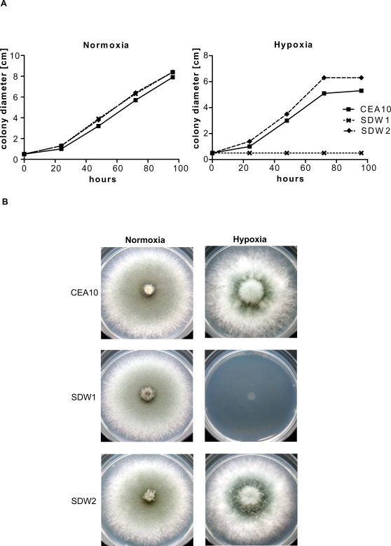
- The ΔsrbA mutant SDW1 showed no detectable hyphal growth under hypoxia (1% O₂, 5% CO₂, 94% N₂), whereas wild-type CEA10 and reconstituted SDW2 grew normally; under normoxia, growth rates were comparable (P>0.01).
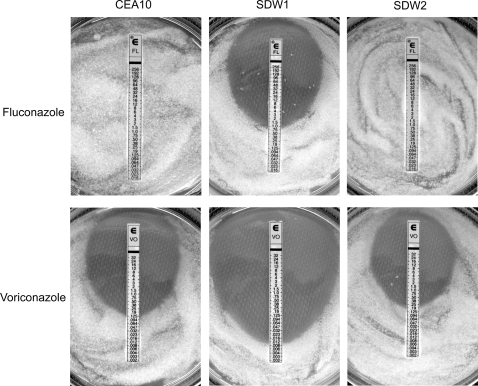
- SDW1 was highly susceptible to fluconazole (MIC 1 µg/ml) and voriconazole (MIC 0.012 µg/ml), while CEA10 and SDW2 were fluconazole-resistant and had voriconazole MICs of 0.125 µg/ml; all strains had equivalent MICs to amphotericin B (0.25 µg/ml) and caspofungin (0.125 µg/ml).
- Microarray profiling in hypoxia identified 87 SrbA-affected genes, including *srbA* itself (38.59-fold), NRPS1 (23.36-fold), ERG25 (15.79-fold), ERG3 (13.57-fold), and ERG24 (11.98-fold) higher in wild type than mutant. GC-MS showed a C-4 methylated sterol-to-ergosterol ratio of 1.94 in SDW1 versus no C-4 methylated sterol accumulation in wild type, suggesting an ERG25 block.
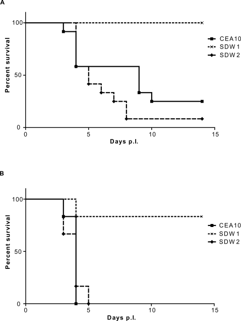
- SDW1 was avirulent in neutropenic CD1 mice (P=0.0002) and significantly attenuated in gp91phox−/− X-CGD mice (P=0.0054/0.005); no differences were found in oxidative stress resistance or RAW264.7 macrophage killing (P>0.01).
Methods
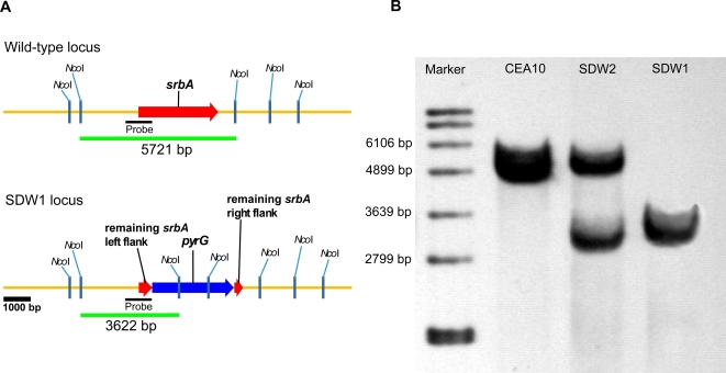
- A *srbA* null mutant (SDW1) was generated in *A. fumigatus* CEA17 by replacing an ~2.2-kb internal *srbA* coding fragment with *A. parasiticus pyrG*; a reconstituted strain (SDW2) was created, and strains were confirmed by Southern blot and PCR.
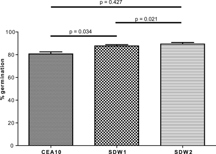
- Phenotypic assays included normoxic/hypoxic growth, microarray transcriptional profiling after 24 h hypoxia, E-test antifungal susceptibility, GC-MS sterol analysis, transmission electron microscopy, conidial germination, H₂O₂ sensitivity, and RAW264.7 macrophage killing.
- Virulence was tested in two murine IPA models: outbred CD1 neutropenic mice (n=12 per strain) and gp91phox−/− X-CGD mice (n=6 per strain), inoculated with 10⁶ conidia; survival was analyzed by log-rank tests, with microarray analysis by SAM using a 2-fold cutoff and zero false discovery rate.
Limitations
- The paper states that the mechanisms behind several phenotypes remain undefined, including how SrbA mediates hypoxia adaptation, azole resistance, and cell polarity; it also notes that no clear SCAP homolog was identified, that S1P/S2P protease mutants did not show expected hypoxic defects (unpublished data), and that SrbA regulation/activation was not elucidated.
- Evident from the text: transcriptional profiling was limited to one hypoxic condition and time point (24 h), so many of the 87 genes may be indirect effects; oxygen tension was not directly measured in infected tissue; and some supporting data are described as unpublished or not shown.
Model-written summary. Check it against the paper before relying on it.
Integrity signals
Registry
Cites
Statistics
Figures
Verification
Claims the paper makes
75 Own result · 55 Own interpretation · 29 Not checked yet · 14 Own method · 2 No basis given
None of the sources cited for this claim have been checked yet.
In the paper
“Interestingly, besides srbA itself, the gene with the highest fold difference in expression in SDW1 is a non-ribosomal peptide synthetase, AFUA_1g10380 (NRPS1 or pes1) [43],[44].”
§ A Sterol-Regulatory Element Binding Protein Is Required for Cell Polarity, Hypoxia Adaptation, Azole Drug Resistance, and Virulence in Aspergillus fumigatus > Results > Transcriptional profiling of SDW1 in response to hypoxia
What the sources say
- Not checked yetcites [43] RA Cramer Jr, 2006, Phylogenomic analysis of non-ribosomal peptide synthetases in the genus Aspergillus., Gene source
- Not checked yetcites [44] EP Reeves, 2006, A nonribosomal peptide synthetase (Pes1) confers protection against oxidative stress in Aspergillus fumigatus., Febs J source
plain- Asserted: passes — The authors put it forward as true.
- Checkable: passes — It can be checked against evidence.
- Load-bearing: unclear — It is unclear whether the paper’s conclusions depend on it.
- Based: passes — The paper gives a reason why it holds.
A result the paper reports from its own data — it cites no other source for it, so there is none to check. The quote shows it is in the paper.
In the paper
“However, no obvious defects in cell wall biosynthesis were observed in the mutant strain”
§ A Sterol-Regulatory Element Binding Protein Is Required for Cell Polarity, Hypoxia Adaptation, Azole Drug Resistance, and Virulence in Aspergillus fumigatus > Results > Transcriptional profiling of SDW1 in response to hypoxia
plain- Asserted: passes — The authors put it forward as true.
- Checkable: passes — It can be checked against evidence.
- Load-bearing: unclear — It is unclear whether the paper’s conclusions depend on it.
- Based: passes — The paper gives a reason why it holds.
A result the paper reports from its own data — it cites no other source for it, so there is none to check. The quote shows it is in the paper.
In the paper
“Several genes previously shown to be involved in ergosterol biosynthesis in fungi were found to be transcriptionally repressed in the absence of SrbA including ERG25, ERG24, and ERG3 (Table 1).”
§ A Sterol-Regulatory Element Binding Protein Is Required for Cell Polarity, Hypoxia Adaptation, Azole Drug Resistance, and Virulence in Aspergillus fumigatus > Results > Transcriptional profiling of SDW1 in response to hypoxia
plaindirect- Asserted: passes — The authors put it forward as true.
- Checkable: passes — It can be checked against evidence.
- Load-bearing: passes — The paper’s conclusions depend on it.
- Based: passes — The paper gives a reason why it holds.
A result the paper reports from its own data — it cites no other source for it, so there is none to check. The quote shows it is in the paper.
In the paper
“After 96 hours of incubation in hypoxia, SDW1 continued to display undetectable growth.”
§ A Sterol-Regulatory Element Binding Protein Is Required for Cell Polarity, Hypoxia Adaptation, Azole Drug Resistance, and Virulence in Aspergillus fumigatus > Results > SrbA is required for hypoxia adaptation in Aspergillus fumigatus
plain- Asserted: passes — The authors put it forward as true.
- Checkable: passes — It can be checked against evidence.
- Load-bearing: passes — The paper’s conclusions depend on it.
- Based: passes — The paper gives a reason why it holds.
A result the paper reports from its own data — it cites no other source for it, so there is none to check. The quote shows it is in the paper.
In the paper
“In addition, a significant number of genes involved in cell wall biosynthesis or homeostasis were observed to be repressed in SDW1 compared to wild type.”
§ A Sterol-Regulatory Element Binding Protein Is Required for Cell Polarity, Hypoxia Adaptation, Azole Drug Resistance, and Virulence in Aspergillus fumigatus > Results > Transcriptional profiling of SDW1 in response to hypoxia
plaindirect- Asserted: passes — The authors put it forward as true.
- Checkable: passes — It can be checked against evidence.
- Load-bearing: unclear — It is unclear whether the paper’s conclusions depend on it.
- Based: passes — The paper gives a reason why it holds.
A result the paper reports from its own data — it cites no other source for it, so there is none to check. The quote shows it is in the paper.
In the paper
“SDW1 and SDW2 both displayed normal hyphal growth rates compared to the wild type strain CEA10 in normoxic conditions on glucose minimal medium (GMM) (Figure 2A) (P>0.01).”
§ A Sterol-Regulatory Element Binding Protein Is Required for Cell Polarity, Hypoxia Adaptation, Azole Drug Resistance, and Virulence in Aspergillus fumigatus > Results > SrbA is required for hypoxia adaptation in Aspergillus fumigatus
plain- Asserted: passes — The authors put it forward as true.
- Checkable: passes — It can be checked against evidence.
- Load-bearing: passes — The paper’s conclusions depend on it.
- Based: passes — The paper gives a reason why it holds.
The paper states this without a source or data behind it here, so there is nothing to check it against. The quote shows it is in the paper.
In the paper
“consequently incapable of causing disease in two distinct murine models of invasive pulmonary aspergillosis (IPA)”
§ A Sterol-Regulatory Element Binding Protein Is Required for Cell Polarity, Hypoxia Adaptation, Azole Drug Resistance, and Virulence in Aspergillus fumigatus > Abstract
plaindirect- Asserted: passes — The authors put it forward as true.
- Checkable: passes — It can be checked against evidence.
- Load-bearing: passes — The paper’s conclusions depend on it.
- Based: passes — The paper gives a reason why it holds.
In the paper
“only one, AFUA_2g01260 (induced 5.02 fold in response to hypoxia), had similarity with a functionally characterized protein, Sre1 from S. pombe [25]”
§ A Sterol-Regulatory Element Binding Protein Is Required for Cell Polarity, Hypoxia Adaptation, Azole Drug Resistance, and Virulence in Aspergillus fumigatus > Results > Identification of srbA in Aspergillus fumigatus
What the sources say
- Not checked yetcites [25] AL Hughes, 2005, SREBP pathway responds to sterols and functions as an oxygen sensor in fission yeast., Cell source
plaindirect- Asserted: passes — The authors put it forward as true.
- Checkable: passes — It can be checked against evidence.
- Load-bearing: passes — The paper’s conclusions depend on it.
- Based: passes — The paper gives a reason why it holds.
A result the paper reports from its own data — it cites no other source for it, so there is none to check. The quote shows it is in the paper.
In the paper
“the SrbA null mutant was highly susceptible to fluconazole and voriconazole”
- Also stated in: § SrbA mediates resistance to the azole class of antifungal drugs · plain
“SDW1 growth was inhibited at the surprisingly low MIC of 1 µg/ml”
- Also stated in: § SrbA mediates resistance to the azole class of antifungal drugs · plain
“SDW1 was significantly more susceptible to Voriconazole and showed a MIC of only 0.012 µg/ml”
§ A Sterol-Regulatory Element Binding Protein Is Required for Cell Polarity, Hypoxia Adaptation, Azole Drug Resistance, and Virulence in Aspergillus fumigatus > Abstract
plaindirect- Asserted: passes — The authors put it forward as true.
- Checkable: passes — It can be checked against evidence.
- Load-bearing: passes — The paper’s conclusions depend on it.
- Based: passes — The paper gives a reason why it holds.
- Also stated in: § SrbA mediates resistance to the azole class of antifungal drugs · plain
A result the paper reports from its own data — it cites no other source for it, so there is none to check. The quote shows it is in the paper.
In the paper
“However, the overall set of genes putatively regulated by SrbA in A. fumigatus is significantly different from data obtained from Sre1 mutants in the yeast S. pombe and C. neoformans.”
§ A Sterol-Regulatory Element Binding Protein Is Required for Cell Polarity, Hypoxia Adaptation, Azole Drug Resistance, and Virulence in Aspergillus fumigatus > Results > Transcriptional profiling of SDW1 in response to hypoxia
plain- Asserted: passes — The authors put it forward as true.
- Checkable: passes — It can be checked against evidence.
- Load-bearing: passes — The paper’s conclusions depend on it.
- Based: passes — The paper gives a reason why it holds.
- It may state two separate claims as one. 30
- Probably not a substantial claim. 35
- The quote only partly states this claim. 1
A result the paper reports from its own data — it cites no other source for it, so there is none to check. The quote shows it is in the paper.
In the paper
“while CEA10 and SDW2 showed resistance to Fluconazole as expected, SDW1 growth was inhibited at the surprisingly low MIC of 1 µg/ml (Figure 3).”
§ A Sterol-Regulatory Element Binding Protein Is Required for Cell Polarity, Hypoxia Adaptation, Azole Drug Resistance, and Virulence in Aspergillus fumigatus > Results > SrbA mediates resistance to the azole class of antifungal drugs
plain- Asserted: passes — The authors put it forward as true.
- Checkable: passes — It can be checked against evidence.
- Load-bearing: passes — The paper’s conclusions depend on it.
- Based: passes — The paper gives a reason why it holds.
It may state two separate claims as one.
A result the paper reports from its own data — it cites no other source for it, so there is none to check. The quote shows it is in the paper.
In the paper
“Addition of exogenous ergosterol or lanosterol did not rescue the SDW1 growth defect or alter wild type growth morphology in hypoxia (data not shown).”
§ A Sterol-Regulatory Element Binding Protein Is Required for Cell Polarity, Hypoxia Adaptation, Azole Drug Resistance, and Virulence in Aspergillus fumigatus > Results > SrbA is required for hypoxia adaptation in Aspergillus fumigatus
plain- Asserted: passes — The authors put it forward as true.
- Checkable: passes — It can be checked against evidence.
- Load-bearing: passes — The paper’s conclusions depend on it.
- Based: passes — The paper gives a reason why it holds.
It may state two separate claims as one.
A result the paper reports from its own data — it cites no other source for it, so there is none to check. The quote shows it is in the paper.
In the paper
“In hypoxia, the wild type strains displayed increased aerial hyphae, decreased conidia production, and consequently exhibited a fluffy colony morphology (Figure 2B).”
§ A Sterol-Regulatory Element Binding Protein Is Required for Cell Polarity, Hypoxia Adaptation, Azole Drug Resistance, and Virulence in Aspergillus fumigatus > Results > SrbA is required for hypoxia adaptation in Aspergillus fumigatus
plaindirect- Asserted: passes — The authors put it forward as true.
- Checkable: passes — It can be checked against evidence.
- Load-bearing: passes — The paper’s conclusions depend on it.
- Based: passes — The paper gives a reason why it holds.
It may state two separate claims as one.
A result the paper reports from its own data — it cites no other source for it, so there is none to check. The quote shows it is in the paper.
In the paper
“Additional BLAST analyses revealed that SrbA is highly conserved amongst the filamentous fungi with putative orthologs found in plant pathogens such as Magnaporthe grisea and Alternaria brassicicola and saprophytic molds such as Neurospora crassa and Aspergillus nidulans.”
§ A Sterol-Regulatory Element Binding Protein Is Required for Cell Polarity, Hypoxia Adaptation, Azole Drug Resistance, and Virulence in Aspergillus fumigatus > Results > Identification of srbA in Aspergillus fumigatus
hedgedcorrelational- Asserted: passes — The authors put it forward as true.
- Checkable: passes — It can be checked against evidence.
- Load-bearing: fails — The paper’s conclusions may not depend on it.
- Based: passes — The paper gives a reason why it holds.
Probably not a substantial claim. The paper’s conclusions may not depend on it.
A result the paper reports from its own data — it cites no other source for it, so there is none to check. The quote shows it is in the paper.
In the paper
“we found that SrbA is required for resistance to Fluconazole and Voriconazole, but not Amphotericin B or Caspofungin”
§ A Sterol-Regulatory Element Binding Protein Is Required for Cell Polarity, Hypoxia Adaptation, Azole Drug Resistance, and Virulence in Aspergillus fumigatus > Results > SrbA mediates resistance to the azole class of antifungal drugs
plaindirect- Asserted: passes — The authors put it forward as true.
- Checkable: passes — It can be checked against evidence.
- Load-bearing: passes — The paper’s conclusions depend on it.
- Based: passes — The paper gives a reason why it holds.
It may state two separate claims as one.
A result the paper reports from its own data — it cites no other source for it, so there is none to check. The quote shows it is in the paper.
In the paper
“All 3 strains showed equivalent minimal inhibitory concentrations (MIC) to Amphotericin B (0.25 µg/ml) and Caspofungin (0.125 µg/ml).”
§ A Sterol-Regulatory Element Binding Protein Is Required for Cell Polarity, Hypoxia Adaptation, Azole Drug Resistance, and Virulence in Aspergillus fumigatus > Results > SrbA mediates resistance to the azole class of antifungal drugs
plain- Asserted: passes — The authors put it forward as true.
- Checkable: passes — It can be checked against evidence.
- Load-bearing: passes — The paper’s conclusions depend on it.
- Based: passes — The paper gives a reason why it holds.
It may state two separate claims as one.
A result the paper reports from its own data — it cites no other source for it, so there is none to check. The quote shows it is in the paper.
In the paper
“Regardless, the majority of X-CGD mice infected with SDW1 survived the infection and displayed no symptoms of IPA by day 14.”
§ A Sterol-Regulatory Element Binding Protein Is Required for Cell Polarity, Hypoxia Adaptation, Azole Drug Resistance, and Virulence in Aspergillus fumigatus > Results > SrbA is required for fungal virulence in two distinct murine models
plain- Asserted: passes — The authors put it forward as true.
- Checkable: passes — It can be checked against evidence.
- Load-bearing: passes — The paper’s conclusions depend on it.
- Based: passes — The paper gives a reason why it holds.
It may state two separate claims as one.
A result the paper reports from its own data — it cites no other source for it, so there is none to check. The quote shows it is in the paper.
In the paper
“Interestingly, this phenotype does not appear to alter the growth rate of the colony, which was comparable to the wild type under normoxic conditions (Figure 2A).”
§ A Sterol-Regulatory Element Binding Protein Is Required for Cell Polarity, Hypoxia Adaptation, Azole Drug Resistance, and Virulence in Aspergillus fumigatus > Results > SrbA is required for cell polarity and hyphal morphogenesis
hedged- Asserted: passes — The authors put it forward as true.
- Checkable: passes — It can be checked against evidence.
- Load-bearing: unclear — It is unclear whether the paper’s conclusions depend on it.
- Based: passes — The paper gives a reason why it holds.
It may state two separate claims as one.
A result the paper reports from its own data — it cites no other source for it, so there is none to check. The quote shows it is in the paper.
In the paper
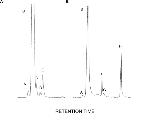
“Interestingly, both strains possessed significant amounts of ergosterol (Figure 7).”
§ A Sterol-Regulatory Element Binding Protein Is Required for Cell Polarity, Hypoxia Adaptation, Azole Drug Resistance, and Virulence in Aspergillus fumigatus > Results > SrbA is required for normal sterol biosynthesis
plain- Asserted: passes — The authors put it forward as true.
- Checkable: passes — It can be checked against evidence.
- Load-bearing: passes — The paper’s conclusions depend on it.
- Based: passes — The paper gives a reason why it holds.
It may state two separate claims as one.
A result the paper reports from its own data — it cites no other source for it, so there is none to check. The quote shows it is in the paper.
In the paper
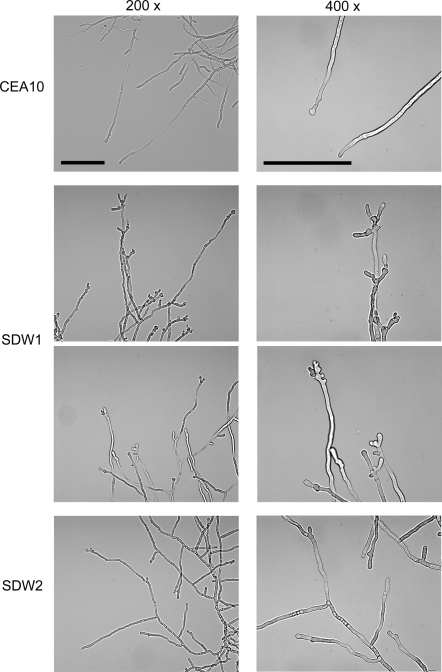
“Our examination of the SrbA null mutant colony morphology subsequently revealed abnormal branching at the hyphal tips in normoxia and an inability of hyphal growth in hypoxia.”
§ A Sterol-Regulatory Element Binding Protein Is Required for Cell Polarity, Hypoxia Adaptation, Azole Drug Resistance, and Virulence in Aspergillus fumigatus > Discussion
plain- Asserted: passes — The authors put it forward as true.
- Checkable: passes — It can be checked against evidence.
- Load-bearing: passes — The paper’s conclusions depend on it.
- Based: passes — The paper gives a reason why it holds.
It may state two separate claims as one.
A result the paper reports from its own data — it cites no other source for it, so there is none to check. The quote shows it is in the paper.
In the paper
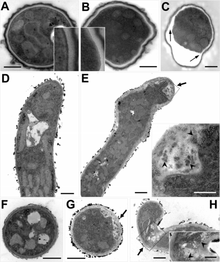
“SDW1 hyphal tips display hyper-branching and a “blunted” abnormal morphological phenotype (Figure 4).”
§ A Sterol-Regulatory Element Binding Protein Is Required for Cell Polarity, Hypoxia Adaptation, Azole Drug Resistance, and Virulence in Aspergillus fumigatus > Results > SrbA is required for cell polarity and hyphal morphogenesis
plain- Asserted: passes — The authors put it forward as true.
- Checkable: passes — It can be checked against evidence.
- Load-bearing: passes — The paper’s conclusions depend on it.
- Based: passes — The paper gives a reason why it holds.
It may state two separate claims as one.
A result the paper reports from its own data — it cites no other source for it, so there is none to check. The quote shows it is in the paper.
In the paper
“The ratio of C-4 methylated sterols to ergosterol in the absence of SrbA is 1.94 whereas no C-4 methylated sterols accumulated in the wild type.”
§ A Sterol-Regulatory Element Binding Protein Is Required for Cell Polarity, Hypoxia Adaptation, Azole Drug Resistance, and Virulence in Aspergillus fumigatus > Results > SrbA is required for normal sterol biosynthesis
plain- Asserted: passes — The authors put it forward as true.
- Checkable: passes — It can be checked against evidence.
- Load-bearing: passes — The paper’s conclusions depend on it.
- Based: passes — The paper gives a reason why it holds.
It may state two separate claims as one.
A result the paper reports from its own data — it cites no other source for it, so there is none to check. The quote shows it is in the paper.
In the paper
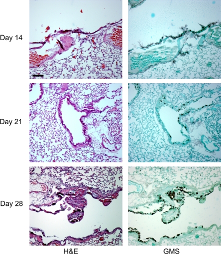
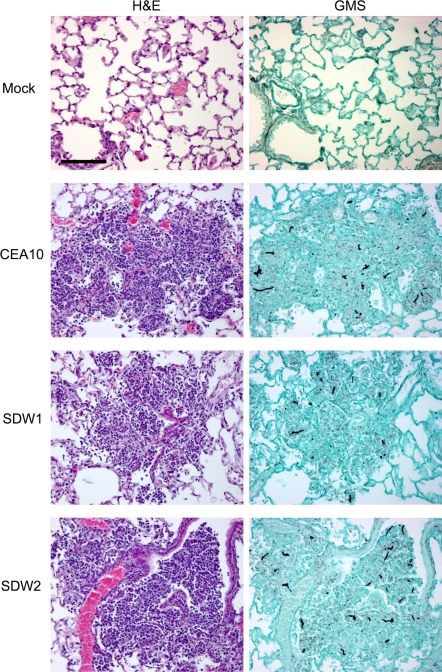
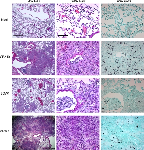
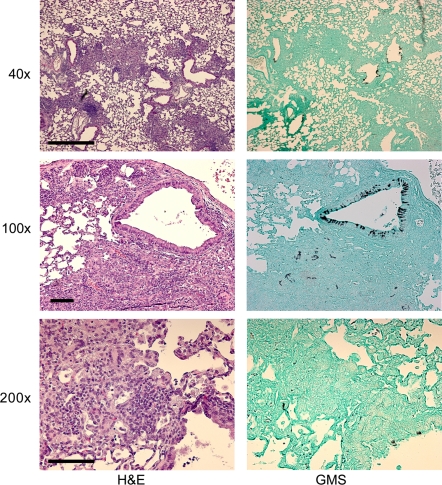
“Histopathology on day 4 of the infection, however, revealed extensive growth and proliferation of the wild type and reconstituted SDW2 strain, but minimal fungal growth and proliferation in mice infected with the SrbA null mutant SDW1 (Figure 11).”
§ A Sterol-Regulatory Element Binding Protein Is Required for Cell Polarity, Hypoxia Adaptation, Azole Drug Resistance, and Virulence in Aspergillus fumigatus > Results > SrbA is required for fungal virulence in two distinct murine models
plain- Asserted: passes — The authors put it forward as true.
- Checkable: passes — It can be checked against evidence.
- Load-bearing: passes — The paper’s conclusions depend on it.
- Based: passes — The paper gives a reason why it holds.
It may state two separate claims as one.
A result the paper reports from its own data — it cites no other source for it, so there is none to check. The quote shows it is in the paper.
In the paper
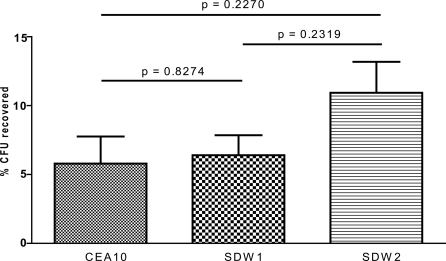
“Lung homogenates from these mice also revealed that viable SDW1 fungus was recoverable from these mice at this time point.”
§ A Sterol-Regulatory Element Binding Protein Is Required for Cell Polarity, Hypoxia Adaptation, Azole Drug Resistance, and Virulence in Aspergillus fumigatus > Results > SrbA is required for fungal virulence in two distinct murine models
plain- Asserted: passes — The authors put it forward as true.
- Checkable: passes — It can be checked against evidence.
- Load-bearing: passes — The paper’s conclusions depend on it.
- Based: passes — The paper gives a reason why it holds.
The quote only partly states this claim.
A result the paper reports from its own data — it cites no other source for it, so there is none to check. The quote shows it is in the paper.
In the paper
“Similar to the results with Fluconazole, SDW1 was significantly more susceptible to Voriconazole and showed a MIC of only 0.012 µg/ml (Figure 3).”
§ A Sterol-Regulatory Element Binding Protein Is Required for Cell Polarity, Hypoxia Adaptation, Azole Drug Resistance, and Virulence in Aspergillus fumigatus > Results > SrbA mediates resistance to the azole class of antifungal drugs
plain- Asserted: passes — The authors put it forward as true.
- Checkable: passes — It can be checked against evidence.
- Load-bearing: passes — The paper’s conclusions depend on it.
- Based: passes — The paper gives a reason why it holds.
It may state two separate claims as one.
A result the paper reports from its own data — it cites no other source for it, so there is none to check. The quote shows it is in the paper.
In the paper
“This phenotype was observed in over 50% of the SDW1 hyphae examined and never observed in the wild type strain.”
§ A Sterol-Regulatory Element Binding Protein Is Required for Cell Polarity, Hypoxia Adaptation, Azole Drug Resistance, and Virulence in Aspergillus fumigatus > Results > SrbA is required for cell polarity and hyphal morphogenesis
plain- Asserted: passes — The authors put it forward as true.
- Checkable: passes — It can be checked against evidence.
- Load-bearing: passes — The paper’s conclusions depend on it.
- Based: passes — The paper gives a reason why it holds.
It may state two separate claims as one.
A result the paper reports from its own data — it cites no other source for it, so there is none to check. The quote shows it is in the paper.
In the paper
“No mock infected animals perished in either murine model in all experiments.”
§ A Sterol-Regulatory Element Binding Protein Is Required for Cell Polarity, Hypoxia Adaptation, Azole Drug Resistance, and Virulence in Aspergillus fumigatus > Materials and Methods > Murine virulence assays
plain- Asserted: passes — The authors put it forward as true.
- Checkable: passes — It can be checked against evidence.
- Load-bearing: fails — The paper’s conclusions may not depend on it.
- Based: passes — The paper gives a reason why it holds.
Probably not a substantial claim. The paper’s conclusions may not depend on it.
A result the paper reports from its own data — it cites no other source for it, so there is none to check. The quote shows it is in the paper.
In the paper
“No difference in results was observed between 24 and 48 h.”
§ A Sterol-Regulatory Element Binding Protein Is Required for Cell Polarity, Hypoxia Adaptation, Azole Drug Resistance, and Virulence in Aspergillus fumigatus > Materials and Methods > Susceptibility testing
plain- Asserted: passes — The authors put it forward as true.
- Checkable: passes — It can be checked against evidence.
- Load-bearing: fails — The paper’s conclusions may not depend on it.
- Based: passes — The paper gives a reason why it holds.
Probably not a substantial claim. The paper’s conclusions may not depend on it.
None of the sources cited for this claim have been checked yet.
In the paper
“Aspergillus fumigatus is a normally benign saprophytic fungus that may cause an often lethal invasive disease in immunocompromised patients, invasive pulmonary aspergillosis (IPA) [1],[2].”
§ A Sterol-Regulatory Element Binding Protein Is Required for Cell Polarity, Hypoxia Adaptation, Azole Drug Resistance, and Virulence in Aspergillus fumigatus > Introduction
What the sources say
hedgedconditional- Asserted: passes — The authors put it forward as true.
- Checkable: passes — It can be checked against evidence.
- Load-bearing: fails — The paper’s conclusions may not depend on it.
- Based: passes — The paper gives a reason why it holds.
Probably not a substantial claim. The paper’s conclusions may not depend on it. It may state two separate claims as one.
None of the sources cited for this claim have been checked yet.
In the paper
“switching from aerobic respiration to various forms of anaerobic respiration to deal with low oxygen levels has been implicated as an important virulence attribute in several prokaryotic pathogens [18],[19]”
§ A Sterol-Regulatory Element Binding Protein Is Required for Cell Polarity, Hypoxia Adaptation, Azole Drug Resistance, and Virulence in Aspergillus fumigatus > Introduction
What the sources say
plaincorrelational- Asserted: passes — The authors put it forward as true.
- Checkable: passes — It can be checked against evidence.
- Load-bearing: fails — The paper’s conclusions may not depend on it.
- Based: passes — The paper gives a reason why it holds.
Probably not a substantial claim. The paper’s conclusions may not depend on it.
In the paper
“Sre1 was found to be required for adaptation to hypoxia and regulated approximately 68% of the genes transcriptionally induced greater than 2-fold in response to anaerobic conditions [31].”
§ A Sterol-Regulatory Element Binding Protein Is Required for Cell Polarity, Hypoxia Adaptation, Azole Drug Resistance, and Virulence in Aspergillus fumigatus > Introduction
What the sources say
- Not checked yetcites [31] BL Todd, 2006, Sterol regulatory element binding protein is a principal regulator of anaerobic gene expression in fission yeast., Mol Cell Biol source
plaindirect- Asserted: passes — The authors put it forward as true.
- Checkable: passes — It can be checked against evidence.
- Load-bearing: passes — The paper’s conclusions depend on it.
- Based: passes — The paper gives a reason why it holds.
It may state two separate claims as one.
None of the sources cited for this claim have been checked yet.
In the paper
“As in fission yeast, the SREBP pathway mediated by Sre1 and Scp1 in C. neoformans was crucial for adaptation to hypoxia and sterol biosynthesis.”
§ A Sterol-Regulatory Element Binding Protein Is Required for Cell Polarity, Hypoxia Adaptation, Azole Drug Resistance, and Virulence in Aspergillus fumigatus > Introduction
What the sources say
- Not checked yetcites [32] YC Chang, 2007, Sre1p, a regulator of oxygen sensing and sterol homeostasis, is required for virulence in Cryptococcus neoformans., Mol Microbiol source
- Not checked yetcites [33] CD Chun, 2007, A Link between virulence and homeostatic responses to hypoxia during infection by the human fungal pathogen Cryptococcus neoformans., PLoS Pathog source
plaindirect- Asserted: passes — The authors put it forward as true.
- Checkable: passes — It can be checked against evidence.
- Load-bearing: fails — The paper’s conclusions may not depend on it.
- Based: passes — The paper gives a reason why it holds.
Probably not a substantial claim. The paper’s conclusions may not depend on it.
A conclusion the paper draws from its own results — it cites no other source for it, so there is none to check. The quote shows it is in the paper.
In the paper
“These pathologic lesions also likely represent areas of poor oxygen availability to the pathogen and host.”
§ A Sterol-Regulatory Element Binding Protein Is Required for Cell Polarity, Hypoxia Adaptation, Azole Drug Resistance, and Virulence in Aspergillus fumigatus > Introduction
hedgedcorrelational- Asserted: passes — The authors put it forward as true.
- Checkable: passes — It can be checked against evidence.
- Load-bearing: passes — The paper’s conclusions depend on it.
- Based: fails — The paper may give no reason why it holds.
Probably not a substantial claim. The paper may give no reason why it holds.
In the paper
“Recently, a novel mechanism of hypoxia adaptation mediated by a highly conserved family of transcription factors, sterol regulatory element-binding proteins (SREBPs), was characterized in fission yeast, Schizosaccharomyces pombe [25].”
§ A Sterol-Regulatory Element Binding Protein Is Required for Cell Polarity, Hypoxia Adaptation, Azole Drug Resistance, and Virulence in Aspergillus fumigatus > Introduction
What the sources say
- Not checked yetcites [25] AL Hughes, 2005, SREBP pathway responds to sterols and functions as an oxygen sensor in fission yeast., Cell source
plaindirect- Asserted: passes — The authors put it forward as true.
- Checkable: passes — It can be checked against evidence.
- Load-bearing: fails — The paper’s conclusions may not depend on it.
- Based: passes — The paper gives a reason why it holds.
Probably not a substantial claim. The paper’s conclusions may not depend on it.
What the paper did — it cites no source for this, so there is none to check. The quote shows it is in the paper.
In the paper
“All strains were rigorously confirmed with Southern blot (Figure 1) and PCR analyses (data not shown).”
§ A Sterol-Regulatory Element Binding Protein Is Required for Cell Polarity, Hypoxia Adaptation, Azole Drug Resistance, and Virulence in Aspergillus fumigatus > Results > SrbA is required for hypoxia adaptation in Aspergillus fumigatus
boosted- Asserted: passes — The authors put it forward as true.
- Checkable: passes — It can be checked against evidence.
- Load-bearing: passes — The paper’s conclusions depend on it.
- Based: passes — The paper gives a reason why it holds.
Probably not a substantial claim.
None of the sources cited for this claim have been checked yet.
In the paper
“these mutants also failed to proliferate in host tissue, failed to cause fatal meningoencephalitis, and displayed hypersensitivity to the azole class of antifungal drugs [32],[33]”
§ A Sterol-Regulatory Element Binding Protein Is Required for Cell Polarity, Hypoxia Adaptation, Azole Drug Resistance, and Virulence in Aspergillus fumigatus > Introduction
What the sources say
- Not checked yetcites [32] YC Chang, 2007, Sre1p, a regulator of oxygen sensing and sterol homeostasis, is required for virulence in Cryptococcus neoformans., Mol Microbiol source
- Not checked yetcites [33] CD Chun, 2007, A Link between virulence and homeostatic responses to hypoxia during infection by the human fungal pathogen Cryptococcus neoformans., PLoS Pathog source
plaindirect- Asserted: passes — The authors put it forward as true.
- Checkable: passes — It can be checked against evidence.
- Load-bearing: passes — The paper’s conclusions depend on it.
- Based: passes — The paper gives a reason why it holds.
Probably not a substantial claim. It may state two separate claims as one.
In the paper
“Yet, the mechanisms utilized by A. fumigatus to survive and cause disease in immunocompromised hosts are not fully understood [4].”
§ A Sterol-Regulatory Element Binding Protein Is Required for Cell Polarity, Hypoxia Adaptation, Azole Drug Resistance, and Virulence in Aspergillus fumigatus > Introduction
What the sources say
- Not checked yetcites [4] TM Hohl, 2007, Aspergillus fumigatus: principles of pathogenesis and host defense., Eukaryot Cell source
plain- Asserted: passes — The authors put it forward as true.
- Checkable: passes — It can be checked against evidence.
- Load-bearing: passes — The paper’s conclusions depend on it.
- Based: passes — The paper gives a reason why it holds.
Probably not a substantial claim.
A conclusion the paper draws from its own results — it cites no other source for it, so there is none to check. The quote shows it is in the paper.
In the paper
“Our results confirm that some roles of SREBPs in filamentous fungi are conserved with yeast including, the response to hypoxia, sterol biosynthesis, and susceptibility to the azole class of antifungal drugs.”
§ A Sterol-Regulatory Element Binding Protein Is Required for Cell Polarity, Hypoxia Adaptation, Azole Drug Resistance, and Virulence in Aspergillus fumigatus > Discussion
boostedcorrelational- Asserted: passes — The authors put it forward as true.
- Checkable: passes — It can be checked against evidence.
- Load-bearing: passes — The paper’s conclusions depend on it.
- Based: passes — The paper gives a reason why it holds.
It may state two separate claims as one.
None of the sources cited for this claim have been checked yet.
In the paper
“Interestingly, C. neoformans lacks an apparent Insig homolog and the Insig homolog in S. pombe does not appear to be required for regulation of SREBP signalling [25],[32].”
§ A Sterol-Regulatory Element Binding Protein Is Required for Cell Polarity, Hypoxia Adaptation, Azole Drug Resistance, and Virulence in Aspergillus fumigatus > Discussion
What the sources say
- Not checked yetcites [25] AL Hughes, 2005, SREBP pathway responds to sterols and functions as an oxygen sensor in fission yeast., Cell source
- Not checked yetcites [32] YC Chang, 2007, Sre1p, a regulator of oxygen sensing and sterol homeostasis, is required for virulence in Cryptococcus neoformans., Mol Microbiol source
hedged- Asserted: passes — The authors put it forward as true.
- Checkable: passes — It can be checked against evidence.
- Load-bearing: fails — The paper’s conclusions may not depend on it.
- Based: passes — The paper gives a reason why it holds.
Probably not a substantial claim. The paper’s conclusions may not depend on it. It may state two separate claims as one.
None of the sources cited for this claim have been checked yet.
In the paper
“In the yeast S. pombe and C. neoformans, it seems clear that Sre1 is regulated post-translationally in response to sterol biosynthesis perturbation that occurs in low oxygen environments.”
§ A Sterol-Regulatory Element Binding Protein Is Required for Cell Polarity, Hypoxia Adaptation, Azole Drug Resistance, and Virulence in Aspergillus fumigatus > Discussion
boosteddirect- Asserted: passes — The authors put it forward as true.
- Checkable: passes — It can be checked against evidence.
- Load-bearing: fails — The paper’s conclusions may not depend on it.
- Based: passes — The paper gives a reason why it holds.
Probably not a substantial claim. The paper’s conclusions may not depend on it.
A conclusion the paper draws from its own results — it cites no other source for it, so there is none to check. The quote shows it is in the paper.
In the paper
“Thus, our finding that the SrbA null mutant in A. fumigatus accumulates 4-methyl sterols may also suggest that these sterols are the trigger for SrbA activation in A. fumigatus.”
§ A Sterol-Regulatory Element Binding Protein Is Required for Cell Polarity, Hypoxia Adaptation, Azole Drug Resistance, and Virulence in Aspergillus fumigatus > Discussion
hedgedconditional- Asserted: passes — The authors put it forward as true.
- Checkable: passes — It can be checked against evidence.
- Load-bearing: fails — The paper’s conclusions may not depend on it.
- Based: passes — The paper gives a reason why it holds.
Probably not a substantial claim. The paper’s conclusions may not depend on it.
A conclusion the paper draws from its own results — it cites no other source for it, so there is none to check. The quote shows it is in the paper.
In the paper
“Thus, we believe that A. fumigatus must overcome significant hypoxia during pulmonary infections, and the inability of the SrbA null mutant to adapt to hypoxic conditions results in rapid cessation of invasive growth and a lack of lethal disease.”
§ A Sterol-Regulatory Element Binding Protein Is Required for Cell Polarity, Hypoxia Adaptation, Azole Drug Resistance, and Virulence in Aspergillus fumigatus > Discussion
hedgeddirect- Asserted: passes — The authors put it forward as true.
- Checkable: passes — It can be checked against evidence.
- Load-bearing: passes — The paper’s conclusions depend on it.
- Based: passes — The paper gives a reason why it holds.
It may state two separate claims as one.
- The authors state this as an assumption: Sites of infection and inflammation in the neutropenic mouse model are hypoxic.
- Rests on an assumption: A. fumigatus must overcome significant hypoxia during pulmonary infections.
A conclusion the paper draws from its own results — it cites no other source for it, so there is none to check. The quote shows it is in the paper.
In the paper
“These two results suggest that growth of the fungus was halted and what immune effector cells present in the immunosuppressed mice were able to clear the infection.”
§ A Sterol-Regulatory Element Binding Protein Is Required for Cell Polarity, Hypoxia Adaptation, Azole Drug Resistance, and Virulence in Aspergillus fumigatus > Discussion
hedgeddirect- Asserted: passes — The authors put it forward as true.
- Checkable: passes — It can be checked against evidence.
- Load-bearing: passes — The paper’s conclusions depend on it.
- Based: passes — The paper gives a reason why it holds.
It may state two separate claims as one.
A conclusion the paper draws from its own results — it cites no other source for it, so there is none to check. The quote shows it is in the paper.
In the paper
“This suggests that SrbA may be transcriptionally regulated in molds.”
§ A Sterol-Regulatory Element Binding Protein Is Required for Cell Polarity, Hypoxia Adaptation, Azole Drug Resistance, and Virulence in Aspergillus fumigatus > Discussion
hedgedconditionalSingle case- Asserted: passes — The authors put it forward as true.
- Checkable: passes — It can be checked against evidence.
- Load-bearing: fails — The paper’s conclusions may not depend on it.
- Based: passes — The paper gives a reason why it holds.
Probably not a substantial claim. The paper’s conclusions may not depend on it.
None of the sources cited for this claim have been checked yet.
In the paper
“A. fumigatus strain CEA17 is a uracil-auxotrophic (pyrG1) mutant of A. fumigatus strain CEA10 [67],[68].”
§ A Sterol-Regulatory Element Binding Protein Is Required for Cell Polarity, Hypoxia Adaptation, Azole Drug Resistance, and Virulence in Aspergillus fumigatus > Materials and Methods > Strains and media
What the sources say
- Not checked yetcites [67] C D'Enfert, 1996, Attenuated virulence of uridine-uracil auxotrophs of Aspergillus fumigatus., Infect Immun source
- Not checked yetcites [68] C d'Enfert, 1996, Selection of multiple disruption events in Aspergillus fumigatus using the orotidine-5′-decarboxylase gene, pyrG, as a unique transformation marker., Curr Genet source
plaindirect- Asserted: passes — The authors put it forward as true.
- Checkable: passes — It can be checked against evidence.
- Load-bearing: fails — The paper’s conclusions may not depend on it.
- Based: passes — The paper gives a reason why it holds.
Probably not a substantial claim. The paper’s conclusions may not depend on it.
A conclusion the paper draws from its own results — it cites no other source for it, so there is none to check. The quote shows it is in the paper.
In the paper
“Once hypoxia is generated during Aspergillus infection, the mutant simply can no longer grow and proliferate, allowing what immune effector cells that remain functional the ability to ultimately clear the infection.”
§ A Sterol-Regulatory Element Binding Protein Is Required for Cell Polarity, Hypoxia Adaptation, Azole Drug Resistance, and Virulence in Aspergillus fumigatus > Discussion
plaindirect- Asserted: passes — The authors put it forward as true.
- Checkable: passes — It can be checked against evidence.
- Load-bearing: passes — The paper’s conclusions depend on it.
- Based: passes — The paper gives a reason why it holds.
It may state two separate claims as one.
- The authors state this as an assumption: Sites of infection and inflammation in the neutropenic mouse model are hypoxic.
- Rests on an assumption: A. fumigatus must overcome significant hypoxia during pulmonary infections.
In the paper
“Interestingly, Takeshita et al. (2008) recently observed that localization of key deposition proteins involved in polarized growth at the hyphal tip requires apical sterol-rich membranes [52].”
§ A Sterol-Regulatory Element Binding Protein Is Required for Cell Polarity, Hypoxia Adaptation, Azole Drug Resistance, and Virulence in Aspergillus fumigatus > Discussion
What the sources say
- Not checked yetcites [52] N Takeshita, 2008, Apical Sterol-rich Membranes Are Essential for Localizing Cell End Markers That Determine Growth Directionality in the Filamentous Fungus Aspergillus nidulans., Mol Biol Cell source
plaindirect- Asserted: passes — The authors put it forward as true.
- Checkable: passes — It can be checked against evidence.
- Load-bearing: passes — The paper’s conclusions depend on it.
- Based: passes — The paper gives a reason why it holds.
Probably not a substantial claim.
None of the sources cited for this claim have been checked yet.
In the paper
“In mammals, Insig is a key regulator of SREBP function where it binds to SCAP and prevents SREBP cleavage in the presence of sterols by maintaining the SREBP-SCAP complex in the endoplasmic reticulum membrane [65],[66].”
§ A Sterol-Regulatory Element Binding Protein Is Required for Cell Polarity, Hypoxia Adaptation, Azole Drug Resistance, and Virulence in Aspergillus fumigatus > Discussion
What the sources say
- Not checked yetcites [65] LJ Engelking, 2006, Severe facial clefting in Insig-deficient mouse embryos caused by sterol accumulation and reversed by lovastatin., J Clin Invest source
- Not checked yetcites [66] T Yang, 2002, Crucial step in cholesterol homeostasis: sterols promote binding of SCAP to INSIG-1, a membrane protein that facilitates retention of SREBPs in ER., Cell source
plaindirect- Asserted: passes — The authors put it forward as true.
- Checkable: passes — It can be checked against evidence.
- Load-bearing: fails — The paper’s conclusions may not depend on it.
- Based: passes — The paper gives a reason why it holds.
Probably not a substantial claim. The paper’s conclusions may not depend on it.
None of the sources cited for this claim have been checked yet.
In the paper
“Currently, it is unclear what specific components of the immune system are affected in this model, but it is clear that differences in the immunosuppression regimen can significantly affect the outcome of infection [56],[57].”
§ A Sterol-Regulatory Element Binding Protein Is Required for Cell Polarity, Hypoxia Adaptation, Azole Drug Resistance, and Virulence in Aspergillus fumigatus > Discussion
What the sources say
- Not checked yetcites [56] V Balloy, 2005, Differences in patterns of infection and inflammation for corticosteroid treatment and chemotherapy in experimental invasive pulmonary aspergillosis., Infect Immun source
- Not checked yetcites [57] SD Stephens-Romero, 2005, The pathogenesis of fatal outcome in murine pulmonary aspergillosis depends on the neutrophil depletion strategy., Infect Immun source
boosteddirect- Asserted: passes — The authors put it forward as true.
- Checkable: passes — It can be checked against evidence.
- Load-bearing: fails — The paper’s conclusions may not depend on it.
- Based: passes — The paper gives a reason why it holds.
Probably not a substantial claim. The paper’s conclusions may not depend on it.
A conclusion the paper draws from its own results — it cites no other source for it, so there is none to check. The quote shows it is in the paper.
In the paper
“However, the SrbA null mutant was virtually avirulent in two distinct murine models of IPA despite a normal growth rate of the fungus in standard laboratory conditions.”
§ A Sterol-Regulatory Element Binding Protein Is Required for Cell Polarity, Hypoxia Adaptation, Azole Drug Resistance, and Virulence in Aspergillus fumigatus > Discussion
plaindirect- Asserted: passes — The authors put it forward as true.
- Checkable: passes — It can be checked against evidence.
- Load-bearing: passes — The paper’s conclusions depend on it.
- Based: passes — The paper gives a reason why it holds.
It may state two separate claims as one.
A conclusion the paper draws from its own results — it cites no other source for it, so there is none to check. The quote shows it is in the paper.
In the paper
“Taken together, these results suggest that while aspects of SrbA signalling in filamentous fungi may be conserved in yeast and mammals, it is likely that significant differences exist in molds that remain to be elucidated.”
§ A Sterol-Regulatory Element Binding Protein Is Required for Cell Polarity, Hypoxia Adaptation, Azole Drug Resistance, and Virulence in Aspergillus fumigatus > Discussion
hedgedcorrelational- Asserted: passes — The authors put it forward as true.
- Checkable: unclear — It is unclear whether it could be checked against evidence.
- Load-bearing: unclear — It is unclear whether the paper’s conclusions depend on it.
- Based: passes — The paper gives a reason why it holds.
It may state two separate claims as one.
None of the sources cited for this claim have been checked yet.
In the paper
“However, HIF1 in humans also responds transcriptionally to hypoxia, but its activity is primarily post-translationally regulated [62],[63].”
§ A Sterol-Regulatory Element Binding Protein Is Required for Cell Polarity, Hypoxia Adaptation, Azole Drug Resistance, and Virulence in Aspergillus fumigatus > Discussion
What the sources say
- Not checked yetcites [62] GL Semenza, 1999, Regulation of mammalian O2 homeostasis by hypoxia-inducible factor 1., Annu Rev Cell Dev Biol source
- Not checked yetcites [63] RS Belaiba, 2007, Hypoxia up-regulates hypoxia-inducible factor-1alpha transcription by involving phosphatidylinositol 3-kinase and nuclear factor kappaB in pulmonary artery smooth muscle cells., Mol Biol Cell source
plaindirect- Asserted: passes — The authors put it forward as true.
- Checkable: passes — It can be checked against evidence.
- Load-bearing: fails — The paper’s conclusions may not depend on it.
- Based: passes — The paper gives a reason why it holds.
Probably not a substantial claim. The paper’s conclusions may not depend on it. It may state two separate claims as one.
None of the sources cited for this claim have been checked yet.
In the paper
“At the apex of hyphae in filamentous fungi, the Spitzenkörper is an accumulation of vesicles that is critical for growth directionality [50],[51].”
§ A Sterol-Regulatory Element Binding Protein Is Required for Cell Polarity, Hypoxia Adaptation, Azole Drug Resistance, and Virulence in Aspergillus fumigatus > Discussion
What the sources say
plaindirect- Asserted: passes — The authors put it forward as true.
- Checkable: passes — It can be checked against evidence.
- Load-bearing: fails — The paper’s conclusions may not depend on it.
- Based: passes — The paper gives a reason why it holds.
Probably not a substantial claim. The paper’s conclusions may not depend on it. It may state two separate claims as one.
None of the sources cited for this claim have been checked yet.
In the paper
“Second, a relationship between mitochondria function, sterol homeostasis, and azole drug resistance has been observed in the yeast S. cerevisiae and Candida glabrata [60],[61].”
§ A Sterol-Regulatory Element Binding Protein Is Required for Cell Polarity, Hypoxia Adaptation, Azole Drug Resistance, and Virulence in Aspergillus fumigatus > Discussion
What the sources say
- Not checked yetcites [60] R Kaur, 2004, Functional genomic analysis of fluconazole susceptibility in the pathogenic yeast Candida glabrata: roles of calcium signaling and mitochondria., Antimicrob Agents Chemother source
- Not checked yetcites [61] DP Kontoyiannis, 2000, Modulation of fluconazole sensitivity by the interaction of mitochondria and erg3p in Saccharomyces cerevisiae., J Antimicrob Chemother source
plaincorrelational- Asserted: passes — The authors put it forward as true.
- Checkable: passes — It can be checked against evidence.
- Load-bearing: fails — The paper’s conclusions may not depend on it.
- Based: passes — The paper gives a reason why it holds.
Probably not a substantial claim. The paper’s conclusions may not depend on it.
A conclusion the paper draws from its own results — it cites no other source for it, so there is none to check. The quote shows it is in the paper.
In the paper
“Thus, even though the ergosterol biosynthesis pathway appears blocked at ERG25 in the SrbA mutant, alternative mechanisms exist for A. fumigatus to produce ergosterol in the absence of SrbA and presumably ERG25 activity.”
§ A Sterol-Regulatory Element Binding Protein Is Required for Cell Polarity, Hypoxia Adaptation, Azole Drug Resistance, and Virulence in Aspergillus fumigatus > Discussion
hedgeddirect- Asserted: passes — The authors put it forward as true.
- Checkable: passes — It can be checked against evidence.
- Load-bearing: passes — The paper’s conclusions depend on it.
- Based: passes — The paper gives a reason why it holds.
It may state two separate claims as one.
What the paper did — it cites no source for this, so there is none to check. The quote shows it is in the paper.
In the paper
“Data for each strain represents six independent experiments and includes three dye swaps.”
§ A Sterol-Regulatory Element Binding Protein Is Required for Cell Polarity, Hypoxia Adaptation, Azole Drug Resistance, and Virulence in Aspergillus fumigatus > Materials and Methods > Microarray-based transcriptional profiling
plain- Asserted: passes — The authors put it forward as true.
- Checkable: passes — It can be checked against evidence.
- Load-bearing: fails — The paper’s conclusions may not depend on it.
- Based: passes — The paper gives a reason why it holds.
Probably not a substantial claim. The paper’s conclusions may not depend on it. It may state two separate claims as one.
What the paper did — it cites no source for this, so there is none to check. The quote shows it is in the paper.
In the paper
“All animal experiments were repeated in duplicate with similar results.”
§ A Sterol-Regulatory Element Binding Protein Is Required for Cell Polarity, Hypoxia Adaptation, Azole Drug Resistance, and Virulence in Aspergillus fumigatus > Results > SrbA is required for fungal virulence in two distinct murine models
plain- Asserted: passes — The authors put it forward as true.
- Checkable: passes — It can be checked against evidence.
- Load-bearing: fails — The paper’s conclusions may not depend on it.
- Based: passes — The paper gives a reason why it holds.
Probably not a substantial claim. The paper’s conclusions may not depend on it.
What the paper did — it cites no source for this, so there is none to check. The quote shows it is in the paper.
In the paper
“Transformants were initially screened by PCR to identify potential homologous recombination events at the srbA locus.”
§ A Sterol-Regulatory Element Binding Protein Is Required for Cell Polarity, Hypoxia Adaptation, Azole Drug Resistance, and Virulence in Aspergillus fumigatus > Materials and Methods > Strain construction
plain- Asserted: passes — The authors put it forward as true.
- Checkable: passes — It can be checked against evidence.
- Load-bearing: fails — The paper’s conclusions may not depend on it.
- Based: passes — The paper gives a reason why it holds.
Probably not a substantial claim. The paper’s conclusions may not depend on it.
What the paper did — it cites no source for this, so there is none to check. The quote shows it is in the paper.
In the paper
“Experiments were performed with triplicate wells and repeated two times for each A. fumigatus strain.”
§ A Sterol-Regulatory Element Binding Protein Is Required for Cell Polarity, Hypoxia Adaptation, Azole Drug Resistance, and Virulence in Aspergillus fumigatus > Materials and Methods > Macrophage assays
plain- Asserted: passes — The authors put it forward as true.
- Checkable: passes — It can be checked against evidence.
- Load-bearing: fails — The paper’s conclusions may not depend on it.
- Based: passes — The paper gives a reason why it holds.
Probably not a substantial claim. The paper’s conclusions may not depend on it.
What the paper did — it cites no source for this, so there is none to check. The quote shows it is in the paper.
In the paper
“Experiments were repeated in duplicates with similar results.”
§ A Sterol-Regulatory Element Binding Protein Is Required for Cell Polarity, Hypoxia Adaptation, Azole Drug Resistance, and Virulence in Aspergillus fumigatus > Materials and Methods > Murine virulence assays
plain- Asserted: passes — The authors put it forward as true.
- Checkable: passes — It can be checked against evidence.
- Load-bearing: fails — The paper’s conclusions may not depend on it.
- Based: passes — The paper gives a reason why it holds.
Probably not a substantial claim. The paper’s conclusions may not depend on it.
What the paper did — it cites no source for this, so there is none to check. The quote shows it is in the paper.
In the paper
“The assay was repeated three times for each concentration.”
§ A Sterol-Regulatory Element Binding Protein Is Required for Cell Polarity, Hypoxia Adaptation, Azole Drug Resistance, and Virulence in Aspergillus fumigatus > Materials and Methods > Oxidative stress assay
plain- Asserted: passes — The authors put it forward as true.
- Checkable: passes — It can be checked against evidence.
- Load-bearing: fails — The paper’s conclusions may not depend on it.
- Based: passes — The paper gives a reason why it holds.
Probably not a substantial claim. The paper’s conclusions may not depend on it.
What the paper did — it cites no source for this, so there is none to check. The quote shows it is in the paper.
In the paper
“A Delta cutoff in SAM that captured the maximum number of significant genes with a false discovery rate of zero was utilized.”
§ A Sterol-Regulatory Element Binding Protein Is Required for Cell Polarity, Hypoxia Adaptation, Azole Drug Resistance, and Virulence in Aspergillus fumigatus > Materials and Methods > Microarray-based transcriptional profiling
plain- Asserted: passes — The authors put it forward as true.
- Checkable: passes — It can be checked against evidence.
- Load-bearing: fails — The paper’s conclusions may not depend on it.
- Based: passes — The paper gives a reason why it holds.
Probably not a substantial claim. The paper’s conclusions may not depend on it.
What the paper did — it cites no source for this, so there is none to check. The quote shows it is in the paper.
In the paper
“The P values we report are always the conservative values.”
§ A Sterol-Regulatory Element Binding Protein Is Required for Cell Polarity, Hypoxia Adaptation, Azole Drug Resistance, and Virulence in Aspergillus fumigatus > Materials and Methods > Statistical analysis
plain- Asserted: passes — The authors put it forward as true.
- Checkable: passes — It can be checked against evidence.
- Load-bearing: fails — The paper’s conclusions may not depend on it.
- Based: passes — The paper gives a reason why it holds.
Probably not a substantial claim. The paper’s conclusions may not depend on it.
What the paper did — it cites no source for this, so there is none to check. The quote shows it is in the paper.
In the paper
“RNA integrity was confirmed with an Agilent Technologies Bioanalyzer.”
§ A Sterol-Regulatory Element Binding Protein Is Required for Cell Polarity, Hypoxia Adaptation, Azole Drug Resistance, and Virulence in Aspergillus fumigatus > Materials and Methods > Isolation of total RNA
plain- Asserted: passes — The authors put it forward as true.
- Checkable: passes — It can be checked against evidence.
- Load-bearing: fails — The paper’s conclusions may not depend on it.
- Based: passes — The paper gives a reason why it holds.
Probably not a substantial claim. The paper’s conclusions may not depend on it.
What the paper did — it cites no source for this, so there is none to check. The quote shows it is in the paper.
In the paper
“To eliminate the chance of heterokaryons, each transformant was streaked with sterile toothpicks a minimum of two times to obtain colonies from single conidia.”
§ A Sterol-Regulatory Element Binding Protein Is Required for Cell Polarity, Hypoxia Adaptation, Azole Drug Resistance, and Virulence in Aspergillus fumigatus > Materials and Methods > Strain construction
plaindirect- Asserted: passes — The authors put it forward as true.
- Checkable: passes — It can be checked against evidence.
- Load-bearing: passes — The paper’s conclusions depend on it.
- Based: passes — The paper gives a reason why it holds.
Probably not a substantial claim. It may state two separate claims as one.
In the paper
“Homologous recombination was confirmed by Southern analysis with the digoxigenin labeling system (Roche Molecular Biochemicals, Mannheim, Germany) as previously described [71].”
§ A Sterol-Regulatory Element Binding Protein Is Required for Cell Polarity, Hypoxia Adaptation, Azole Drug Resistance, and Virulence in Aspergillus fumigatus > Materials and Methods > Strain construction
What the sources say
- Not checked yetcites [71] RA Cramer, 2003, Cloning of a gene encoding an Alt a 1 isoallergen differentially expressed by the necrotrophic fungus Alternaria brassicicola during Arabidopsis infection., Appl Environ Microbiol source
plain- Asserted: passes — The authors put it forward as true.
- Checkable: passes — It can be checked against evidence.
- Load-bearing: passes — The paper’s conclusions depend on it.
- Based: passes — The paper gives a reason why it holds.
Probably not a substantial claim.
What the paper did — it cites no source for this, so there is none to check. The quote shows it is in the paper.
In the paper
“Counting was repeated three times for each strain and the mean and standard deviation are reported.”
§ A Sterol-Regulatory Element Binding Protein Is Required for Cell Polarity, Hypoxia Adaptation, Azole Drug Resistance, and Virulence in Aspergillus fumigatus > Materials and Methods > Conidia Germination assay
plain- Asserted: passes — The authors put it forward as true.
- Checkable: passes — It can be checked against evidence.
- Load-bearing: fails — The paper’s conclusions may not depend on it.
- Based: passes — The paper gives a reason why it holds.
Probably not a substantial claim. The paper’s conclusions may not depend on it. It may state two separate claims as one.
What the paper did — it cites no source for this, so there is none to check. The quote shows it is in the paper.
In the paper
“In those cases, we found that both the t test and Mann-Whitney test indicated the same results (i.e., both indicated significance or insignificance); however, typically one test gave a more conservative (larger, but still <0.01) P value.”
§ A Sterol-Regulatory Element Binding Protein Is Required for Cell Polarity, Hypoxia Adaptation, Azole Drug Resistance, and Virulence in Aspergillus fumigatus > Materials and Methods > Statistical analysis
plain- Asserted: passes — The authors put it forward as true.
- Checkable: passes — It can be checked against evidence.
- Load-bearing: fails — The paper’s conclusions may not depend on it.
- Based: passes — The paper gives a reason why it holds.
Probably not a substantial claim. The paper’s conclusions may not depend on it.
The authors state this as an assumption.
Sites of infection and inflammation in the neutropenic mouse model are hypoxic.
“We hypothesize that these sites of infection and inflammation in this model are hypoxic.”
The authors state this as an assumption.
The hyper-inflammatory response in X-CGD mice generates significant hypoxia in the lung.
“with the hypothesis that the hyper-inflammatory response would generate significant hypoxia in the lung”
The paper relies on this without support.
A. fumigatus must overcome significant hypoxia during pulmonary infections.
“we believe that A. fumigatus must overcome significant hypoxia during pulmonary infections”
Claims other papers make about this one
Each is checked against this paper’s own text: does it say what the citing paper says it does?
In the citing paper
“In A. fumigatus and C. neoformans, expression of cyp51A /ERG11 in response to azole exposure is regulated by the sterol regulatory element-binding protein (SREBP), SrbA and Sre1, respectively28,29.”
From Molecular mechanisms governing antifungal drug resistance (2023) · cites this paper as [28]
Self-check
Percentages, sums and reported differences re-computed from the paper’s own numbers.
Each contradiction between two passages listed here was confirmed by an independent check.
Rated minor: it would not touch a result — a value no result rests on, a p-value that stays on the same side of the significance level, or a conflict that is clerical, resolved elsewhere in the paper, or outside its own findings.
In the paper
§ A Sterol-Regulatory Element Binding Protein Is Required for Cell Polarity, Hypoxia Adaptation, Azole Drug Resistance, and Virulence in Aspergillus fumigatus > Results > SrbA is required for normal sterol biosynthesis
“the accumulation of 4-methylfecosterol and 4,4-dimethylergosta-8,24(28)-dien-3β-ol in the absence of SrbA”
§ A Sterol-Regulatory Element Binding Protein Is Required for Cell Polarity, Hypoxia Adaptation, Azole Drug Resistance, and Virulence in Aspergillus fumigatus > Results > SrbA is required for normal sterol biosynthesis
“H- 4,4-demethylergosta-8,24(28)-dien-3β-ol.”
Rated minor: it would not touch a result — a value no result rests on, a p-value that stays on the same side of the significance level, or a conflict that is clerical, resolved elsewhere in the paper, or outside its own findings.
In the paper
§ A Sterol-Regulatory Element Binding Protein Is Required for Cell Polarity, Hypoxia Adaptation, Azole Drug Resistance, and Virulence in Aspergillus fumigatus > Introduction
“the transcriptional repressors Rox1p and Mot3p, that down-regulate genes required for hypoxia adaptation [21].”
§ A Sterol-Regulatory Element Binding Protein Is Required for Cell Polarity, Hypoxia Adaptation, Azole Drug Resistance, and Virulence in Aspergillus fumigatus > Introduction
“However, in hypoxic conditions, Rox1p and Mot3p are expressed and this leads to transcriptional induction of genes involved in hypoxia adaptation [22].”
Citations
Cites
THE USE OF LEAD CITRATE AT HIGH pH AS AN ELECTRON-OPAQUE STAIN IN ELECTRON MICROSCOPY
Reynolds, E. S. (1963) · The Journal of Cell Biology · doi:10.1083/jcb.17.1.208 · Cited by 24,938
In silico prediction of protein-protein interactions in human macrophages
Souiai, O., Guerfali, F. Z., Miled, S. B., Brun, C., & Benkahla, A. (2014) · BMC Research Notes · doi:10.1186/1756-0500-7-157 · Cited by 10,785
Significance analysis of microarrays applied to the ionizing radiation response
Tusher, V. G., Tibshirani, R., & Chu, G. (2001) · Proceedings of the National Academy of Sciences · doi:10.1073/pnas.091062498 · Cited by 10,568
Aspergillus fumigatus and Aspergillosis
Latgé, J.-P. (1999) · Clinical Microbiology Reviews · doi:10.1128/cmr.12.2.310 · Cited by 2,307
Regulation of Mammalian O 2 Homeostasis by Hypoxia-Inducible Factor 1
Semenza, G. L. (1999) · Annual Review of Cell and Developmental Biology · doi:10.1146/annurev.cellbio.15.1.551 · Cited by 1,914 · cited 1×
“in response to hypoxia (induced >5 fold). This suggests that SrbA may be transcriptionally regulated in molds. However, HIF1 in humans also responds transcriptionally to hypoxia, but its activity is primarily post-translationally regulated”
BackgroundAspergillus fumigatus and Aspergillosis in 2019
Latgé, J.-P., & Chamilos, G. (2019) · Clinical Microbiology Reviews · doi:10.1128/cmr.00140-18 · Cited by 1,400
Genomic sequence of the pathogenic and allergenic filamentous fungus Aspergillus fumigatus
Nierman, W. C., Pain, A., Anderson, M. J., Wortman, J. R., Kim, H. S., Arroyo, J., Berriman, M., Abe, K., Archer, D. B., Bermejo, C., et al. (2005) · Nature · doi:10.1038/nature04332 · Cited by 1,391
Clinical, Cellular, and Molecular Factors That Contribute to Antifungal Drug Resistance
White, T. C., Marr, K. A., & Bowden, R. A. (1998) · Clinical Microbiology Reviews · doi:10.1128/cmr.11.2.382 · Cited by 1,353
Mycobacterium tuberculosis Pathogenesis and Molecular Determinants of Virulence
Smith, I. (2003) · Clinical Microbiology Reviews · doi:10.1128/cmr.16.3.463-496.2003 · Cited by 1,154
Wenger, R. H. (2002) · The FASEB Journal · doi:10.1096/fj.01-0944rev · Cited by 1,135
Cited by
Sorted by citations
Aspergillus fumigatus and Aspergillosis in 2019
Latgé, J.-P., & Chamilos, G. (2019) · Clinical Microbiology Reviews · doi:10.1128/cmr.00140-18 · Cited by 1,400
Pathogenesis of Aspergillus fumigatus in Invasive Aspergillosis
Dagenais, T. R. T., & Keller, N. P. (2009) · Clinical Microbiology Reviews · doi:10.1128/cmr.00055-08 · Cited by 993
Regulatory circuitry governing fungal development, drug resistance, and disease.
Shapiro, R. S., Robbins, N., & Cowen, L. E. (2011) · Microbiology and Molecular Biology Reviews · doi:10.1128/mmbr.00045-10 · Cited by 576
Antifungal drug resistance: evolution, mechanisms and impact
Revie, N. M., Iyer, K. R., Robbins, N., & Cowen, L. E. (2018) · Current Opinion in Microbiology · doi:10.1016/j.mib.2018.02.005 · Cited by 552
Aspergillus fumigatus morphology and dynamic host interactions
van de Veerdonk, F. L., Gresnigt, M. S., Romani, L., Netea, M. G., & Latgé, J.-P. (2017) · Nature Reviews Microbiology · doi:10.1038/nrmicro.2017.90 · Cited by 490
Metadata
- Authors
- Willger, S. D., Puttikamonkul, S., Kim, K.-H., Burritt, J. B., Grahl, N., Metzler, L. J., Barbuch, R., Bard, M., Lawrence, C. B., & Cramer, R. A.
- Affiliations
- Montana State University (US)
- Virginia Tech (US)
- Indiana University – Purdue University Indianapolis (US)
- Corresponding author
- Cramer, Robert A.
- ORCID
- Venue
- PLoS Pathogens
- Publisher
- Public Library of Science (PLoS)
- Published
- 2008-11-07
- Volume
- 4
- Issue
- 11
- Pages
- e1000200
- ISSN
- 1553-7366 (ISSN-L)
- 1553-7374
- PMC
- PMC2572145
- OpenAlex
- W1973084729
- Semantic Scholar
- 802ec4d21922ca7d880b430703b49d264f0f8e35
- Cited by
- 345
- References
- 84
- Citations by year
- 2026: 18 · 2025: 15 · 2024: 17 · 2023: 14 · 2022: 15 · 2021: 27 · 2020: 23 · 2019: 23 · 2018: 17 · 2017: 31 · 2016: 28 · 2015: 15 · 2014: 19 · 2013: 22 · 2012: 22
- FWCI
- 5.21
- Type
- journal article
- Language
- en
- Open access
- gold
- doi.org
- Locations
- PLoS Pathogens (publishedVersion, cc-by)
- PubMed Central (publishedVersion, cc-by)
- PubMed (publishedVersion)
- DOAJ (DOAJ: Directory of Open Access Journals) (submittedVersion, cc-by-sa)
- IUScholarWorks (Indiana University) (submittedVersion, cc-by)
- VTechWorks (Virginia Tech) (submittedVersion)
- Publisher
- Funders
- Montana State University
- National Institutes of Health (RR 020185)
- National Institute of General Medical Sciences (R01GM062104, GM 62104)
- National Institute of Allergy and Infectious Diseases (L40 AI084526)
- National Center for Research Resources (P20RR020185, RR 020185)
- Topics
- Life Sciences › Biochemistry, Genetics and Molecular Biology › Molecular Biology › Fungal and yeast genetics research
- Health Sciences › Medicine › Infectious Diseases › Antifungal resistance and susceptibility
- Health Sciences › Medicine › Infectious Diseases › Fungal Infections and Studies
- Keywords
- Aspergillus fumigatus, SREBP, hypoxia adaptation, cell polarity, fluconazole, voriconazole, ergosterol biosynthesis, sterol biosynthesis, hyphal morphology, invasive pulmonary aspergillosis, fungal virulence, gene expression profiling, filamentous fungi
- MeSH terms
- Adaptation, Physiological, Animals, Hypoxia, Aspergillus fumigatus, Azoles, Drug Resistance, Fungal Proteins, Mice, Inbred Strains, Transcription, Genetic, Virulence, Gene Expression Regulation, Fungal, Cell Polarity, Gene Expression Profiling, Mice, Sterol Regulatory Element Binding Proteins
About these checks
Registry, retraction, Cites and statistics results are computed, not judged, with one exception: a printed reference that no index lists is read once by a model before it counts as a citation error. Claims are extracted by a model and kept only when their quote appears verbatim in the text; each cited source is then checked, with a model’s help, for whether it says what the claim relies on. Those verdicts can be wrong — read the claim’s quote and the cited source before relying on one.
A language model drew the claims from the paper on 2026-10-09, under version 3 of the claim definition (model deepseek-v4.1-flash, prompt extract-v3).
A second model labelled each claim on 2026-10-09 (model jev-1.13-20260917, definition revision gold-guideline-v0.2).
Statistics and arithmetic are recomputed, not judged.
Confidence bands are shown only for fields calibrated on our annotated test set. None is calibrated yet, so no band is shown.
What counts as a claim ›How we measure confidence ›How we check this ›
Are you an author, or do you see a problem with this page? Request a review
