Cited by 72

The DOI matches its Crossref record; 6 of the 6 references it cites were found in a registry or an index.

Analysis of Multiple HPV E6 PDZ Interactions Defines Type-Specific PDZ Fingerprints That Predict Oncogenic Potential

Thomas, Miranda, Myers, Michael P., Massimi, Paola, Guarnaccia, Corrado, Banks, Lawrence·2016·PLOS Pathogens

Overview

The claims and statistics of this paper haven’t been checked yet.

Run the check — free, no account needed. It reads the open-access text and checks the sources it cites.

A few free checks a day · papers already verified open instantly · results are public

Integrity signals

Registry

Crossref · 10.1371/journal.ppat.1005766

Cites

6 of 6 references verified0 citation errors · 0 not checkable

Statistics

Figures

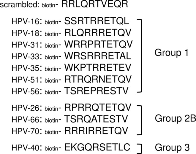
Fig 1 The biotinylated peptides used in this study; the peptides correspond to the 10 C-terminal amino acid residues from the E6 protein of each virus type. The scrambled peptide has the same amino acid composition as the HPV-18 peptide. Group 1 HPV types are defined as definitely carcinogenic in humans; Group 2B as possibly carcinogenic in humans, and Group 3 as no risk (IARC Monograph 100B).
Fig 1 The biotinylated peptides used in this study; the peptides correspond to the 10 C-terminal amino acid residues from the E6 protein of each virus type. The scrambled peptide has the same amino acid composition as the HPV-18 peptide. Group 1 HPV types are defined as definitely carcinogenic in humans; Group 2B as possibly carcinogenic in humans, and Group 3 as no risk (IARC Monograph 100B).CC BY · PubMed Central
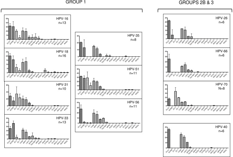
Fig 2 Peptide pulldown assays analysed by mass spectroscopy. Mass spectroscopy results of the pulldown assays from HaCat cells. Each histogram shows the numbers of peptides from each cellular PDZ domain–containing protein that was pulled down from HaCat extracts; n = total number of PDZ proteins pulled down by the E6 C-terminal peptide from HPV types 16, 18, 31, 33, 35, 51, 56 (Group 1); HPV types 26, 66, 70 (Group 2B); HPV type 40 (Group 3). The histograms were drawn using the Prism program, error bars show Standard error of the Mean. The scrambled peptide pulled down no PDZ domain-containing proteins in any assay.
Fig 2 Peptide pulldown assays analysed by mass spectroscopy. Mass spectroscopy results of the pulldown assays from HaCat cells. Each histogram shows the numbers of peptides from each cellular PDZ domain–containing protein that was pulled down from HaCat extracts; n = total number of PDZ proteins pulled down by the E6 C-terminal peptide from HPV types 16, 18, 31, 33, 35, 51, 56 (Group 1); HPV types 26, 66, 70 (Group 2B); HPV type 40 (Group 3). The histograms were drawn using the Prism program, error bars show Standard error of the Mean. The scrambled peptide pulled down no PDZ domain-containing proteins in any assay.CC BY · PubMed Central
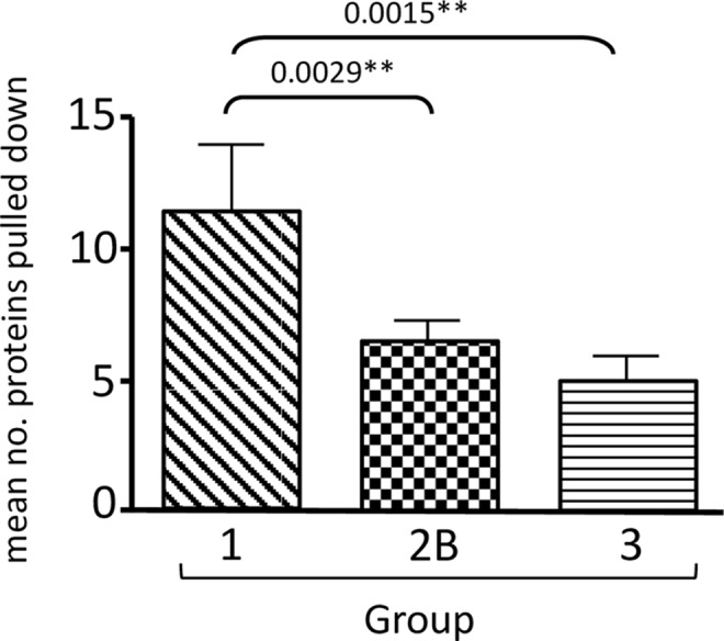
Fig 3 The mean numbers of PDZ domain-containing proteins pulled down by peptides from the Group 1 HPV types was significantly greater than the number pulled down by Group 2B (P = 0.0029) or Group 3 (P = 0.0015) types. Numbers were analysed using the Prism software and standard deviations are shown. The scrambled peptide pulled down no PDZ domain-containing proteins in any assay.
Fig 3 The mean numbers of PDZ domain-containing proteins pulled down by peptides from the Group 1 HPV types was significantly greater than the number pulled down by Group 2B (P = 0.0029) or Group 3 (P = 0.0015) types. Numbers were analysed using the Prism software and standard deviations are shown. The scrambled peptide pulled down no PDZ domain-containing proteins in any assay.CC BY · PubMed Central
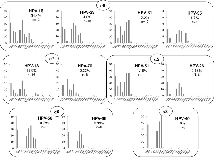
Fig 4 The number of cellular PDZ proteins targeted by the E6 PBMs increases with the cancer-causing potential of the virus type. The histograms show the number of PDZ domain-containing protein peptides pulled down by each E6 PBM, adjusted for the molecular weight of the protein concerned. The histograms are arranged by HPV phylogenetic group (boxes) and by the percentage of total invasive cervical cancers associated with each HPV type (highest on the left); the percentages are shown.
Fig 4 The number of cellular PDZ proteins targeted by the E6 PBMs increases with the cancer-causing potential of the virus type. The histograms show the number of PDZ domain-containing protein peptides pulled down by each E6 PBM, adjusted for the molecular weight of the protein concerned. The histograms are arranged by HPV phylogenetic group (boxes) and by the percentage of total invasive cervical cancers associated with each HPV type (highest on the left); the percentages are shown.CC BY · PubMed Central
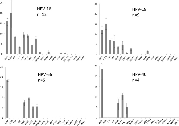
Fig 5 Mass spectroscopy results of the pulldown assays from Normal Immortal Keratinocytes (NIKS), using E6 C-terminal peptides from HPV types representing Group 1 (HPV-16, HPV-18), Group 2 (HPV-66) and Group 3 (HPV-40). The histograms show the numbers of peptides from each cellular PDZ domain–containing protein that was pulled down from NIKS cell extracts; n = total number of PDZ proteins pulled down by each E6 C-terminal peptide. The histograms were analysed using the Prism program and standard deviations are shown. The scrambled peptide pulled down no PDZ domain-containing proteins in any assay.
Fig 5 Mass spectroscopy results of the pulldown assays from Normal Immortal Keratinocytes (NIKS), using E6 C-terminal peptides from HPV types representing Group 1 (HPV-16, HPV-18), Group 2 (HPV-66) and Group 3 (HPV-40). The histograms show the numbers of peptides from each cellular PDZ domain–containing protein that was pulled down from NIKS cell extracts; n = total number of PDZ proteins pulled down by each E6 C-terminal peptide. The histograms were analysed using the Prism program and standard deviations are shown. The scrambled peptide pulled down no PDZ domain-containing proteins in any assay.CC BY · PubMed Central
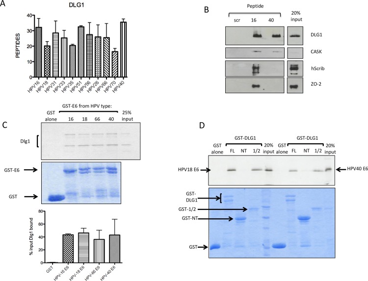
Fig 6 DLG1 is a common target of the E6 proteins, regardless of oncogenic potential. A. DLG1 binds to the E6 PBMs of both oncogenic and non-oncogenic HPV types; the histogram shows mean peptide numbers from the mass spectroscopy analysis of all pulldowns, analysed using Prism software, error bars show Standard Error of the Mean. B. The non-oncogenic HPV-40 E6 PBM binds DLG1 to a level similar to the HPV-16 E6 PBM. A peptide pulldown from HaCat cell extract, using the control (scr), HPV-16 and HPV-40 peptides, was analysed by western blot and probed for DLG1, CASK, hScrib and ZO-2, as indicated. C. DLG1 binds to the E6 proteins of both oncogenic and non-oncogenic HPV types. In vitro translated radiolabelled DLG1 was incubated with the full-length HPV-66 and HPV-40 E6 proteins expressed as GST fusion proteins. GST alone, GST-HPV-16E6 and HPV-18-E6 were included as controls. The upper panel shows the autoradiograph of bound DLG1 and the lower panel shows the Coomassie stained gel. Arrows indicate the GST-E6 proteins. The histogram shows the quantitation of at least three binding assays, analysed by the ImageJ and Prism programmes. Standard deviations are shown. D. The non-oncogenic HPV-40 E6 protein binds to DLG1 via its PDZ domains. A GST-pulldown assay, using in vitro translated radiolabelled HPV-18 and HPV-40 E6 proteins, together with GST fusion proteins with full-length DLG1(FL), the N-terminus of DLG1, lacking any PDZ domain (NT), and the first 401 amino acid residues of DLG1, containing the PDZ domains 1 and 2 (1/2). The upper panel shows the autoradiograph of the bound E6 proteins, the lower panel shows the Coomassie stained gel. Arrows indicate the GST fusion proteins.
Fig 6 DLG1 is a common target of the E6 proteins, regardless of oncogenic potential. A. DLG1 binds to the E6 PBMs of both oncogenic and non-oncogenic HPV types; the histogram shows mean peptide numbers from the mass spectroscopy analysis of all pulldowns, analysed using Prism software, error bars show Standard Error of the Mean. B. The non-oncogenic HPV-40 E6 PBM binds DLG1 to a level similar to the HPV-16 E6 PBM. A peptide pulldown from HaCat cell extract, using the control (scr), HPV-16 and HPV-40 peptides, was analysed by western blot and probed for DLG1, CASK, hScrib and ZO-2, as indicated. C. DLG1 binds to the E6 proteins of both oncogenic and non-oncogenic HPV types. In vitro translated radiolabelled DLG1 was incubated with the full-length HPV-66 and HPV-40 E6 proteins expressed as GST fusion proteins. GST alone, GST-HPV-16E6 and HPV-18-E6 were included as controls. The upper panel shows the autoradiograph of bound DLG1 and the lower panel shows the Coomassie stained gel. Arrows indicate the GST-E6 proteins. The histogram shows the quantitation of at least three binding assays, analysed by the ImageJ and Prism programmes. Standard deviations are shown. D. The non-oncogenic HPV-40 E6 protein binds to DLG1 via its PDZ domains. A GST-pulldown assay, using in vitro translated radiolabelled HPV-18 and HPV-40 E6 proteins, together with GST fusion proteins with full-length DLG1(FL), the N-terminus of DLG1, lacking any PDZ domain (NT), and the first 401 amino acid residues of DLG1, containing the PDZ domains 1 and 2 (1/2). The upper panel shows the autoradiograph of the bound E6 proteins, the lower panel shows the Coomassie stained gel. Arrows indicate the GST fusion proteins.CC BY · PubMed Central
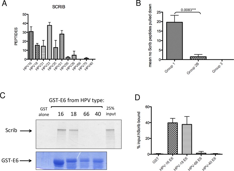
Fig 7 hScrib is preferentially bound by cancer-causing HPV E6 proteins. A. The histogram shows the mean numbers of hScrib peptides pulled down by each E6 C-terminus peptide, detected by mass spectroscopy. Standard Errors of the Mean are shown. B. Comparison of the numbers of hScrib peptides pulled down by the Group 1 and Group 2B peptides shows a statistically significant (P = 0.0083) difference. Numbers were analysed by Prism software and standard deviations are shown. C. hScrib is bound by the E6 proteins of oncogenic HPV types. In vitro translated radiolabelled hScrib was incubated with the full-length GST-HPV-16E6, HPV-18-E6, HPV-66-E6 and HPV-40-E6 proteins. The upper panel shows the autoradiograph of bound hScrib and the lower panel shows the Coomassie stained gel. Arrows indicate the GST-E6 proteins; the GST alone is not visible on a gel of low enough percentage acrylamide to allow hScrib visualisation, but was equivalent to that in Fig 2C and 2D. D. The histogram shows the quantitation of at least three binding assays, analysed by the Image J and Prism programmes. Standard deviations are shown.
Fig 7 hScrib is preferentially bound by cancer-causing HPV E6 proteins. A. The histogram shows the mean numbers of hScrib peptides pulled down by each E6 C-terminus peptide, detected by mass spectroscopy. Standard Errors of the Mean are shown. B. Comparison of the numbers of hScrib peptides pulled down by the Group 1 and Group 2B peptides shows a statistically significant (P = 0.0083) difference. Numbers were analysed by Prism software and standard deviations are shown. C. hScrib is bound by the E6 proteins of oncogenic HPV types. In vitro translated radiolabelled hScrib was incubated with the full-length GST-HPV-16E6, HPV-18-E6, HPV-66-E6 and HPV-40-E6 proteins. The upper panel shows the autoradiograph of bound hScrib and the lower panel shows the Coomassie stained gel. Arrows indicate the GST-E6 proteins; the GST alone is not visible on a gel of low enough percentage acrylamide to allow hScrib visualisation, but was equivalent to that in Fig 2C and 2D. D. The histogram shows the quantitation of at least three binding assays, analysed by the Image J and Prism programmes. Standard deviations are shown.CC BY · PubMed Central
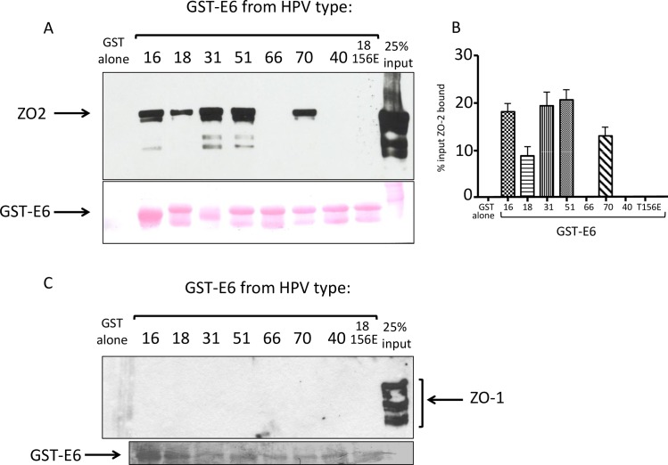
Fig 8 Novel interacting partners of HPV E6 proteins. ZO-2 binds to HPV E6 proteins in a PBM-dependent manner. (A). Extract of 293 cells transfected with ZO-2-expressing plasmid was incubated with GST-E6 proteins from HPV types 16, 18, 31, 51, 66, 70, 40 and the HPV18 T156E mutant, which ablates PDZ binding. Above: bound proteins detected by western blot probed with anti-HA antibody. Below: Ponceau staining of the nitrocellulose membranes, showing the GST-E6 proteins (arrowed). (B). The histogram shows the collated results of at least 3 assays. Standard deviations are shown. (C). No binding is seen with HA-ZO-1. Extract of 293 cells transfected with ZO-1-expressing plasmid was incubated with GST-E6 proteins from HPV types 16, 18, 31, 51, 66, 70, 40 and the HPV18 T156E mutant, which ablates PDZ binding. Above: bound proteins detected by western blot probed with anti-HA antibody. Below: Ponceau staining of the nitrocellulose membranes, showing the GST-E6 proteins (arrowed).
Fig 8 Novel interacting partners of HPV E6 proteins. ZO-2 binds to HPV E6 proteins in a PBM-dependent manner. (A). Extract of 293 cells transfected with ZO-2-expressing plasmid was incubated with GST-E6 proteins from HPV types 16, 18, 31, 51, 66, 70, 40 and the HPV18 T156E mutant, which ablates PDZ binding. Above: bound proteins detected by western blot probed with anti-HA antibody. Below: Ponceau staining of the nitrocellulose membranes, showing the GST-E6 proteins (arrowed). (B). The histogram shows the collated results of at least 3 assays. Standard deviations are shown. (C). No binding is seen with HA-ZO-1. Extract of 293 cells transfected with ZO-1-expressing plasmid was incubated with GST-E6 proteins from HPV types 16, 18, 31, 51, 66, 70, 40 and the HPV18 T156E mutant, which ablates PDZ binding. Above: bound proteins detected by western blot probed with anti-HA antibody. Below: Ponceau staining of the nitrocellulose membranes, showing the GST-E6 proteins (arrowed).CC BY · PubMed Central
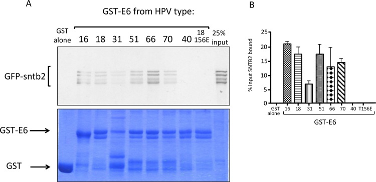
Fig 9 Novel interacting partners of HPV E6 proteins. Beta-2-syntrophin (SNTB2) binds to HPV E6 proteins in a PBM-dependent manner. (A). Autoradiograph of a GST pulldown assay using in vitro translated, radiolabelled SNTB2, together with GST-E6 fusion proteins from HPV types 16, 18, 31, 51, 66, 70, 40 and the 18E6T156E mutant. The Coomassie stained gel shows the GST fusion protein input (arrowed). (B). The histogram shows the collated results of at least 3 assays, standard deviations are shown.
Fig 9 Novel interacting partners of HPV E6 proteins. Beta-2-syntrophin (SNTB2) binds to HPV E6 proteins in a PBM-dependent manner. (A). Autoradiograph of a GST pulldown assay using in vitro translated, radiolabelled SNTB2, together with GST-E6 fusion proteins from HPV types 16, 18, 31, 51, 66, 70, 40 and the 18E6T156E mutant. The Coomassie stained gel shows the GST fusion protein input (arrowed). (B). The histogram shows the collated results of at least 3 assays, standard deviations are shown.CC BY · PubMed Central
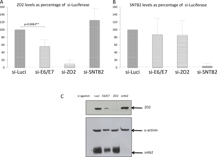
Fig 10 ZO-2 levels are increased upon HPV E6 expression. Western blot analysis of (A). ZO-2 and (B). SNTB2 levels in HeLa cells. si-ablation of E6/E7 expression results in a significant (p = 0.0063) reduction in ZO-2 levels. HeLa cells were transfected with siRNA to Luciferase (Luci) as control, with si-HPV-18 E6/E7 to ablate E6 expression, with si-ZO-2 or si-SNTB2. ZO-2 and SNTB2 levels were determined by Western Blot after 48h. The histograms show the results of at least three independent assays, analysed by Image J and Prism programmes and standard deviations are shown. (C). A representative western blot assay.
Fig 10 ZO-2 levels are increased upon HPV E6 expression. Western blot analysis of (A). ZO-2 and (B). SNTB2 levels in HeLa cells. si-ablation of E6/E7 expression results in a significant (p = 0.0063) reduction in ZO-2 levels. HeLa cells were transfected with siRNA to Luciferase (Luci) as control, with si-HPV-18 E6/E7 to ablate E6 expression, with si-ZO-2 or si-SNTB2. ZO-2 and SNTB2 levels were determined by Western Blot after 48h. The histograms show the results of at least three independent assays, analysed by Image J and Prism programmes and standard deviations are shown. (C). A representative western blot assay.CC BY · PubMed Central
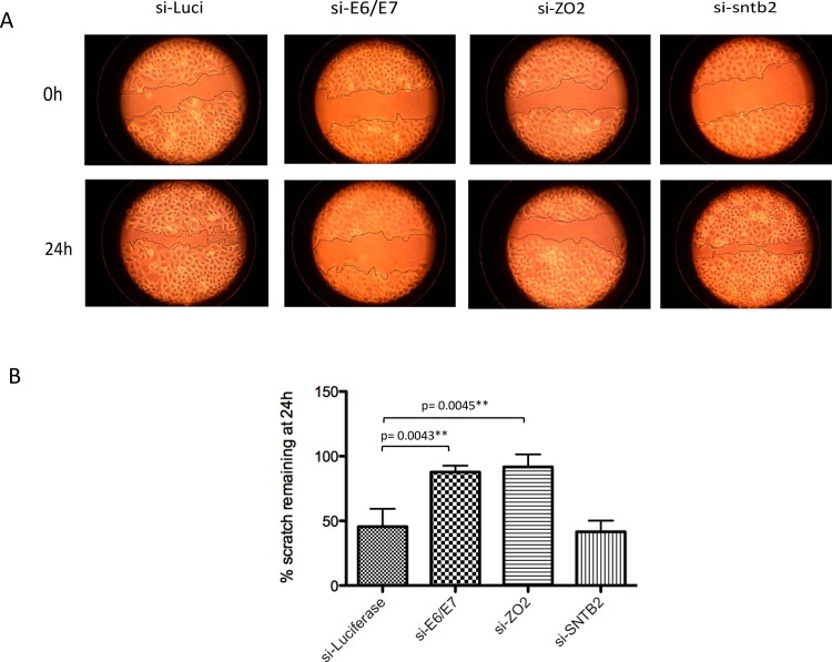
Fig 11 Increased ZO-2 levels promote cell migration. Ablation of ZO-2 expression reduces wound healing ability in HeLa cells, to a similar degree as ablation of E6 expression. (A). Confluent HeLa cells were scratched with a plastic pipette tip 48h after being transfected with si-RNA to luciferase, HPV 18 E6/E7, ZO-2 or SNTB2. The cells were photographed immediately post-scratch (0h) and after 24h. (B). The histogram shows the percentage area of the scratch remaining at 24h, from three independent experiments and standard deviations are shown.
Fig 11 Increased ZO-2 levels promote cell migration. Ablation of ZO-2 expression reduces wound healing ability in HeLa cells, to a similar degree as ablation of E6 expression. (A). Confluent HeLa cells were scratched with a plastic pipette tip 48h after being transfected with si-RNA to luciferase, HPV 18 E6/E7, ZO-2 or SNTB2. The cells were photographed immediately post-scratch (0h) and after 24h. (B). The histogram shows the percentage area of the scratch remaining at 24h, from three independent experiments and standard deviations are shown.CC BY · PubMed Central
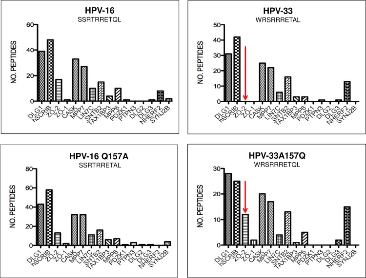
Fig 12 The molecular basis for substrate selection. Histograms representing the mass spectroscopy results of peptide pulldowns from HaCat cell extract, using peptides representing the C-termini of HPV-16 and HPV-33, and peptides in which the penultimate amino acid residue was swapped: HPV-16Q150A and HPV-33A148Q. The most marked effects of the change are arrowed. The histograms were drawn using the Prism software, the peptide sequences are shown on each histogram.
Fig 12 The molecular basis for substrate selection. Histograms representing the mass spectroscopy results of peptide pulldowns from HaCat cell extract, using peptides representing the C-termini of HPV-16 and HPV-33, and peptides in which the penultimate amino acid residue was swapped: HPV-16Q150A and HPV-33A148Q. The most marked effects of the change are arrowed. The histograms were drawn using the Prism software, the peptide sequences are shown on each histogram.CC BY · PubMed Central

Verification

Claims the paper makes

The claims and statistics of this paper haven’t been checked yet.

Run the check — free, no account needed. It reads the open-access text and checks the sources it cites.

A few free checks a day · papers already verified open instantly · results are public

Claims other papers make about this one

Each is checked against this paper’s own text: does it say what the citing paper says it does?

Self-check

Percentages, sums and reported differences re-computed from the paper’s own numbers.

The self-check needs the paper’s full text.

Citations

Cites

Show all 6 references

Research works printed but not linked: 39

  1. IARC monographs on the evaluation of carcinogenic risks to humans. Volume 100(B): Biological agents. 2012. WHO Press, ISBN13 9789283213192
  2. M Thomas, 2008, Human papillomaviruses, cervical cancer and cell polarity, Oncogene, 27, 7018, 10.1038/onc.2008.351 · DOI · Paper page
  3. K Münger, 1989, Complex formation of human papillomavirus E7 proteins with the retinoblastoma tumor suppressor gene product, EMBO J, 8, 4099, 10.1002/j.1460-2075.1989.tb08594.x · DOI · Paper page
  4. SN Boyer, 1996, E7 protein of human papilloma virus-16 induces degradation of retinoblastoma protein through the ubiquitin-proteasome pathway, Cancer Res, 56, 4620
  5. M Scheffner, 1990, The E6 oncoprotein encoded by human papillomavirus types 16 and 18 promotes the degradation of p53, Cell, 63, 1129, 10.1016/0092-8674(90)90409-8 · DOI · Paper page
  6. M Thomas, 1998, Inhibition of Bak-induced apoptosis by HPV-18 E6, Oncogene, 17, 2943, 10.1038/sj.onc.1202223 · DOI · Paper page
  7. M Thomas, 1999, Human papillomavirus (HPV) E6 interactions with Bak are conserved amongst E6 proteins from high and low risk HPV types, J Gen Virol, 80, 1513, 10.1099/0022-1317-80-6-1513 · DOI · Paper page
  8. ML Nguyen, 2003, The PDZ ligand domain of the human papillomavirus type 16 E6 protein is required for E6's induction of epithelial hyperplasia in vivo, J Virol, 77, 6957, 10.1128/JVI.77.12.6957-6964.2003 · DOI
  9. C Lee, 2004, Role of the PDZ domain-binding motif of the oncoprotein E6 in the pathogenesis of human papillomavirus type 31, J Virol, 78, 12366, 10.1128/JVI.78.22.12366-12377.2004 · DOI · Paper page
  10. L Nicolaides, 2011, Stabilization of HPV16 E6 protein by PDZ proteins, and potential implications for genome maintenance, Virology, 414, 137, 10.1016/j.virol.2011.03.017 · DOI · Paper page
  11. CP Delury, 2013, The role of protein kinase A regulation of the E6 PDZ-binding domain during the differentiation-dependent life cycle of human papillomavirus type 18, J Virol, 87, 9463, 10.1128/JVI.01234-13 · DOI
  12. N Brimer, 2014, Papillomavirus E6 PDZ interactions can be replaced by repression of p53 to promote episomal human papillomavirus genome maintenance, J Virol, 88, 3027, 10.1128/JVI.02360-13 · DOI
  13. T Kiyono, 1997, Binding of high-risk human papillomavirus E6 oncoproteins to the human homologue of the Drosophila discs large tumor suppressor protein, Proc Natl Acad Sci U S A, 94, 11612, 10.1073/pnas.94.21.11612 · DOI · Paper page
  14. RA Watson, 2003, Activity of the human papillomavirus E6 PDZ-binding motif correlates with an enhanced morphological transformation of immortalized human keratinocytes, J Cell Sci, 116, 4925, 10.1242/jcs.00809 · DOI
  15. A Shai, 2010, E6-associated protein is required for human papillomavirus type 16 E6 to cause cervical cancer in mice, Cancer Res, 70, 5064, 10.1158/0008-5472.CAN-09-3307 · DOI
  16. VK Subbaiah, 2011, PDZ domains: the building blocks regulating tumorigenesis, Biochem J, 439, 195, 10.1042/BJ20110903 · DOI · Paper page
  17. R Javier, 2008, Cell polarity proteins: common targets for tumorigenic human viruses, Oncogene, 27, 7031, 10.1038/onc.2008.352 · DOI · Paper page
  18. D Pim, 2012, Human papillomaviruses and the specificity of PDZ domain targeting, FEBS J, 279, 3530, 10.1111/j.1742-4658.2012.08709.x · DOI · Paper page
  19. Y Zhang, 2007, Structures of a human papillomavirus (HPV) E6 polypeptide bound to MAGUK proteins: mechanisms of targeting tumor suppressors by a high-risk HPV oncoprotein, J Virol, 81, 3618, 10.1128/JVI.02044-06 · DOI
  20. E Belotti, 2013, The human PDZome: a gateway to PSD95-Disc large-zonula occludens (PDZ)-mediated functions, Mol Cell Proteomics, 12, 2587, 10.1074/mcp.O112.021022 · DOI
  21. R Vincentelli, 2015, Quantifying domain-ligand affinities and specificities by high-throughput holdup assay, Nat Methods, 12, 787, 10.1038/nmeth.3438 · DOI
  22. M Thomas, 2008, Analysis of specificity determinants in the interactions of different HPV E6 proteins with their PDZ domain-containing substrates, Virology, 376, 371, 10.1016/j.virol.2008.03.021 · DOI
  23. K Luck, 2012, The emerging contribution of sequence context to the specificity of protein interactions mediated by PDZ domains, FEBS Lett, 586, 2648, 10.1016/j.febslet.2012.03.056 · DOI
  24. K Van Doorslaer, 2015, Degradation of human PDZ-proteins by human alphapapillomaviruses represents an evolutionary adaptation to a novel cellular niche, PLoS Pathog, 11, e1004980, 10.1371/journal.ppat.1004980 · DOI
  25. S Fournane, 2011, Surface plasmon resonance analysis of the binding of high-risk mucosal HPV E6 oncoproteins to the PDZ1 domain of the tight junction protein MAGI-1, J Mol Recognit, 24, 511, 10.1002/jmr.1056 · DOI
  26. SS Lee, 1997, Binding of human virus oncoproteins to hDlg/SAP97, a mammalian homolog of the Drosophila discs large tumor suppressor protein, Proc Natl Acad Sci U S A, 94, 6670, 10.1073/pnas.94.13.6670 · DOI · Paper page
  27. D Gardiol, 1999, Oncogenic human papillomavirus E6 proteins target the discs large tumour suppressor for proteasome-mediated degradation, Oncogene, 18, 5487, 10.1038/sj.onc.1202920 · DOI · Paper page
  28. S Nakagawa, 2000, Human scribble (Vartul) is targeted for ubiquitin-mediated degradation by the high-risk papillomavirus E6 proteins and the E6AP ubiquitin-protein ligase, Mol Cell Biol, 20, 8244, 10.1128/MCB.20.21.8244-8253.2000 · DOI · Paper page
  29. M Thomas, 2005, The hScrib/Dlg apico-basal control complex is differentially targeted by HPV-16 and HPV-18 E6 proteins, Oncogene, 24, 6222, 10.1038/sj.onc.1208757 · DOI · Paper page
  30. J Hernández-Monge, 2013, Papillomavirus E6 oncoprotein up-regulates occludin and ZO-2 expression in ovariectomized mice epidermis, Exp Cell Res, 319, 2588, 10.1016/j.yexcr.2013.07.028 · DOI
  31. G Halec, 2014, Pathogenic role of the eight probably/possibly carcinogenic HPV types 26, 53, 66, 67, 68, 70, 73 and 82 in cervical cancer, J Pathol, 234, 441, 10.1002/path.4405 · DOI
  32. P Massimi, 2012, Differential regulation of cell-cell contact, invasion and anoikis by hScrib and hDlg in keratinocytes, PLoS One
  33. LC Young, 2013, An MRAS, SHOC2, and SCRIB complex coordinates ERK pathway activation with polarity and tumorigenic growth, Mol Cell, 52, 679, 10.1016/j.molcel.2013.10.004 · DOI
  34. IA Elsum, 2014, Scrib heterozygosity predisposes to lung cancer and cooperates with KRas hyperactivation to accelerate lung cancer progression in vivo, Oncogene, 33, 5523, 10.1038/onc.2013.498 · DOI · Paper page
  35. C Kranjec, 2016, The high-risk HPV E6 target scribble (hScrib) is required for HPV E6 expression in cervical tumour-derived cell lines, Papillomavirus Res, 2, 70, 10.1016/j.pvr.2016.04.001 · DOI
  36. M Wigler, 1979, Transformation of mammalian cells with genes from procaryotes and eucaryotes, Cell, 16, 777, 10.1016/0092-8674(79)90093-X · DOI
  37. ER Flores, 1999, Establishment of the human papillomavirus type 16 (HPV-16) life cycle in an immortalized human foreskin keratinocyte cell line, Virology, 262, 344, 10.1006/viro.1999.9868 · DOI
  38. BL Allen-Hoffmann, 2000, Normal growth and differentiation in a spontaneously immortalised near-diploid human keratinocyte cell line, NIKS, J Invest Dermatol, 114, 444, 10.1046/j.1523-1747.2000.00869.x · DOI
  39. V Tomaić, 2009, Human and primate tumour viruses use PDZ binding as an evolutionarily conserved mechanism of targeting cell polarity regulators, Oncogene, 28, 1, 10.1038/onc.2008.365 · DOI

Cited by

See all 72

Metadata

Authors
Thomas, M., Myers, M. P., Massimi, P., Guarnaccia, C., & Banks, L.
Venue
PLOS Pathogens
Published
2016-08-02
Volume
12
Issue
8
Pages
e1005766
ISSN
1553-7374
Cited by
72
References
6
Language
en

About these checks

Registry, retraction, Cites and statistics results are computed, not judged, with one exception: a printed reference that no index lists is read once by a model before it counts as a citation error. Claims are extracted by a model and kept only when their quote appears verbatim in the text; each cited source is then checked, with a model’s help, for whether it says what the claim relies on. Those verdicts can be wrong — read the claim’s quote and the cited source before relying on one.

Statistics and arithmetic are recomputed, not judged.

Confidence bands are shown only for fields calibrated on our annotated test set. None is calibrated yet, so no band is shown.

What counts as a claim ›How we measure confidence ›How we check this ›

Are you an author, or do you see a problem with this page? Request a review

How to cite

Cite CoChat for the checks on this page, and the paper for its own findings.

This page
CoChat. (2026). Citation check: Analysis of Multiple HPV E6 PDZ Interactions Defines Type-Specific PDZ Fingerprints That Predict Oncogenic Potential. https://app.cochat.ai/papers/10.1371/journal.ppat.1005766
The paper
Thomas, M., Myers, M. P., Massimi, P., Guarnaccia, C., & Banks, L. (2016). Analysis of Multiple HPV E6 PDZ Interactions Defines Type-Specific PDZ Fingerprints That Predict Oncogenic Potential. PLOS Pathogens. https://doi.org/10.1371/journal.ppat.1005766