

The DOI matches its Crossref record; 62 of the 62 references it cites were found in a registry or an index.
Systematic, multiparametric analysis of Mycobacterium tuberculosis intracellular infection offers insight into coordinated virulence
Open accessAbstract
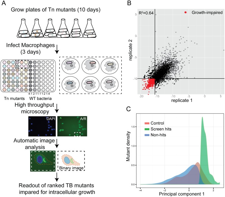
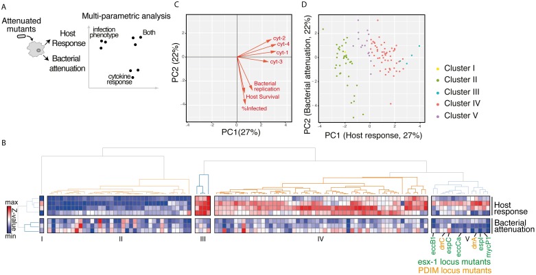
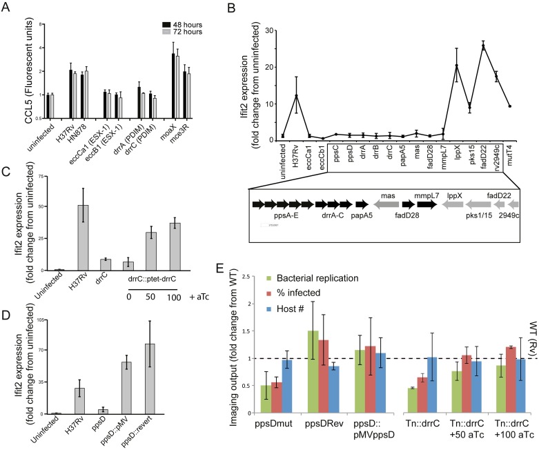
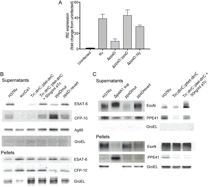
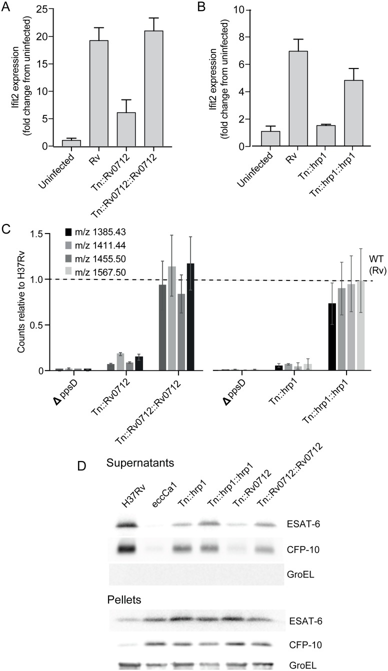
A key to the pathogenic success of Mycobacterium tuberculosis (Mtb), the causative agent of tuberculosis, is the capacity to survive within host macrophages. Although several factors required for this survival have been identified, a comprehensive knowledge of such factors and how they work together to manipulate the host environment to benefit bacterial survival are not well understood. To systematically identify Mtb factors required for intracellular growth, we screened an arrayed, non-redundant Mtb transposon mutant library by high-content imaging to characterize the mutant-macrophage interaction. Based on a combination of imaging features, we identified mutants impaired for intracellular survival. We then characterized the phenotype of infection with each mutant by profiling the induced macrophage cytokine response. Taking a systems-level approach to understanding the biology of identified mutants, we performed a multiparametric analysis combining pathogen and host phenotypes to predict functional relationships between mutants based on clustering. Strikingly, mutants defective in two well-known virulence factors, the ESX-1 protein secretion system and the virulence lipid phthiocerol dimycocerosate (PDIM), clustered together. Building upon the shared phenotype of loss of the macrophage type I interferon (IFN) response to infection, we found that PDIM production and export are required for coordinated secretion of ESX-1-substrates, for phagosomal permeabilization, and for downstream induction of the type I IFN response. Multiparametric clustering also identified two novel genes that are required for PDIM production and induction of the type I IFN response. Thus, multiparametric analysis combining host and pathogen infection phenotypes can be used to identify novel functional relationships between genes that play a role in infection.
Overview
The claims and statistics of this paper haven’t been checked yet.
Run the check — free, no account needed. It reads the open-access text and checks the sources it cites.
Integrity signals
Registry
Cites
Statistics
Figures
Verification
Claims the paper makes
The claims and statistics of this paper haven’t been checked yet.
Run the check — free, no account needed. It reads the open-access text and checks the sources it cites.
Claims other papers make about this one
Each is checked against this paper’s own text: does it say what the citing paper says it does?
The cited source says what the claim attributes to it.
In the citing paper
“The DosR system activates transcription of hrp1, a PDIM synthesis factor113, during short-term and long-term oxygen deprivation57,115.”
From The mycobacterial cell envelope — a moving target (2020) · cites this paper as [113]
The cited source says what the claim attributes to it.
In the citing paper
“PDIM may also facilitate phagosomal rupture and induction of the infection-promoting type I interferon response113,114.”
From The mycobacterial cell envelope — a moving target (2020) · cites this paper as [113]
In the citing paper
“However, ESX-1 is not the only factor involved in the process; it has been shown that besides ESX-1, the mycobacterial virulence lipids phthiocerol dimycocerosates also contribute to phagosomal rupture (76)(77)(78).”
From ESX/Type VII Secretion Systems—An Important Way Out for Mycobacterial Proteins (2019) · cites this paper as [78]
Self-check
Percentages, sums and reported differences re-computed from the paper’s own numbers.
The self-check needs the paper’s full text.
Citations
Cites
CellProfiler: image analysis software for identifying and quantifying cell phenotypes
Carpenter, A. E., Jones, T. R., Lamprecht, M. R., Clarke, C., Kang, I. H., Friman, O., Guertin, D. A., Chang, J. H., Lindquist, R. A., Moffat, J., et al. (2006) · Genome biology · doi:10.1186/gb-2006-7-10-r100 · Cited by 5,212
Self-citationMycobacterial persistence requires the utilization of host cholesterol
Pandey, A. K., & Sassetti, C. M. (2008) · Proceedings of the National Academy of Sciences · doi:10.1073/pnas.0711159105 · Cited by 1,064
Inhibition of Respiration by Nitric Oxide Induces a Mycobacterium tuberculosis Dormancy Program
Voskuil, M. I., Schnappinger, D., Visconti, K. C., Harrell, M. I., Dolganov, G., Sherman, D. R., & Schoolnik, G. K. (2003) · The Journal of Experimental Medicine · doi:10.1084/jem.20030205 · Cited by 975
Host-directed therapy of tuberculosis based on interleukin-1 and type I interferon crosstalk
Mayer-Barber, K. D., Andrade, B. B., Oland, S. D., Amaral, E. P., Barber, D. L., Gonzales, J., Derrick, S. C., Shi, R., Kumar, N. P., Wei, W., et al. (2014) · Nature · doi:10.1038/nature13489 · Cited by 757
Complex lipid determines tissue-specific replication of Mycobacterium tuberculosis in mice
Cox, J. S., Chen, B., McNeil, M., & Jacobs, W. R. (1999) · Nature · doi:10.1038/47042 · Cited by 746
Regulation of the Mycobacterium tuberculosis hypoxic response gene encoding α-crystallin
Sherman, D. R., Voskuil, M. I., Schnappinger, D., Liao, R., Harrell, M. I., & Schoolnik, G. K. (2001) · Proceedings of the National Academy of Sciences · doi:10.1073/pnas.121172498 · Cited by 735
Pym, A. S., Brodin, P., Brosch, R., Huerre, M., & Cole, S. T. (2002) · Molecular Microbiology · doi:10.1046/j.1365-2958.2002.03237.x · Cited by 729
Park, H., Guinn, K. M., Harrell, M. I., Liao, R., Voskuil, M. I., Tompa, M., Schoolnik, G. K., & Sherman, D. R. (2003) · Molecular Microbiology · doi:10.1046/j.1365-2958.2003.03474.x · Cited by 712
Strong, M., Sawaya, M. R., Wang, S., Phillips, M. L., Cascio, D., & Eisenberg, D. S. (2006) · Proceedings of the National Academy of Sciences · doi:10.1073/pnas.0602606103 · Cited by 704
Genome-wide requirements for Mycobacterium tuberculosis adaptation and survival in macrophages
Rengarajan, J., Bloom, B. R., & Rubin, E. J. (2005) · Proceedings of the National Academy of Sciences · doi:10.1073/pnas.0503272102 · Cited by 679
Cited by
Sorted by citations
Immune evasion and provocation by Mycobacterium tuberculosis
Chandra, P., Grigsby, S. J., & Philips, J. A. (2022) · Nature Reviews Microbiology · doi:10.1038/s41579-022-00763-4 · Cited by 652
The mycobacterial cell envelope — a moving target
Dulberger, C. L., Rubin, E. J., & Boutte, C. C. (2020) · Nature Reviews Microbiology · doi:10.1038/s41579-019-0273-7 · Cited by 365
Mycobacterium tuberculosis infection of host cells in space and time
Bussi, C., & Gutierrez, M. G. (2019) · FEMS Microbiology Reviews · doi:10.1093/femsre/fuz006 · Cited by 356
Allué‐Guardia, A., García, J. I., & Torrelles, J. B. (2021) · Frontiers in Microbiology · doi:10.3389/fmicb.2021.612675 · Cited by 233
The Macrophage: A Disputed Fortress in the Battle against Mycobacterium tuberculosis
Queval, C. J., Brosch, R., & Simeone, R. (2017) · Frontiers in Microbiology · doi:10.3389/fmicb.2017.02284 · Cited by 215
Cholesterol and fatty acids grease the wheels of Mycobacterium tuberculosis pathogenesis
Wilburn, K. M., Fieweger, R. A., & VanderVen, B. C. (2018) · Pathogens and Disease · doi:10.1093/femspd/fty021 · Cited by 203
Macrophage: A Cell With Many Faces and Functions in Tuberculosis
Ahmad, F., Rani, A., Alam, A., Zarin, S., Pandey, S., Singh, H., Hasnain, S. E., & Ehtesham, N. Z. (2022) · Frontiers in Immunology · doi:10.3389/fimmu.2022.747799 · Cited by 207
Tuberculosis and the art of macrophage manipulation
Upadhyay, S., Mittal, E., & Philips, J. A. (2018) · Pathogens and Disease · doi:10.1093/femspd/fty037 · Cited by 186
Immunometabolic crosstalk during bacterial infection
Rosenberg, G., Riquelme, S. A., Prince, A. S., & Avraham, R. (2022) · Nature Microbiology · doi:10.1038/s41564-022-01080-5 · Cited by 152
Self-citationLerner, T. R., Queval, C. J., Fearns, A., Repnik, U., Griffiths, G. W., & Gutierrez, M. G. (2018) · BMC Biology · doi:10.1186/s12915-017-0471-6 · Cited by 118
Mittal, E., Skowyra, M. L., Uwase, G., Tinaztepe, E., Mehra, A., Köster, S., Hanson, P. I., & Philips, J. A. (2018) · mBio · doi:10.1128/mbio.01765-18 · Cited by 110
Howard, N. C., Marín, N. D., Ahmed, M., Rosa, B. A., Martin, J. C., Bambousková, M., Sergushichev, A., Loginicheva, E., Kurepina, N. E., Rangel‐Moreno, J., et al. (2018) · Nature Microbiology · doi:10.1038/s41564-018-0245-0 · Cited by 108
Evolution of virulence in the Mycobacterium tuberculosis complex
Orgeur, M., & Brosch, R. (2018) · Current Opinion in Microbiology · doi:10.1016/j.mib.2017.11.021 · Cited by 107
Lipid metabolism and its implication in mycobacteria–host interaction
Gago, G., Diacovich, L., & Gramajo, H. C. (2017) · Current Opinion in Microbiology · doi:10.1016/j.mib.2017.11.020 · Cited by 98
Host Cell Targets of Released Lipid and Secreted Protein Effectors of Mycobacterium tuberculosis
Augenstreich, J., & Briken, V. (2020) · Frontiers in Cellular and Infection Microbiology · doi:10.3389/fcimb.2020.595029 · Cited by 79
Aylan, B., Bernard, E. M., Pellegrino, E., Botella, L., Fearns, A., Athanasiadi, N., Bussi, C., Santucci, P., & Gutierrez, M. G. (2023) · Nature Microbiology · doi:10.1038/s41564-023-01335-9 · Cited by 78
White, D. W., Elliott, S. R., Odean, E. J., Bemis, L., & Tischler, A. D. (2018) · mBio · doi:10.1128/mbio.00778-18 · Cited by 77
Self-citationThe structure of the endogenous ESX-3 secretion system
Poweleit, N., Czudnochowski, N., Nakagawa, R. M., Trinidad, D., Murphy, K. C., Sassetti, C. M., & Rosenberg, O. S. (2019) · eLife · doi:10.7554/elife.52983 · Cited by 76
Mycobacterium tuberculosis biology, pathogenicity and interaction with the host
Warner, D. F., Barczak, A. K., Gutierrez, M. G., & Mizrahi, V. (2025) · Nature Reviews Microbiology · doi:10.1038/s41579-025-01201-x · Cited by 81
Self-citationSpreading of a mycobacterial cell-surface lipid into host epithelial membranes promotes infectivity
Cambier, C. J., Banik, S. M., Buonomo, J. A., & Bertozzi, C. R. (2020) · eLife · doi:10.7554/elife.60648 · Cited by 70
Osman, M. M., Shanahan, J. K., Chu, F., Takaki, K. K., Pinckert, M. L., Pagán, A. J., Brosch, R., Conrad, W. H., & Ramakrishnan, L. (2022) · Proceedings of the National Academy of Sciences · doi:10.1073/pnas.2122161119 · Cited by 60
Path‐seq identifies an essential mycolate remodeling program for mycobacterial host adaptation
Peterson, E. J., Bailo, R., Rothchild, A. C., Arrieta‐Ortiz, M. L., Kaur, A., Pan, M., Mai, D., Abidi, A. A., Cooper, C., Aderem, A., et al. (2019) · Molecular Systems Biology · doi:10.15252/msb.20188584 · Cited by 63
M. tuberculosisinfection of human iPSDM reveals complex membrane dynamics during xenophagy evasion
Bernard, E. M., Fearns, A., Bussi, C., Santucci, P., Peddie, C. J., Lai, R., Collinson, L., & Gutierrez, M. G. (2020) · Journal of Cell Science · doi:10.1242/jcs.252973 · Cited by 60
Tiwari, S., Casey, R., Goulding, C. W., Hingley‐Wilson, S., & Jacobs, W. R. (2019) · Microbiology Spectrum · doi:10.1128/microbiolspec.bai-0024-2019 · Cited by 61
Roles for phthiocerol dimycocerosate lipids in Mycobacterium tuberculosis pathogenesis
Rens, C., Chao, J. D., Sexton, D. L., Tocheva, E. I., & Av‐Gay, Y. (2021) · Microbiology · doi:10.1099/mic.0.001042 · Cited by 60
Osman, M. M., Pagán, A. J., Shanahan, J. K., & Ramakrishnan, L. (2020) · PLoS ONE · doi:10.1371/journal.pone.0233252 · Cited by 48
ESX/Type VII Secretion Systems—An Important Way Out for Mycobacterial Proteins
Vaziri, F., & Brosch, R. (2019) · Microbiology Spectrum · doi:10.1128/microbiolspec.psib-0029-2019 · Cited by 50
Perspectives and Advances in the Understanding of Tuberculosis
Kinsella, R. L., Zhu, D. X., Harrison, G. A., Bridwell, A. E. M., Prusa, J., Chavez, S. M., & Stallings, C. L. (2021) · Annual Review of Pathology Mechanisms of Disease · doi:10.1146/annurev-pathol-042120-032916 · Cited by 51
Mycobacterial Evolution Intersects With Host Tolerance
Saelens, J. W., Viswanathan, G., & Tobin, D. M. (2019) · Frontiers in Immunology · doi:10.3389/fimmu.2019.00528 · Cited by 52
Augenstreich, J., Haanappel, E., Sayes, F., Siméone, R., Guillet, V., Mazères, S., Chalut, C., Mourey, L., Brosch, R., Guilhot, C., et al. (2020) · Frontiers in Cellular and Infection Microbiology · doi:10.3389/fcimb.2020.00420 · Cited by 46
Mycobacterium tuberculosis virulence lipid PDIM inhibits autophagy in mice
Mittal, E., Prasad, G. V. R. K., Upadhyay, S., Sadadiwala, J., Olive, A. J., Yang, G., Sassetti, C. M., & Philips, J. A. (2024) · Nature Microbiology · doi:10.1038/s41564-024-01797-5 · Cited by 42
Phan, T. H., van Leeuwen, L. M., Kuijl, C. P., Ummels, R., van Stempvoort, G., Rubio-Canalejas, A., Piersma, S. R., Jiménez, C. R., van der Sar, A. M., Houben, E. N. G., et al. (2018) · PLoS Pathogens · doi:10.1371/journal.ppat.1007247 · Cited by 47
Oxidative damage and delayed replication allow viable Mycobacterium tuberculosis to go undetected
Saito, K., Mishra, S., Warrier, T., Cicchetti, N., Mi, J., Weber, E., Jiang, X., Roberts, J., Gouzy, A., Kaplan, E., et al. (2021) · Science Translational Medicine · doi:10.1126/scitranslmed.abg2612 · Cited by 41
High-Content Imaging to Phenotype Antimicrobial Effects on Individual Bacteria at Scale
Sridhar, S., Forrest, S., Warne, B., Maes, M., Baker, S. G., Dougan, G., & Scott, J. B. (2021) · mSystems · doi:10.1128/msystems.00028-21 · Cited by 40
Modeling Tubercular ESX-1 Secretion Using Mycobacterium marinum
Chirakos, A. E., Balaram, A., Conrad, W. H., & Champion, P. A. D. (2020) · Microbiology and Molecular Biology Reviews · doi:10.1128/mmbr.00082-19 · Cited by 44
Breaching the phagosome, the case of the tuberculosis agent
Siméone, R., Sayes, F., Lawarée, E., & Brosch, R. (2021) · Cellular Microbiology · doi:10.1111/cmi.13344 · Cited by 41
Multi-Omics Technologies Applied to Tuberculosis Drug Discovery
Goff, A., Cantillon, D. M., Wildner, L. M., & Waddell, S. J. (2020) · Applied Sciences · doi:10.3390/app10134629 · Cited by 38
Uptake‐independent killing of macrophages by extracellular Mycobacterium tuberculosis aggregates
Toniolo, C., Dhar, N., & McKinney, J. D. (2023) · The EMBO Journal · doi:10.15252/embj.2023113490 · Cited by 36
Mitochondria: Powering the Innate Immune Response to Mycobacterium tuberculosis Infection
Patrick, K. L., & Watson, R. O. (2021) · Infection and Immunity · doi:10.1128/iai.00687-20 · Cited by 38
Sanchez, K. G., Ferrell, M. J., Chirakos, A. E., Nicholson, K. R., Abramovitch, R. B., Champion, M. M., & Champion, P. A. D. (2020) · mBio · doi:10.1128/mbio.02807-19 · Cited by 38
Santucci, P., Aylan, B., Botella, L., Bernard, E. M., Bussi, C., Pellegrino, E., Athanasiadi, N., & Gutierrez, M. G. (2022) · mBio · doi:10.1128/mbio.00117-22 · Cited by 33
Parbhoo, T., Mouton, J., & Sampson, S. L. (2022) · Frontiers in Cellular and Infection Microbiology · doi:10.3389/fcimb.2022.956607 · Cited by 39
Rapidly moving new bacteria to model-organism status
Liu, H., & Deutschbauer, A. M. (2018) · Current Opinion in Biotechnology · doi:10.1016/j.copbio.2017.12.006 · Cited by 32
Circulation of M. tuberculosis Beijing genotype in Latin America and the Caribbean
Irene, C.-C. M., Rodríguez–Castillo, J. G., Hernández‐Pando, R., & Murcia, M. I. (2019) · Pathogens and Global Health · doi:10.1080/20477724.2019.1710066 · Cited by 32
Pooled genetic screens with image‐based profiling
Walton, R. T., Singh, A. K., & Blainey, P. C. (2022) · Molecular Systems Biology · doi:10.15252/msb.202110768 · Cited by 29
Conserved ESX-1 Substrates EspE and EspF Are Virulence Factors That Regulate Gene Expression
Chirakos, A. E., Nicholson, K. R., Huffman, A., & Champion, P. A. D. (2020) · Infection and Immunity · doi:10.1128/iai.00289-20 · Cited by 28
A Small Protein but with Diverse Roles: A Review of EsxA in Mycobacterium–Host Interaction
Bao, Y., Wang, L., & Sun, J. (2021) · Cells · doi:10.3390/cells10071645 · Cited by 28
Chugh, S., Tiwari, P., Suri, C., Gupta, S. K., Singh, P., Bouzeyen, R., Kidwai, S., Srivastava, M., Rameshwaram, N. R., Kumar, Y., et al. (2024) · Proceedings of the National Academy of Sciences · doi:10.1073/pnas.2309664121 · Cited by 26
Butler, R. E., Smith, A. A., Mendum, T. A., Chandran, A., Wu, H., Lefrançois, L. H., Chambers, M. A., Soldati, T., & Stewart, G. R. (2020) · The ISME Journal · doi:10.1038/s41396-019-0572-z · Cited by 28
Mittal, E., Roth, A. T., Seth, A., Singamaneni, S., Beatty, W. L., & Philips, J. A. (2023) · eLife · doi:10.7554/elife.85416 · Cited by 24
Comparing the Metabolic Capabilities of Bacteria in the Mycobacterium tuberculosis Complex
Fieweger, R. A., Wilburn, K. M., & VanderVen, B. C. (2019) · Microorganisms · doi:10.3390/microorganisms7060177 · Cited by 28
A guide to Mycobacterium mutagenesis
Borgers, K., Vandewalle, K., Festjens, N., & Callewaert, N. (2019) · FEBS Journal · doi:10.1111/febs.15041 · Cited by 21
Hinman, A. E., Jani, C., Pringle, S. C., Zhang, W. R., Jain, N., Martinot, A. J., & Barczak, A. K. (2021) · eLife · doi:10.7554/elife.73984 · Cited by 25
Self-citationHe, X., Jiang, H., Chen, H., Zhang, H., Liu, Y., Xu, Z., Wu, F., Guo, S., Hou, J., Yang, M., et al. (2017) · Molecular & Cellular Proteomics · doi:10.1074/mcp.ra117.000296 · Cited by 20
Advances in the development of molecular genetic tools for Mycobacterium tuberculosis
Chhotaray, C., Tan, Y., Mugweru, J., Islam, M. M., Hameed, H. M. A., Wang, S., Lu, Z., Wang, C., Li, X., Tan, S., et al. (2018) · Journal of genetics and genomics/Journal of Genetics and Genomics · doi:10.1016/j.jgg.2018.06.003 · Cited by 17
Mycobacterium marinum as a model for understanding principles of mycobacterial pathogenesis
Menon, A. R., Prest, R. J., Tobin, D. M., & Champion, P. A. D. (2025) · Journal of Bacteriology · doi:10.1128/jb.00047-25 · Cited by 23
Lefrançois, L. H., Nitschke, J., Wu, H., Panis, G., Prados, J., Butler, R. E., Mendum, T. A., Hanna, N., Stewart, G. R., & Soldati, T. (2024) · mSystems · doi:10.1128/msystems.01326-23 · Cited by 17
Quezada, L. A. L., Smith, R. L., Lupoli, T. J., Edoo, Z., Li, X., Gold, B. S., Roberts, J., Ling, Y., Park, S. W., Nguyen, Q., et al. (2020) · Frontiers in Microbiology · doi:10.3389/fmicb.2020.01248 · Cited by 18
Self-citationMalik, A. A., Shariq, M., Sheikh, J. A., Jaiswal, U., Fayaz, H., Shrivastava, G., Ehtesham, N. Z., & Hasnain, S. E. (2024) · Critical Reviews in Biochemistry and Molecular Biology · doi:10.1080/10409238.2024.2411264 · Cited by 16
Jones, B. S., Hu, D. D., Nicholson, K. R., Cronin, R. M., Weaver, S. D., Champion, M. M., & Champion, P. A. D. (2024) · mSphere · doi:10.1128/msphere.00005-24 · Cited by 13
Xander, C. J., Saranathan, R., Jacobs, W. R., & Braunstein, M. (2024) · Infection and Immunity · doi:10.1128/iai.00217-24 · Cited by 17
Jani, C., Jain, N., Marsh, A. K., Uchil, P., Doan, T., Hudspith, M., Glover, O. T., Baskir, Z. R., Boucau, J., Root, D. E., et al. (2025) · Science Advances · doi:10.1126/sciadv.adr6166 · Cited by 16
Self-citationKoiri, D., Nandi, M., Abik Hameem, P. M., Bhausaheb, A. J., Kumar, A., Behura, A., Meher, G., Choudhary, V., Choubey, S., & Saleem, M. (2025) · Cell Reports · doi:10.1016/j.celrep.2025.115328 · Cited by 17
Heyman, O., Yehezkel, D., Mattioli, C. C., Blumberger, N., Rosenberg, G., Solomon, A., Hoffman, D., Moshe, N. B. B., & Avraham, R. (2023) · Proceedings of the National Academy of Sciences · doi:10.1073/pnas.2218812120 · Cited by 13
Self-citationLan, S., Li, Z., Hao, H., Liu, S., Huang, Z., Bai, Y., Li, Y., Yan, X., Gao, P., Chen, S., et al. (2023) · The FASEB Journal · doi:10.1096/fj.202300678r · Cited by 14
Harnessing the power of microscopy images to accelerate drug discovery: what are the possibilities?
Boyd, J. D., Fennell, M., & Carpenter, A. E. (2020) · Expert Opinion on Drug Discovery · doi:10.1080/17460441.2020.1743675 · Cited by 14
Self-citationUnraveling Mycobacterium tuberculosis acid resistance and pH homeostasis mechanisms
Laudouze, J., Canaan, S., Gouzy, A., & Santucci, P. (2025) · FEBS Letters · doi:10.1002/1873-3468.70023 · Cited by 15
Elliott, S. R., White, D. W., & Tischler, A. D. (2018) · Infection and Immunity · doi:10.1128/iai.00660-18 · Cited by 14
Self-citationAylan, B., Botella, L., Gutierrez, M. G., & Santucci, P. (2022) · FEBS Open Bio · doi:10.1002/2211-5463.13537 · Cited by 14
Targeting infection-specific peptides in immunopeptidomics studies for vaccine target discovery
Leddy, O. K., Yuki, Y., Carrington, M. N., Bryson, B. D., & White, F. M. (2025) · The Journal of Experimental Medicine · doi:10.1084/jem.20250444 · Cited by 11
Radhakrishnan, S. K., & Sundaramurthy, V. (2026) · mBio · doi:10.1128/mbio.03959-25 · Cited by 13
Malik, A. A., Shariq, M., Sheikh, J. A., Zarin, S., Ahuja, Y., Fayaz, H., Alam, A., Ehtesham, N. Z., & Hasnain, S. E. (2024) · Critical Reviews in Microbiology · doi:10.1080/1040841x.2024.2321494 · Cited by 10
Shankaran, D., Arumugam, P., Vasanthakumar, R. P., Singh, A., Bothra, A., Gandotra, S., & Rao, V. (2022) · The Journal of Immunology · doi:10.4049/jimmunol.2101029 · Cited by 10
Jani, C., Solomon, S. L., Peters, J. M., Pringle, S. C., Hinman, A. E., Boucau, J., Bryson, B. D., & Barczak, A. K. (2023) · mSystems · doi:10.1128/msystems.00052-23 · Cited by 10
Self-citationYabaji, S. M., Chatterjee, S., Waligurski, E., Gimelbrant, A. A., & Kramnik, I. (2022) · STAR Protocols · doi:10.1016/j.xpro.2022.101241 · Cited by 9
Tirap controls Mycobacterium tuberculosis phagosomal acidification
Belhaouane, I., Pochet, A., Chatagnon, J., Hoffmann, E., Queval, C. J., Deboosère, N., Boidin‐Wichlacz, C., Majlessi, L., Sencio, V., Heumel, S., et al. (2023) · PLoS Pathogens · doi:10.1371/journal.ppat.1011192 · Cited by 8
Blanco, F. C., Bigi, M. M., García, E. A., Elola, M. T., Vázquez, C. L., & Bigi, F. (2023) · Pathogens · doi:10.3390/pathogens12091159 · Cited by 8
Bobba, S., Chauhan, K. S., Akter, S., Das, S., Mittal, E., Mathema, B., Philips, J. A., & Khader, S. A. (2024) · PLoS Pathogens · doi:10.1371/journal.ppat.1012137 · Cited by 7
ESX/Type VII Secretion Systems-An Important Way Out for Mycobacterial Proteins
Vaziri, F., & Brosch, R. (2019) · ASM Press eBooks · doi:10.1128/9781683670285.ch28 · Cited by 8
Weaver, S. D., Hu, D. D., Jones, B. S., Champion, P. A. D., & Champion, M. M. (2025) · Microbiology Resource Announcements · doi:10.1128/mra.01161-25 · Cited by 3
pH-responsive substrate switching in mycobacterial type VII ESX secretion
Collars, O. A., Hernandez, R. L., Weaver, S. D., Prest, R. J., Manu, C., Viswanathan, G., Cronin, R. M., Jones, B. S., Tobin, D. M., Champion, M. M., et al. (2026) · mSphere · doi:10.1128/msphere.00056-26 · Cited by 4
Tiwari, S., Casey, R., Goulding, C. W., Hingley‐Wilson, S., & Jacobs, W. R. (2020) · John Wiley & Sons, Inc. eBooks · doi:10.1128/9781683670261.ch8 · Cited by 5
Dynamic Architecture of Mycobacterial Outer Membranes Revealed by All-Atom SimulationsPreprint
Brown, T. P., Chavent, M. G., & Im, W. (2025) · eLife · doi:10.7554/elife.108644.1 · Cited by 4
Protein-Mediated Virulence in Mycobacterium tuberculosis
Farnia, P., Velayati, A. A., Ghanavi, J., & Farnia, P. (2025) · Advances in experimental medicine and biology · doi:10.1007/978-3-031-96883-9_5 · Cited by 3
Multi-Omics Approaches in Drug Discovery
Rakshit, G., Komal, K., Dagur, P., & Jayaprakash, V. (2023) · Interdisciplinary biotechnological advances · doi:10.1007/978-981-99-1316-9_4 · Cited by 3
Jones, B. S., Pareek, V., Hu, D. D., Weaver, S. D., Syska, C., Galfano, G., Champion, M. M., & Champion, P. A. D. (2025) · Proceedings of the National Academy of Sciences · doi:10.1073/pnas.2502577122 · Cited by 3
Mycobacterial Genetic Technologies for Probing the Host-Pathogen Microenvironment
Adefisayo, O. O., Curtis, E. R., & Smith, C. M. (2023) · Infection and Immunity · doi:10.1128/iai.00430-22 · Cited by 3
NINJ1 mediates necrosis of Mycobacterium tuberculosis infected human macrophagesPreprint
Sætra, R. S., Hansen, M., Kappelhoff, S., Bugge, M., Marstad, A., Ryan, L., Devant, P., Kagan, J. C., Freude, K., Beckwith, K. S., et al. (2025) · bioRxiv (Cold Spring Harbor Laboratory) · doi:10.1101/2025.11.04.685750 · Cited by 2
Perret, A., Michard, C., Moreau, D., & Soldati, T. (2026) · doi:10.1002/9781394232413.ch7 · Cited by 3
The role of macrophages in tuberculosis
Sikder, M. H., Hossen, M. J., Matin, M. A., Rahman, M., Ahmed, M. S., Asadazzuaman, M., & Islam, A. (2022) · Elsevier eBooks · doi:10.1016/b978-0-12-822368-0.00017-7 · Cited by 2
Koiri, D., Nandi, M., Hameem, P. M. A., Bhausaheb, A. J., Meher, G., Behura, A., Kumar, A., Choudhary, V., Choubey, S., & Saleem, M. (2024) · bioRxiv (Cold Spring Harbor Laboratory) · doi:10.1101/2024.04.19.590309 · Cited by 2
Leddy, O. K., Yuki, Y., Carrington, M. N., Bryson, B. D., & White, F. M. (2024) · bioRxiv (Cold Spring Harbor Laboratory) · doi:10.1101/2024.09.11.612454 · Cited by 2
Dynamic architecture of mycobacterial outer membranes revealed by all-atom simulations
Brown, T. P., Chavent, M. G., & Im, W. (2026) · eLife · doi:10.7554/elife.108644.3 · Cited by 1
Wang, H., Li, D., Li, S., Chao Hui, Z. H. O. U., Huang, X., Huang, W., Niu, C., Ding, C., Song, J., Zhou, S., et al. (2025) · International Journal of Biological Macromolecules · doi:10.1016/j.ijbiomac.2025.146193 · Cited by 2
Ge, G., Cheng, Z., Xu, J., Yue, B., Hou, H., Liu, Y., Zhu, C., Ge, H., Shi, S., Li, J., et al. (2026) · BMC Genomics · doi:10.1186/s12864-026-12540-1 · Cited by 1
Blanco, F. L., Vázquez, C. L., Bigi, M. M., Rocha, R. V., García, E. A., & Bigi, F. (2026) · International Journal of Molecular Sciences · doi:10.3390/ijms27094099 · Cited by 1
Application and progress of highcontent imaging in molecular biology
Boyang, H., Yangyanqiu, W., Wenting, R., Chenxin, Y., Chu, J., Zhanbo, Q., Yanjun, Y., Qiang, Y., & Han, S. (2023) · Biotechnology Journal · doi:10.1002/biot.202300170 · Cited by 2
Zhao, X., Bao, Y., Wang, L., Qian, W. P., & Sun, J. (2019) · bioRxiv (Cold Spring Harbor Laboratory) · doi:10.1101/611434 · Cited by 1
High-content imaging to phenotype antimicrobial effects on individual bacteria at scalePreprint
Sridhar, S., Forrest, S., Warne, B., Maes, M., Baker, S. G., Gordon Dougan, B., & Scott, J. B. (2021) · bioRxiv (Cold Spring Harbor Laboratory) · doi:10.1101/2021.01.11.426307 · Cited by 1
Wang, H., Wen, X., Huang, W., Li, D., Huang, X., Zhou, C., Niu, C., Du, D., Song, J., Song, Y., et al. (2020) · SSRN Electronic Journal · doi:10.2139/ssrn.3741239 · Cited by 1
Metadata
- Authors
- Barczak, A. K., Avraham, R., Singh, S., Luo, S. S., Zhang, W. R., Bray, M.-A., Hinman, A. E., Thompson, M., Nietupski, R. M., Golas, A., Montgomery, P., Fitzgerald, M., Smith, R. S., White, D. W., Tischler, A. D., Carpenter, A. E., & Hung, D. T.
- Affiliations
- Broad Institute (US)
- Harvard University (US)
- University of Minnesota (US)
- Corresponding author
- Hung, Deborah T.
- ORCID
- Venue
- PLOS Pathogens
- Publisher
- Public Library of Science (PLoS)
- Published
- 2017-05-15
- Volume
- 13
- Issue
- 5
- Pages
- e1006363
- ISSN
- 1553-7366 (ISSN-L)
- 1553-7374
- PMC
- PMC5444860
- OpenAlex
- W2615762114
- Cited by
- 120
- References
- 62
- Citations by year
- 2026: 16 · 2025: 14 · 2024: 11 · 2023: 14 · 2022: 12 · 2021: 10 · 2020: 14 · 2019: 13 · 2018: 12 · 2017: 4
- FWCI
- 9.05
- Type
- journal article
- Language
- en
- Update notices
- new_version (2017-05-25)
- Open access
- gold
- doi.org
- Licence
- Locations
- PLoS Pathogens (publishedVersion, cc-by)
- Europe PMC (PubMed Central) (publishedVersion, cc-by)
- PubMed Central (publishedVersion, cc-by)
- PubMed (publishedVersion)
- Digital Access to Scholarship at Harvard (DASH) (Harvard University) (submittedVersion, cc-by)
- DOAJ (DOAJ: Directory of Open Access Journals) (submittedVersion, cc-by-sa)
- Figshare (submittedVersion, cc-by)
- Funders
- National Institute of Allergy and Infectious Diseases (K08-AI080944)
- Massachusetts General Hospital (Claflin Award)
- Ragon Institute (TB Seed Grant)
- Broad Institute Tuberculosis Donor Group and Pershing Square Foundation
- Harvard University Center for AIDS Research (US) (P30-AI060354)
- Massachusetts General Hospital Department of Medicine (Pingree Transformative Scholar Award)
- Topics
- Health Sciences › Medicine › Infectious Diseases › Tuberculosis Research and Epidemiology
- Health Sciences › Medicine › Epidemiology › Mycobacterium research and diagnosis
- Health Sciences › Medicine › Infectious Diseases › Infectious Diseases and Tuberculosis
- Keywords
- Mycobacterium tuberculosis, intracellular survival, macrophages, virulence factors, ESX-1 secretion system, phthiocerol dimycocerosate, type I interferon response, transposon mutant library, high-content imaging, macrophage cytokine response, tuberculosis pathogenesis
- MeSH terms
- Animals, Antigens, Bacterial, Bacterial Proteins, Cell Line, Macrophages, Mutation, Mycobacterium tuberculosis, Phagosomes, Phenotype, Tuberculosis, Virulence, Gene Library, Cytokines, Mice, Host-Pathogen Interactions
About these checks
Registry, retraction, Cites and statistics results are computed, not judged, with one exception: a printed reference that no index lists is read once by a model before it counts as a citation error. Claims are extracted by a model and kept only when their quote appears verbatim in the text; each cited source is then checked, with a model’s help, for whether it says what the claim relies on. Those verdicts can be wrong — read the claim’s quote and the cited source before relying on one.
Statistics and arithmetic are recomputed, not judged.
Confidence bands are shown only for fields calibrated on our annotated test set. None is calibrated yet, so no band is shown.
What counts as a claim ›How we measure confidence ›How we check this ›
Are you an author, or do you see a problem with this page? Request a review
