Cited by 40

The DOI matches its Crossref record.

The HPV-18 E7 CKII phospho acceptor site is required for maintaining the transformed phenotype of cervical tumour-derived cells

Basukala, Om, Mittal, Suruchi, Massimi, Paola, Bestagno, Marco, Banks, Lawrence·2019·PLOS Pathogens

Overview

The claims and statistics of this paper haven’t been checked yet.

Run the check — free, no account needed. It reads the open-access text and checks the sources it cites.

A few free checks a day · papers already verified open instantly · results are public

Integrity signals

Registry

Crossref · 10.1371/journal.ppat.1007769

Cites

Statistics

Figures

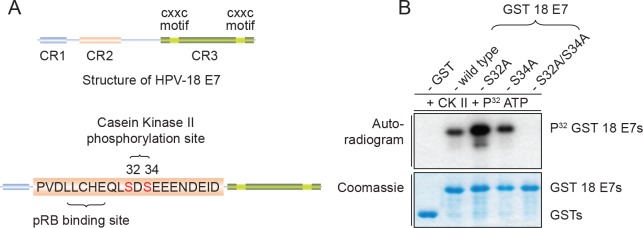
Fig 1 Mutation of HPV-18 E7 wild type to S32A/S34A abolishes phosphorylation by casein kinase II (CKII). (A) Schematic representation of HPV-18 E7 conserved regions (CR), the pRB binding site and CKII phosphorylation site in the CR2 domain. (B) In vitro phosphorylation of GST HPV-18 E7 fusion proteins. Purified GST 18E7 wild type and mutants, as indicated, were incubated with CKII and 32P-γATP. After extensive washing, bound proteins were subjected to SDS-PAGE and autoradiographic analysis. The upper panel represents the autoradiogram and the lower panel represents the Coomassie Blue-stained gel.
Fig 1 Mutation of HPV-18 E7 wild type to S32A/S34A abolishes phosphorylation by casein kinase II (CKII). (A) Schematic representation of HPV-18 E7 conserved regions (CR), the pRB binding site and CKII phosphorylation site in the CR2 domain. (B) In vitro phosphorylation of GST HPV-18 E7 fusion proteins. Purified GST 18E7 wild type and mutants, as indicated, were incubated with CKII and 32P-γATP. After extensive washing, bound proteins were subjected to SDS-PAGE and autoradiographic analysis. The upper panel represents the autoradiogram and the lower panel represents the Coomassie Blue-stained gel.CC BY · PubMed Central
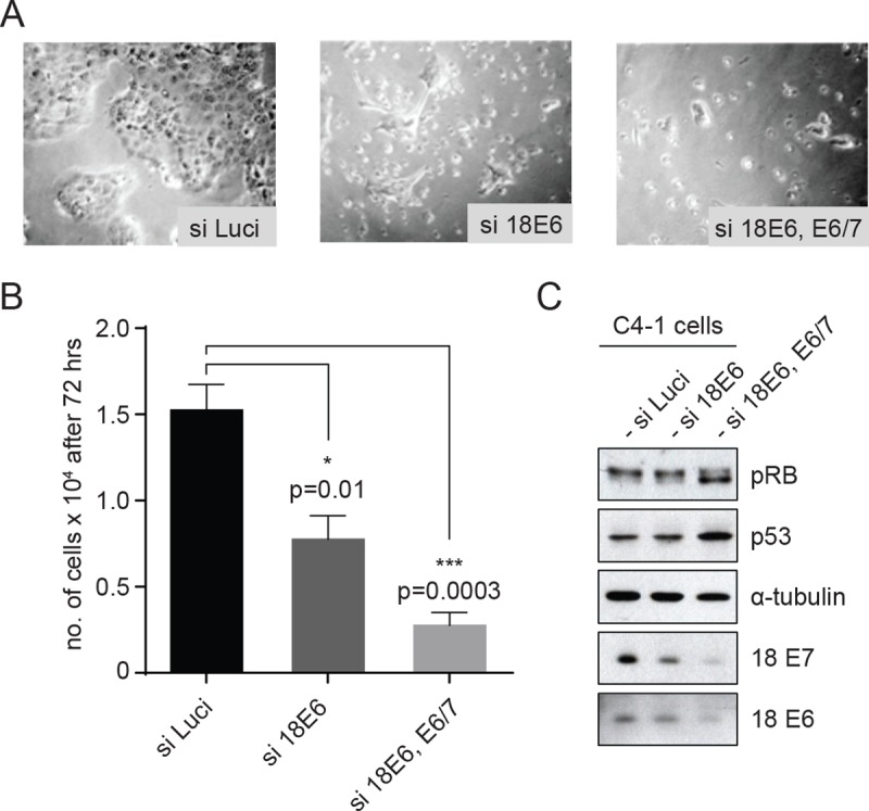
Fig 2 C4-1 cells are dependent on continued expression of E6 and E7 for growth and proliferation. (A) Micrograph of the cells after 72h of siRNA treatment. C4-1 cells were transfected with si-Luciferase (si-Luci), si-HPV18 E6, si-HPV18E6 and E6/7 together, using RNAimax transfection reagent. (B) After 72h incubation, cells were counted to assess the remaining cell number and the graph shows the quantification after the different siRNA transfections. Asterisks indicate the p-value (*—p<0.01, ***—p<0.0003) of mean cell number in siRNA transfections compared with siLuci, using an unpaired t-test. (C) Western blots showing levels of pRB, p53, α-tubulin, 18E7 and 18E6 after transfection with the indicated siRNAs.
Fig 2 C4-1 cells are dependent on continued expression of E6 and E7 for growth and proliferation. (A) Micrograph of the cells after 72h of siRNA treatment. C4-1 cells were transfected with si-Luciferase (si-Luci), si-HPV18 E6, si-HPV18E6 and E6/7 together, using RNAimax transfection reagent. (B) After 72h incubation, cells were counted to assess the remaining cell number and the graph shows the quantification after the different siRNA transfections. Asterisks indicate the p-value (*—p<0.01, ***—p<0.0003) of mean cell number in siRNA transfections compared with siLuci, using an unpaired t-test. (C) Western blots showing levels of pRB, p53, α-tubulin, 18E7 and 18E6 after transfection with the indicated siRNAs.CC BY · PubMed Central
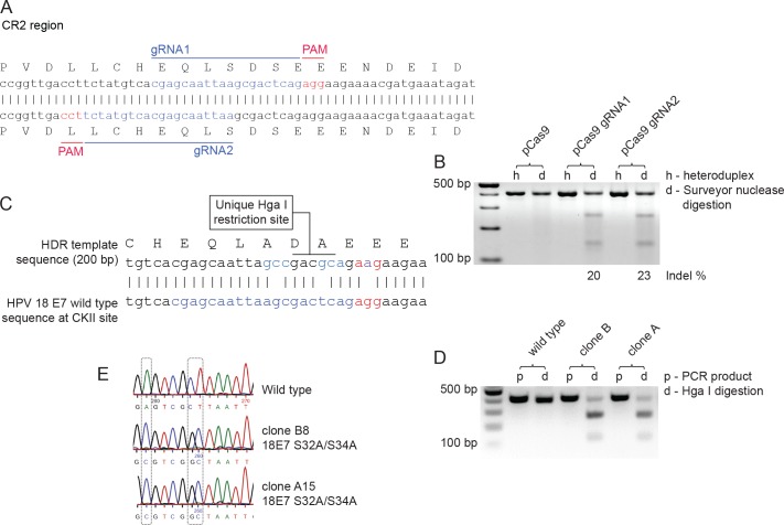
Fig 3 Strategy for genome editing HPV-18 E7 S32A/S34A in C4-1 cells. (A) Design of gRNA targeting the CKII phosphorylation site in the HPV-18 E7 CR2 region. gRNAs targeting the CKII phosphorylation site in HPV-18 E7 genomic region were designed using the online gRNA design platform–CRISPR MultiTargeter (http://www.multicrispr.net/). gRNA oligos were then annealed and cloned into the BbsI restriction site in pSpCas9(BB)-2A-GFP (PX458). (B) Surveyor assay for assessing efficiency of gRNAs. C4-1 cells were transfected with pSpCas9(BB)-2A-GFP or with the same construct plus gRNA1 or gRNA2. After 72h, cells were FACS-sorted to collect GFP-positive cells. Genomic DNA was extracted to PCR-amplify the specific region spanning E7 (414bp). Amplicons were denatured and reannealed to allow DNA heteroduplex formation, then treated with Surveyor nuclease and Surveyor enhancer (IDT) and analyzed on a 2% agarose gel. Lanes h are heteroduplex PCR products and lanes d are Surveyor nuclease digestion products for respective Cas9 and gRNA treatment constructs. (C) Design of single-stranded DNA oligonucleotide (ssODN) as donor template for homology-directed repair (HDR). ssODN was designed as a 100bp homology arm, flanking the predicted double strand break site to change the serine 32 to alanine (agc to gcc) and the serine 34 to alanine (tca to gca). The mutagenesis was designed to insert a unique HgaI restriction site, to allow screening of the edited clones by genomic DNA isolation, PCR amplification of the edited region and restriction digestion. (D) Representative agarose gel electrophoresis of Hga I digestion of 18E7 PCR amplicons from wild type and mutant clones. (E) Validation of the genome editing with Sanger sequencing.
Fig 3 Strategy for genome editing HPV-18 E7 S32A/S34A in C4-1 cells. (A) Design of gRNA targeting the CKII phosphorylation site in the HPV-18 E7 CR2 region. gRNAs targeting the CKII phosphorylation site in HPV-18 E7 genomic region were designed using the online gRNA design platform–CRISPR MultiTargeter (http://www.multicrispr.net/). gRNA oligos were then annealed and cloned into the BbsI restriction site in pSpCas9(BB)-2A-GFP (PX458). (B) Surveyor assay for assessing efficiency of gRNAs. C4-1 cells were transfected with pSpCas9(BB)-2A-GFP or with the same construct plus gRNA1 or gRNA2. After 72h, cells were FACS-sorted to collect GFP-positive cells. Genomic DNA was extracted to PCR-amplify the specific region spanning E7 (414bp). Amplicons were denatured and reannealed to allow DNA heteroduplex formation, then treated with Surveyor nuclease and Surveyor enhancer (IDT) and analyzed on a 2% agarose gel. Lanes h are heteroduplex PCR products and lanes d are Surveyor nuclease digestion products for respective Cas9 and gRNA treatment constructs. (C) Design of single-stranded DNA oligonucleotide (ssODN) as donor template for homology-directed repair (HDR). ssODN was designed as a 100bp homology arm, flanking the predicted double strand break site to change the serine 32 to alanine (agc to gcc) and the serine 34 to alanine (tca to gca). The mutagenesis was designed to insert a unique HgaI restriction site, to allow screening of the edited clones by genomic DNA isolation, PCR amplification of the edited region and restriction digestion. (D) Representative agarose gel electrophoresis of Hga I digestion of 18E7 PCR amplicons from wild type and mutant clones. (E) Validation of the genome editing with Sanger sequencing.CC BY · PubMed Central
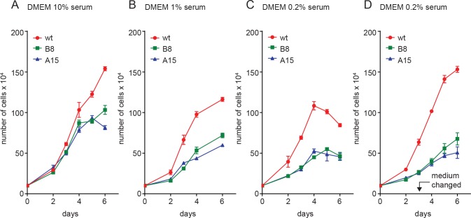
Fig 4 Growth of CKII site mutant cells is reduced in low nutrient conditions. (A) C4-1 wild type and mutant lines were seeded at 3 x 10⁵ cells per 60 mm petri dish, in DMEM with 10% fetal bovine serum and counted over a 6 day period. For determination of growth in low serum, the assay was repeated in DMEM with (B) 1% serum, (C) 0.2% serum and (D) 0.2% serum with medium replaced on the third day. The results show the mean of at least three independent assays and the error bars show standard deviations.
Fig 4 Growth of CKII site mutant cells is reduced in low nutrient conditions. (A) C4-1 wild type and mutant lines were seeded at 3 x 10⁵ cells per 60 mm petri dish, in DMEM with 10% fetal bovine serum and counted over a 6 day period. For determination of growth in low serum, the assay was repeated in DMEM with (B) 1% serum, (C) 0.2% serum and (D) 0.2% serum with medium replaced on the third day. The results show the mean of at least three independent assays and the error bars show standard deviations.CC BY · PubMed Central
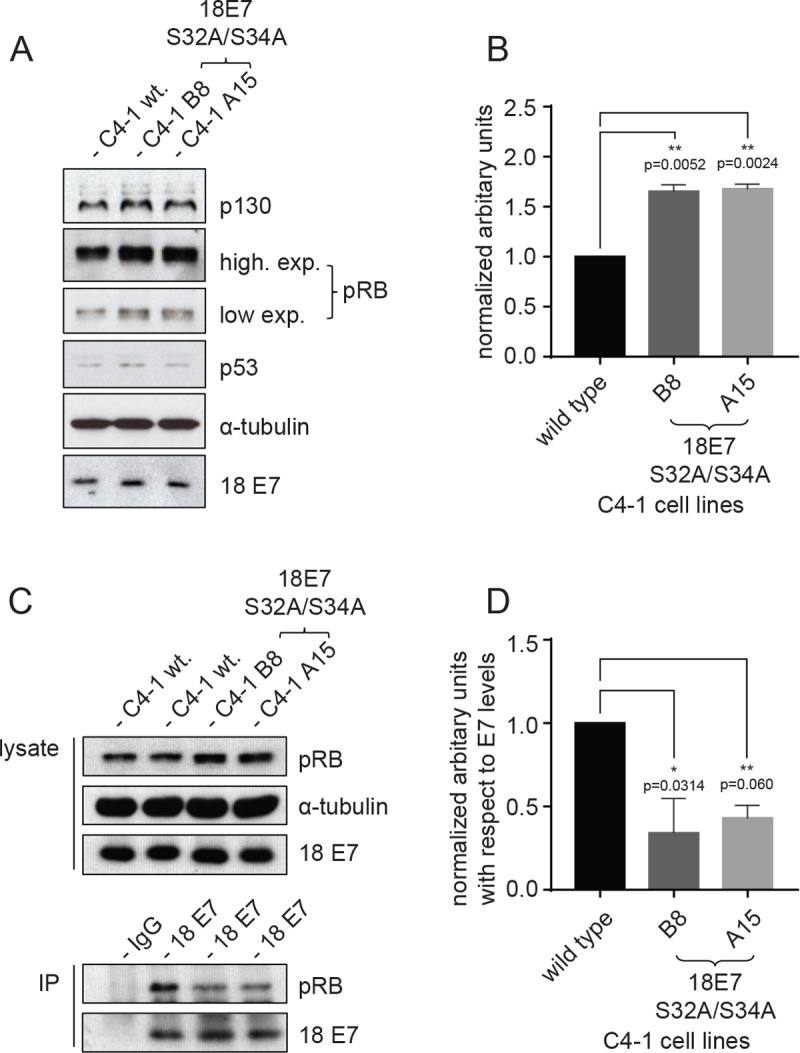
Fig 5 Steady-state levels of HPV-18 E7 and pocket proteins in the C4-1 cell lines. (A) Western blot analysis of total cell extracts obtained from the wild type and mutant C4-1 lines. These were probed for p130, pRb, p53, HPV-18 E7, and α-tubulin was used as a loading control. (B) Quantification of levels of pRB normalized with respect to α-tubulin shown as bars with the standard error of mean. Asterisks indicate the p-value of mean levels of pRB in mutant cells compared with wildtype cells. (C) Cells lysates from wildtype and CKII mutant C4-1 cells were immunoprecipitated with anti-HPV-18 E7 antibody overnight and then incubated with protein A beads for 90 minutes at 4°C. Immunoprecipitated complexes were then washed with lysis buffer and analysed by Western blotting for pRB. Mouse monoclonal anti-GFP antibody was used as control IgG. The upper panels show the protein inputs for the immunoprecipitations, with α-tubulin serving as a loading control and the results of immunoprecipitation are shown in the lower two panels. (D) Quantification of the levels of pRB immunoprecipitated with respect to levels of E7 shown as bars with the standard error of mean. Asterisks indicate the p-value of mean levels of pRB immunoprecipitated in mutant cells compared with wildtype cells.
Fig 5 Steady-state levels of HPV-18 E7 and pocket proteins in the C4-1 cell lines. (A) Western blot analysis of total cell extracts obtained from the wild type and mutant C4-1 lines. These were probed for p130, pRb, p53, HPV-18 E7, and α-tubulin was used as a loading control. (B) Quantification of levels of pRB normalized with respect to α-tubulin shown as bars with the standard error of mean. Asterisks indicate the p-value of mean levels of pRB in mutant cells compared with wildtype cells. (C) Cells lysates from wildtype and CKII mutant C4-1 cells were immunoprecipitated with anti-HPV-18 E7 antibody overnight and then incubated with protein A beads for 90 minutes at 4°C. Immunoprecipitated complexes were then washed with lysis buffer and analysed by Western blotting for pRB. Mouse monoclonal anti-GFP antibody was used as control IgG. The upper panels show the protein inputs for the immunoprecipitations, with α-tubulin serving as a loading control and the results of immunoprecipitation are shown in the lower two panels. (D) Quantification of the levels of pRB immunoprecipitated with respect to levels of E7 shown as bars with the standard error of mean. Asterisks indicate the p-value of mean levels of pRB immunoprecipitated in mutant cells compared with wildtype cells.CC BY · PubMed Central
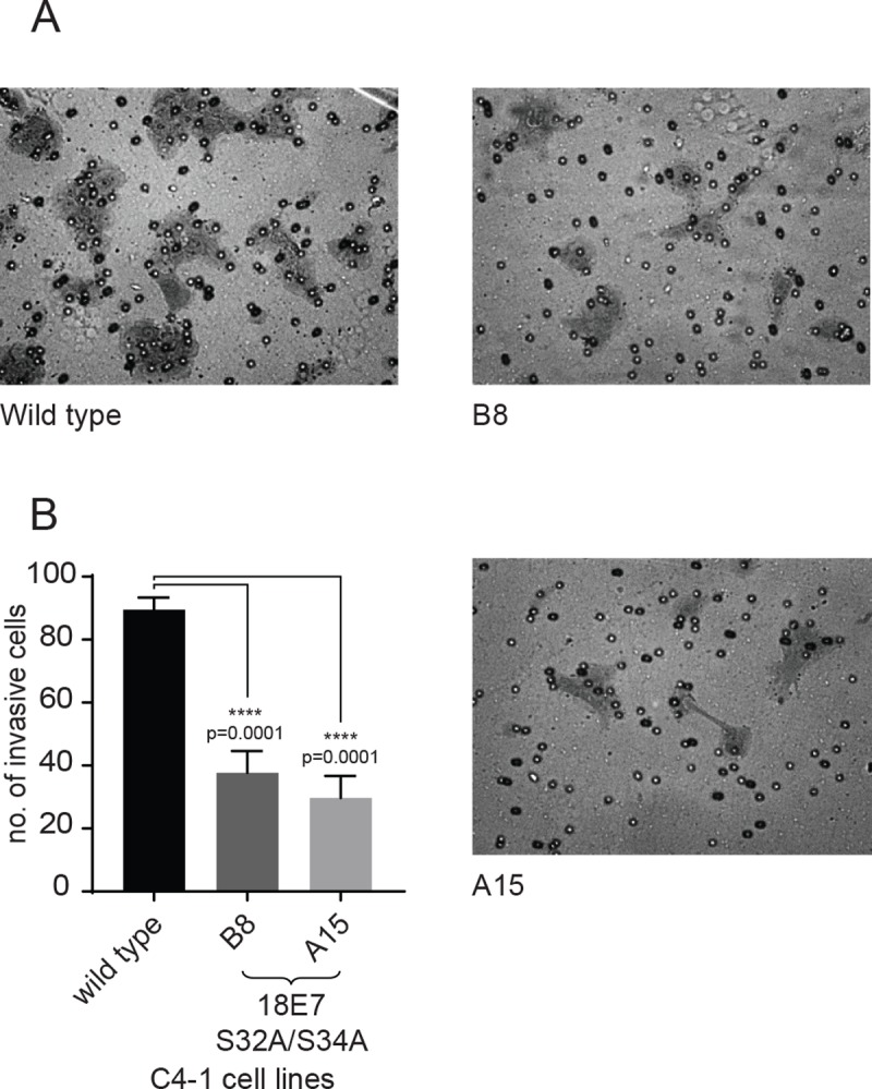
Fig 6 CKII site mutant C4-1 cells show defects in matrigel invasion. Equal numbers of wild type and mutant C4-1 cell lines were seeded in the upper part of Matrigel invasion chambers, in DMEM without serum. DMEM with 2% serum was used as a chemoattractant in the lower chamber. After 22h, cells that had invaded the lower chamber were fixed and stained with 0.5% Crystal Violet in 5% glutaraldehyde for 10 mins. (A) Representative micrographs showing invasive cells from each cell line as indicated. (B) Quantification of the mean number invasive cells shown as bars with the standard error of mean. Asterisks indicate the p-value of mean numbers of invasive cells, compared with the wild type cells.
Fig 6 CKII site mutant C4-1 cells show defects in matrigel invasion. Equal numbers of wild type and mutant C4-1 cell lines were seeded in the upper part of Matrigel invasion chambers, in DMEM without serum. DMEM with 2% serum was used as a chemoattractant in the lower chamber. After 22h, cells that had invaded the lower chamber were fixed and stained with 0.5% Crystal Violet in 5% glutaraldehyde for 10 mins. (A) Representative micrographs showing invasive cells from each cell line as indicated. (B) Quantification of the mean number invasive cells shown as bars with the standard error of mean. Asterisks indicate the p-value of mean numbers of invasive cells, compared with the wild type cells.CC BY · PubMed Central
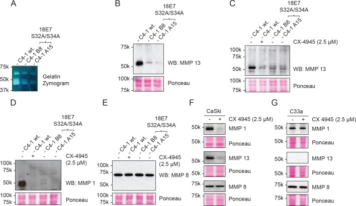
Fig 7 Matrix metalloproteases MMP 1 and MMP 13 are downregulated in CKII site mutant C4-1 cell lines. (A) Confluent wild type and CKII site mutant C4-1 cell lines were incubated in a serum-free medium for 48 hours. The conditioned media from the cell lines were concentrated and equal amounts of protein run on non-reducing 10% SDS-PAGE containing 0.1% gelatin, The gel was then renatured and incubated overnight at 37°C for gelatin hydrolysis. After incubation, the gel was stained with Coomassie Blue to visualize the bands of hydrolyzed substrate. (B) Equal amounts of concentrated conditioned media from the indicated cell lines were analysed by western blotting for MMP13 (upper panel). The lower panel shows the Ponceau stain of the nitrocellulose to confirm equal protein loading. Panels (C, D, E, F, G) Cells were processed as in panel B, but with the addition of 2.5 μM of CX-4945, as indicated, to specifically inhibit CKII activity. Conditioned media from wildtype C4-1 and mutant cells, plus CaSki and C33a cells, were then processed as before and analysed by western blotting for MMP13, MMP1 and MMP8 (upper panels), with the lower panels in each case showing the Ponceau stain of the nitrocellulose membranes.
Fig 7 Matrix metalloproteases MMP 1 and MMP 13 are downregulated in CKII site mutant C4-1 cell lines. (A) Confluent wild type and CKII site mutant C4-1 cell lines were incubated in a serum-free medium for 48 hours. The conditioned media from the cell lines were concentrated and equal amounts of protein run on non-reducing 10% SDS-PAGE containing 0.1% gelatin, The gel was then renatured and incubated overnight at 37°C for gelatin hydrolysis. After incubation, the gel was stained with Coomassie Blue to visualize the bands of hydrolyzed substrate. (B) Equal amounts of concentrated conditioned media from the indicated cell lines were analysed by western blotting for MMP13 (upper panel). The lower panel shows the Ponceau stain of the nitrocellulose to confirm equal protein loading. Panels (C, D, E, F, G) Cells were processed as in panel B, but with the addition of 2.5 μM of CX-4945, as indicated, to specifically inhibit CKII activity. Conditioned media from wildtype C4-1 and mutant cells, plus CaSki and C33a cells, were then processed as before and analysed by western blotting for MMP13, MMP1 and MMP8 (upper panels), with the lower panels in each case showing the Ponceau stain of the nitrocellulose membranes.CC BY · PubMed Central
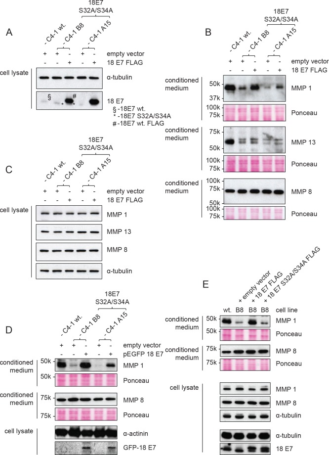
Fig 8 Exogenous expression of wild type HPV-18 E7 can rescue the downregulation of MMP 1 and MMP 13 in CKII site mutant lines. Wild type C4-1 cells were transfected transiently with empty pCMV vector and mutant cell lines with pCMV vector expressing FLAG-tagged wild type HPV-18 E7. (A) The cells were then harvested directly in 2X sample buffer and analysed by SDS-PAGE and Western blotting for levels of 18E7 and α-tubulin. The position of endogenous wild type E7 (§), the S32A/S34A mutant (*) and the wild type FLAG-tagged E7 (#) is also shown. (B) Concentrated conditioned medium from the cells was analysed by western blotting for MMP1, MMP13 and MMP8 (upper panels). The lower panels show the Ponceau stain of the nitrocellulose membranes to confirm equal levels of protein input. (C) Western blot analysis of total cell extracts for MMP1, MMP13 and MMP8 with α-tubulin used as the loading control. (D) C4-1 cells were transfected with pEGFP 18E7 wildtype plasmid or empty vector as indicated and selected by FACS sorting and G418 selection to obtain stable cells stably expressing GFP E7. The cells were analysed for MMP1 and MMP8 in the conditioned medium and expression of GFP 18E7 was ascertained in the cell lysate by western blotting. (E) C4-1 B8 CKII mutant cells were transfected with pCMV empty or pCMV 18E7 wildtype or pCMV 18E7 S32A/S34A mutant expressing plasmids as indicated and selected with G418 to obtain stable cell lines. The cells were then analysed for MMP secretion in the conditioned medium and the cell lysate by western blotting.
Fig 8 Exogenous expression of wild type HPV-18 E7 can rescue the downregulation of MMP 1 and MMP 13 in CKII site mutant lines. Wild type C4-1 cells were transfected transiently with empty pCMV vector and mutant cell lines with pCMV vector expressing FLAG-tagged wild type HPV-18 E7. (A) The cells were then harvested directly in 2X sample buffer and analysed by SDS-PAGE and Western blotting for levels of 18E7 and α-tubulin. The position of endogenous wild type E7 (§), the S32A/S34A mutant (*) and the wild type FLAG-tagged E7 (#) is also shown. (B) Concentrated conditioned medium from the cells was analysed by western blotting for MMP1, MMP13 and MMP8 (upper panels). The lower panels show the Ponceau stain of the nitrocellulose membranes to confirm equal levels of protein input. (C) Western blot analysis of total cell extracts for MMP1, MMP13 and MMP8 with α-tubulin used as the loading control. (D) C4-1 cells were transfected with pEGFP 18E7 wildtype plasmid or empty vector as indicated and selected by FACS sorting and G418 selection to obtain stable cells stably expressing GFP E7. The cells were analysed for MMP1 and MMP8 in the conditioned medium and expression of GFP 18E7 was ascertained in the cell lysate by western blotting. (E) C4-1 B8 CKII mutant cells were transfected with pCMV empty or pCMV 18E7 wildtype or pCMV 18E7 S32A/S34A mutant expressing plasmids as indicated and selected with G418 to obtain stable cell lines. The cells were then analysed for MMP secretion in the conditioned medium and the cell lysate by western blotting.CC BY · PubMed Central
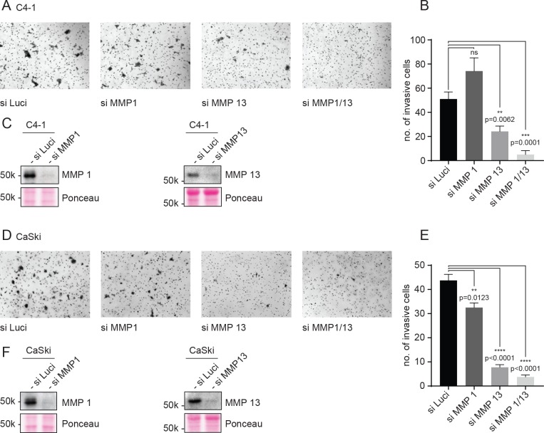
Fig 9 Knockdown of MMP1/13 abrogates invasion of C4-1 and CaSki cells. C4-1 and CaSki cells were transfected with indicated siRNAs. After 48 hours, equal number of cells were seeded in the upper part of Matrigel invasion chambers, in DMEM without serum. DMEM with 10% of serum was used as a chemoattractant in the lower chamber. After 22h, cells that had invaded the lower chamber were fixed and stained with 0.5% Crystal Violet in 5% glutaraldehyde for 10 mins. (A and D) Representative micrographs showing invasive cells from each siRNA treatment as indicated. (B and E) Quantification of the mean number of invasive cells per field shown as bars with the standard error of mean obtained from at least 4 separate fields. Asterisks indicate the p-value of mean numbers of invasive cells, compared with the wild type cells. (C and F) Western blot analysis of concentrated conditioned medium from indicated siRNA treatments for MMP1 and MMP13.
Fig 9 Knockdown of MMP1/13 abrogates invasion of C4-1 and CaSki cells. C4-1 and CaSki cells were transfected with indicated siRNAs. After 48 hours, equal number of cells were seeded in the upper part of Matrigel invasion chambers, in DMEM without serum. DMEM with 10% of serum was used as a chemoattractant in the lower chamber. After 22h, cells that had invaded the lower chamber were fixed and stained with 0.5% Crystal Violet in 5% glutaraldehyde for 10 mins. (A and D) Representative micrographs showing invasive cells from each siRNA treatment as indicated. (B and E) Quantification of the mean number of invasive cells per field shown as bars with the standard error of mean obtained from at least 4 separate fields. Asterisks indicate the p-value of mean numbers of invasive cells, compared with the wild type cells. (C and F) Western blot analysis of concentrated conditioned medium from indicated siRNA treatments for MMP1 and MMP13.CC BY · PubMed Central
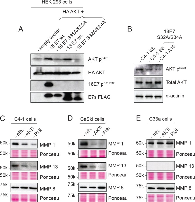
Fig 10 Activation of AKT is perturbed in the absence of CKII phosphorylation of E7. (A) HEK 293 cells were transfected with HA-AKT together with HPV-16 E7 FLAG-HA, wildtype or 16 E7 S31A/S32A, HPV-18 E7 FLAG, wildtype or 18 E7 S32A/S34A or empty vector. After 24 hours, the cells were serum starved for 16 hours and harvested directly with SDS PAGE sample buffer and analysed by Western blotting for phospho-AKT, total AKT (HA), phoshpo 16E7 and total E7 (FLAG), as indicated. (B) Confluent C4-1 wildtype and CKII mutant cells were serum starved for 48 hours and total levels of AKT, p-AKT, and α-actinin were analysed by Western blotting. (C, D, E) Confluent C4-1, CaSki and C33a cells were either treated with AKT inhibitor or PI3K inhibitor in serum free medium for 48 hours. Conditioned medium from the treatments were analysed by Western blotting for MMP1, MMP13 and MMP8. In each case the lower panels show the Ponceau stain of the nitrocellulose membranes.
Fig 10 Activation of AKT is perturbed in the absence of CKII phosphorylation of E7. (A) HEK 293 cells were transfected with HA-AKT together with HPV-16 E7 FLAG-HA, wildtype or 16 E7 S31A/S32A, HPV-18 E7 FLAG, wildtype or 18 E7 S32A/S34A or empty vector. After 24 hours, the cells were serum starved for 16 hours and harvested directly with SDS PAGE sample buffer and analysed by Western blotting for phospho-AKT, total AKT (HA), phoshpo 16E7 and total E7 (FLAG), as indicated. (B) Confluent C4-1 wildtype and CKII mutant cells were serum starved for 48 hours and total levels of AKT, p-AKT, and α-actinin were analysed by Western blotting. (C, D, E) Confluent C4-1, CaSki and C33a cells were either treated with AKT inhibitor or PI3K inhibitor in serum free medium for 48 hours. Conditioned medium from the treatments were analysed by Western blotting for MMP1, MMP13 and MMP8. In each case the lower panels show the Ponceau stain of the nitrocellulose membranes.CC BY · PubMed Central

Verification

Claims the paper makes

The claims and statistics of this paper haven’t been checked yet.

Run the check — free, no account needed. It reads the open-access text and checks the sources it cites.

A few free checks a day · papers already verified open instantly · results are public

Claims other papers make about this one

Each is checked against this paper’s own text: does it say what the citing paper says it does?

Self-check

Percentages, sums and reported differences re-computed from the paper’s own numbers.

The self-check needs the paper’s full text.

Citations

Cites

No references could be resolved yet.

Research works printed but not linked: 44

  1. H zur Hausen, 2009, Papillomaviruses in the causation of human cancers—a brief historical account, Virology, 384, 260, 10.1016/j.virol.2008.11.046 · DOI · Paper page
  2. J Doorbar, 2012, The biology and life-cycle of human papillomaviruses, Vaccine, 30, F55, 10.1016/j.vaccine.2012.06.083 · DOI
  3. A Roman, 2013, The papillomavirus E7 proteins, Virology, 445, 138, 10.1016/j.virol.2013.04.013 · DOI
  4. SB Vande Pol, 2013, Papillomavirus E6 oncoproteins, Virology, 445, 115, 10.1016/j.virol.2013.04.026 · DOI
  5. M Yoshinouchi, 2003, In vitro and in vivo growth suppression of human papillomavirus 16-positive cervical cancer cells by E6 siRNA, Molecular therapy: the journal of the American Society of Gene Therapy, 8, 762, 10.1016/j.ymthe.2003.08.004 · DOI · Paper page
  6. K Yamato, 2008, New highly potent and specific E6 and E7 siRNAs for treatment of HPV16 positive cervical cancer, Cancer gene therapy, 15, 140, 10.1038/sj.cgt.7701118 · DOI · Paper page
  7. K Butz, 2003, siRNA targeting of the viral E6 oncogene efficiently kills human papillomavirus-positive cancer cells, Oncogene, 22, 5938, 10.1038/sj.onc.1206894 · DOI · Paper page
  8. K Huh, 2007, Human papillomavirus type 16 E7 oncoprotein associates with the cullin 2 ubiquitin ligase complex, which contributes to degradation of the retinoblastoma tumor suppressor, Journal of virology, 81, 9737, 10.1128/JVI.00881-07 · DOI · Paper page
  9. A Szalmas, 2017, The PTPN14 Tumor Suppressor Is a Degradation Target of Human Papillomavirus E7, Journal of virology, 91, 10.1128/JVI.00057-17 · DOI · Paper page
  10. EA White, 2016, High-Risk Human Papillomavirus E7 Proteins Target PTPN14 for Degradation, mBio, 7, 10.1128/mBio.01530-16 · DOI · Paper page
  11. J Poirson, 2017, Mapping the interactome of HPV E6 and E7 oncoproteins with the Ubiquitin-Proteasome System, The FEBS journal
  12. KW Huh, 2005, Association of the human papillomavirus type 16 E7 oncoprotein with the 600-kDa retinoblastoma protein-associated factor, p600, Proceedings of the National Academy of Sciences of the United States of America, 102, 11492, 10.1073/pnas.0505337102 · DOI · Paper page
  13. MS Barbosa, 1990, The region of the HPV E7 oncoprotein homologous to adenovirus E1a and Sv40 large T antigen contains separate domains for Rb binding and casein kinase II phosphorylation, The EMBO journal, 9, 153, 10.1002/j.1460-2075.1990.tb08091.x · DOI
  14. JM Firzlaff, 1989, The E7 protein of human papillomavirus type 16 is phosphorylated by casein kinase II, The New biologist, 1, 44
  15. JM Firzlaff, 1991, Negative charge at the casein kinase II phosphorylation site is important for transformation but not for Rb protein binding by the E7 protein of human papillomavirus type 16, Proceedings of the National Academy of Sciences of the United States of America, 88, 5187, 10.1073/pnas.88.12.5187 · DOI
  16. P Massimi, 1996, HPV-16 E7 and adenovirus E1a complex formation with TATA box binding protein is enhanced by casein kinase II phosphorylation, Oncogene, 12, 2325
  17. FA Dick, 2002, Three regions of the pRB pocket domain affect its inactivation by human papillomavirus E7 proteins, Journal of virology, 76, 6224, 10.1128/JVI.76.12.6224-6234.2002 · DOI
  18. M Singh, 2005, Molecular determinants for the complex formation between the retinoblastoma protein and LXCXE sequences, The Journal of biological chemistry, 280, 37868, 10.1074/jbc.M504877200 · DOI
  19. LB Chemes, 2010, Targeting mechanism of the retinoblastoma tumor suppressor by a prototypical viral oncoprotein. Structural modularity, intrinsic disorder and phosphorylation of human papillomavirus E7, The FEBS journal, 277, 973, 10.1111/j.1742-4658.2009.07540.x · DOI
  20. A Zine El Abidine, 2017, A naturally occurring variant of HPV-16 E7 exerts increased transforming activity through acquisition of an additional phospho-acceptor site, Virology, 500, 218, 10.1016/j.virol.2016.10.023 · DOI
  21. MO Nogueira, 2017, Monitoring HPV-16 E7 phosphorylation events, Virology, 503, 70, 10.1016/j.virol.2016.12.030 · DOI
  22. A Psyrri, 2004, Role of the retinoblastoma pathway in senescence triggered by repression of the human papillomavirus E7 protein in cervical carcinoma cells, Cancer research, 64, 3079, 10.1158/0008-5472.CAN-03-3739 · DOI
  23. A Schneider-Gadicke, 1986, Different human cervical carcinoma cell lines show similar transcription patterns of human papillomavirus type 18 early genes, The EMBO journal, 5, 2285, 10.1002/j.1460-2075.1986.tb04496.x · DOI
  24. DL Jones, 1997, Destabilization of the RB tumor suppressor protein and stabilization of p53 contribute to HPV type 16 E7-induced apoptosis, Virology, 239, 97, 10.1006/viro.1997.8851 · DOI · Paper page
  25. NJ Genovese, 2008, Casein kinase II motif-dependent phosphorylation of human papillomavirus E7 protein promotes p130 degradation and S-phase induction in differentiated human keratinocytes, Journal of virology, 82, 4862, 10.1128/JVI.01202-07 · DOI
  26. K Srivastava, 2017, p63 drives invasion in keratinocytes expressing HPV16 E6/E7 genes through regulation of Src-FAK signalling, Oncotarget, 8, 16202, 10.18632/oncotarget.3892 · DOI · Paper page
  27. WM Chien, 2000, Casein kinase II phosphorylation of the human papillomavirus-18 E7 protein is critical for promoting S-phase entry, Cell growth & differentiation: the molecular biology journal of the American Association for Cancer Research, 11, 425
  28. O Marin, 1994, Design and synthesis of two new peptide substrates for the specific and sensitive monitoring of casein kinases-1 and -2, Biochemical and biophysical research communications, 198, 898, 10.1006/bbrc.1994.1128 · DOI
  29. S Sarno, 1996, Protein kinase CK2 mutants defective in substrate recognition. Purification and kinetic analysis, The Journal of biological chemistry, 271, 10595, 10.1074/jbc.271.18.10595 · DOI
  30. D Pim, 2005, Activation of the protein kinase B pathway by the HPV-16 E7 oncoprotein occurs through a mechanism involving interaction with PP2A, Oncogene, 24, 7830, 10.1038/sj.onc.1208935 · DOI · Paper page
  31. H Wang, 2014, Matrix metalloproteinase-14 expression and its prognostic value in cervical carcinoma, Cell biochemistry and biophysics, 70, 729, 10.1007/s12013-014-9974-8 · DOI · Paper page
  32. Y Zhai, 2005, Expression of membrane type 1 matrix metalloproteinase is associated with cervical carcinoma progression and invasion, Cancer research, 65, 6543, 10.1158/0008-5472.CAN-05-0231 · DOI · Paper page
  33. J Kaewprag, 2013, HPV16 oncoproteins promote cervical cancer invasiveness by upregulating specific matrix metalloproteinases, PloS one, 8, e71611, 10.1371/journal.pone.0071611 · DOI · Paper page
  34. D Zhu, 2015, E6/E7 oncoproteins of high risk HPV-16 upregulate MT1-MMP, MMP-2 and MMP-9 and promote the migration of cervical cancer cells, International journal of clinical and experimental pathology, 8, 4981
  35. CW Menges, 2006, Human papillomavirus type 16 E7 up-regulates AKT activity through the retinoblastoma protein, Cancer research, 66, 5555, 10.1158/0008-5472.CAN-06-0499 · DOI · Paper page
  36. N Johansson, 1999, Collagenase-3 (MMP-13) is expressed by tumor cells in invasive vulvar squamous cell carcinomas, The American journal of pathology, 154, 469, 10.1016/S0002-9440(10)65293-5 · DOI
  37. M Balbin, 2003, Loss of collagenase-2 confers increased skin tumor susceptibility to male mice, Nature genetics, 35, 252, 10.1038/ng1249 · DOI
  38. B Akgul, 2006, Expression of matrix metalloproteinase (MMP)-2, MMP-9, MMP-13, and MT1-MMP in skin tumors of human papillomavirus type 8 transgenic mice, Experimental dermatology, 15, 35, 10.1111/j.0906-6705.2005.00387.x · DOI
  39. JT Korpi, 2008, Collagenase-2 (matrix metalloproteinase-8) plays a protective role in tongue cancer, British journal of cancer, 98, 766, 10.1038/sj.bjc.6604239 · DOI
  40. A Haukioja, 2017, Persistent Oral Human Papillomavirus (HPV) Infection is Associated with Low Salivary Levels of Matrix Metalloproteinase 8 (MMP-8), Journal of clinical virology: the official publication of the Pan American Society for Clinical Virology, 97, 4, 10.1016/j.jcv.2017.10.011 · DOI
  41. R Tian, 2018, Identification and validation of the role of matrix metalloproteinase-1 in cervical cancer, International journal of oncology, 52, 1198
  42. SL Gonzalez, 2001, Degradation of the retinoblastoma tumor suppressor by the human papillomavirus type 16 E7 oncoprotein is important for functional inactivation and is separable from proteasomal degradation of E7, Journal of virology, 75, 7583, 10.1128/JVI.75.16.7583-7591.2001 · DOI · Paper page
  43. FA Ran, 2013, Genome engineering using the CRISPR-Cas9 system, Nature protocols, 8, 2281, 10.1038/nprot.2013.143 · DOI · Paper page
  44. P Massimi, 2012, Differential regulation of cell-cell contact, invasion and anoikis by hScrib and hDlg in keratinocytes, PloS one, 7, e40279, 10.1371/journal.pone.0040279 · DOI

Metadata

Authors
Basukala, O., Mittal, S., Massimi, P., Bestagno, M., & Banks, L.
Venue
PLOS Pathogens
Published
2019-05-22
Volume
15
Issue
5
Pages
e1007769
ISSN
1553-7374
Cited by
40
References
0
Language
en

About these checks

Registry, retraction, Cites and statistics results are computed, not judged, with one exception: a printed reference that no index lists is read once by a model before it counts as a citation error. Claims are extracted by a model and kept only when their quote appears verbatim in the text; each cited source is then checked, with a model’s help, for whether it says what the claim relies on. Those verdicts can be wrong — read the claim’s quote and the cited source before relying on one.

Statistics and arithmetic are recomputed, not judged.

Confidence bands are shown only for fields calibrated on our annotated test set. None is calibrated yet, so no band is shown.

What counts as a claim ›How we measure confidence ›How we check this ›

Are you an author, or do you see a problem with this page? Request a review

How to cite

Cite CoChat for the checks on this page, and the paper for its own findings.

This page
CoChat. (2026). Citation check: The HPV-18 E7 CKII phospho acceptor site is required for maintaining the transformed phenotype of cervical tumour-derived cells. https://app.cochat.ai/papers/10.1371/journal.ppat.1007769
The paper
Basukala, O., Mittal, S., Massimi, P., Bestagno, M., & Banks, L. (2019). The HPV-18 E7 CKII phospho acceptor site is required for maintaining the transformed phenotype of cervical tumour-derived cells. PLOS Pathogens. https://doi.org/10.1371/journal.ppat.1007769