

The DOI matches its Crossref record; 52 of the 52 references it cites were found in a registry or an index.
Control of Amygdala Circuits by 5-HT Neurons via 5-HT and Glutamate Cotransmission
Open accessAbstract
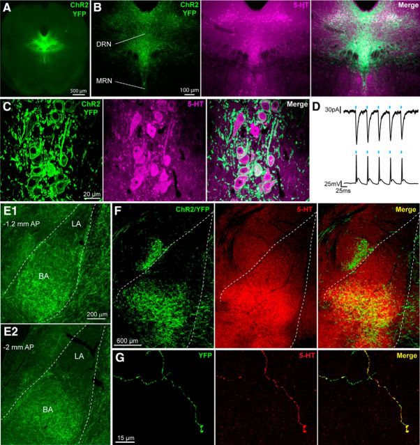
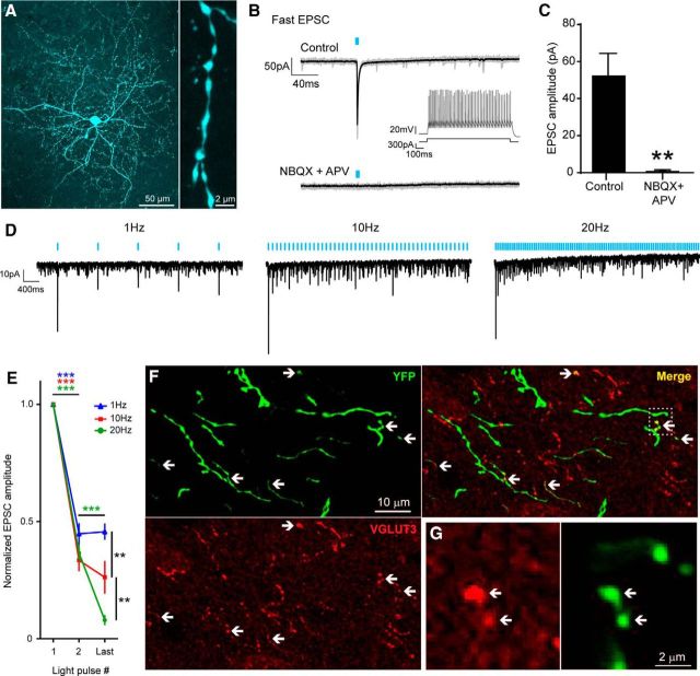
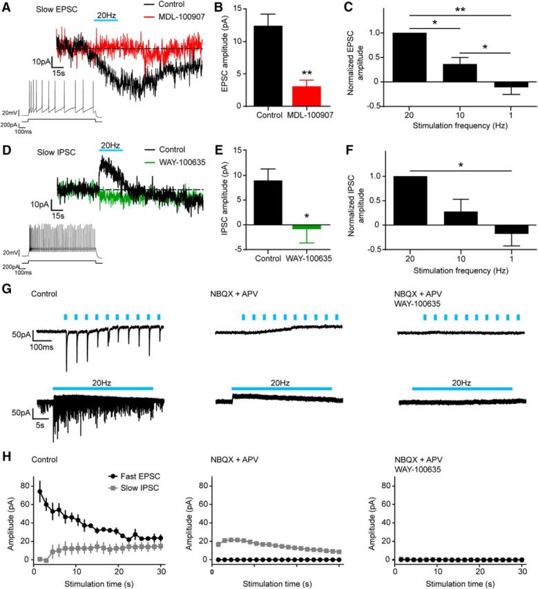
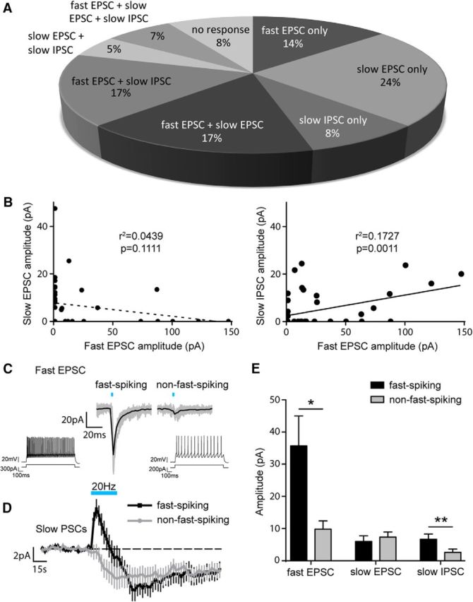
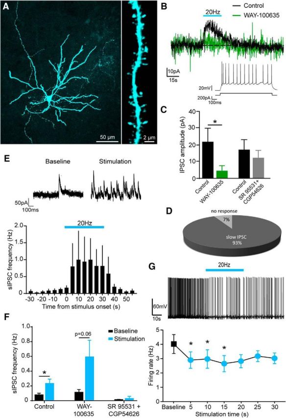
The serotonin (5-HT) system and the amygdala are key regulators of emotional behavior. Several lines of evidence suggest that 5-HT transmission in the amygdala is implicated in the susceptibility and drug treatment of mood disorders. Therefore, elucidating the physiological mechanisms through which midbrain 5-HT neurons modulate amygdala circuits could be pivotal in understanding emotional regulation in health and disease. To shed light on these mechanisms, we performed patch-clamp recordings from basal amygdala (BA) neurons in brain slices from mice with channelrhodopsin genetically targeted to 5-HT neurons. Optical stimulation of 5-HT terminals at low frequencies (≤1 Hz) evoked a short-latency excitation of BA interneurons (INs) that was depressed at higher frequencies. Pharmacological analysis revealed that this effect was mediated by glutamate and not 5-HT because it was abolished by ionotropic glutamate receptor antagonists. Optical stimulation of 5-HT terminals at higher frequencies (10–20 Hz) evoked both slow excitation and slow inhibition of INs. These effects were mediated by 5-HT because they were blocked by antagonists of 5-HT2Aand 5-HT1Areceptors, respectively. These fast glutamate- and slow 5-HT-mediated responses often coexisted in the same neuron. Interestingly, fast-spiking and non-fast-spiking INs displayed differential modulation by glutamate and 5-HT. Furthermore, optical stimulation of 5-HT terminals did not evoke glutamate release onto BA principal neurons, but inhibited these cells directly via activation of 5-HT1Areceptors and indirectly via enhanced GABA release. Collectively, these findings suggest that 5-HT neurons exert a frequency-dependent, cell-type-specific control over BA circuitry via 5-HT and glutamate co-release to inhibit the BA output. SIGNIFICANCE STATEMENTThe modulation of the amygdala by serotonin (5-HT) is important for emotional regulation and is implicated in the pathogenesis and treatment of affective disorders. Therefore, it is essential to determine the physiological mechanisms through which 5-HT neurons in the dorsal raphe nuclei modulate amygdala circuits. Here, we combined optogenetic, electrophysiological, and pharmacological approaches to study the effects of activation of 5-HT axons in the basal nucleus of the amygdala (BA). We found that 5-HT neurons co-release 5-HT and glutamate onto BA neurons in a cell-type-specific and frequency-dependent manner. Therefore, we suggest that theories on the contribution of 5-HT neurons to amygdala function should be revised to incorporate the concept of 5-HT/glutamate cotransmission.
Overview
The claims and statistics of this paper haven’t been checked yet.
Run the check — free, no account needed. It reads the open-access text and checks the sources it cites.
Integrity signals
Registry
Cites
Statistics
Figures
Verification
Claims the paper makes
The claims and statistics of this paper haven’t been checked yet.
Run the check — free, no account needed. It reads the open-access text and checks the sources it cites.
Claims other papers make about this one
Each is checked against this paper’s own text: does it say what the citing paper says it does?
In the citing paper
“~60% of DR serotonin neurons co-express the vesicular glutamate transporter Vglut3 in the rat (Gras et al., 2002) and co-release glutamate in the mouse (Liu et al., 2014; Sengupta et al., 2017).”
From Anatomically defined and functionally distinct dorsal raphe serotonin sub-systems (2018) · cites this paper as Sengupta et al., 2017
Self-check
Percentages, sums and reported differences re-computed from the paper’s own numbers.
The self-check needs the paper’s full text.
Citations
Cites
Steinbusch, H. W. M. (1981) · Neuroscience · doi:10.1016/0306-4522(81)90146-9 · Cited by 2,415
From circuits to behaviour in the amygdala
Janak, P. H., & Tye, K. M. (2015) · Nature · doi:10.1038/nature14188 · Cited by 2,011 · cited 1×
“The amygdala is a brain region that comprises multiple nuclei that collectively encode many aspects of emotionality including fear learning and memory (Duvarci and Pare, 2014; Janak and Tye, 2015).”
BackgroundPetilla terminology: nomenclature of features of GABAergic interneurons of the cerebral cortex
The Petilla Interneuron Nomenclature Group (PING) (2008) · Nature Reviews Neuroscience · doi:10.1038/nrn2402 · Cited by 1,491
Amygdala circuitry mediating reversible and bidirectional control of anxiety
Tye, K. M., Prakash, R., Kim, S.-Y., Fenno, L. E., Grosenick, L., Zarabi, H., Thompson, K. R., Gradinaru, V., Ramakrishnan, C., & Deisseroth, K. (2011) · Nature · doi:10.1038/nature09820 · Cited by 1,289
West, M. J. (1999) · Trends in Neurosciences · doi:10.1016/s0166-2236(98)01362-9 · Cited by 820
Amygdala Microcircuits Controlling Learned Fear
Duvarci, S., & Pare, D. (2014) · Neuron · doi:10.1016/j.neuron.2014.04.042 · Cited by 751
A third vesicular glutamate transporter expressed by cholinergic and serotoninergic neurons
Gras, C., Herzog, E., Bellenchi, G. C., Bernard, V., Ravassard, P., Pohl, M., Gasnier, B., Giros, B., & El Mestikawy, S. (2002) · The Journal of Neuroscience · doi:10.1523/jneurosci.22-13-05442.2002 · Cited by 639
A circuit mechanism for differentiating positive and negative associations
Namburi, P., Beyeler, A., Yorozu, S., Calhoon, G. G., Halbert, S. A., Wichmann, R., Holden, S. S., Mertens, K. L., Anahtar, M., Felix-Ortiz, A. C., et al. (2015) · Nature · doi:10.1038/nature14366 · Cited by 614
Long-range connectivity defines behavioral specificity of amygdala neurons
Senn, V., Wolff, S. B. E., Herry, C., Grenier, F., Ehrlich, I., Gründemann, J., Fadok, J. P., Müller, C., Letzkus, J. J., & Lüthi, A. (2014) · Neuron · doi:10.1016/j.neuron.2013.11.006 · Cited by 522
Amygdala interneuron subtypes control fear learning through disinhibition
Wolff, S. B. E., Gründemann, J., Tovote, P., Krabbe, S., Jacobson, G. A., Müller, C. R., Herry, C., Ehrlich, I., Friedrich, R. W., Letzkus, J. J., et al. (2014) · Nature · doi:10.1038/nature13258 · Cited by 515
Cited by
Sorted by citations
Anatomically defined and functionally distinct dorsal raphe serotonin sub-systems
Ren, J., Friedmann, D., Xiong, J., Liu, C. D., Ferguson, B. R., Weerakkody, T., DeLoach, K. E., Ran, C., Pun, A., Sun, Y., et al. (2018) · Cell · doi:10.1016/j.cell.2018.07.043 · Cited by 498
“, 2002) and corelease glutamate in the mouse (Liu et al., 2014; Sengupta et al., 2017).”
Embracing diversity in the 5-HT neuronal system
Okaty, B. W., Commons, K. G., & Dymecki, S. M. (2019) · Nature Reviews Neuroscience · doi:10.1038/s41583-019-0151-3 · Cited by 319
Ren, J., Isakova, A., Friedmann, D., Zeng, J., Grutzner, S. M., Pun, A., Zhao, G. Q., Kolluru, S. S., Wang, R., Lin, R., et al. (2019) · eLife · doi:10.7554/elife.49424 · Cited by 304
“, 2019), and basolateral amygdala (Sengupta et al., 2017).”
ResultBarnes, N. M., Ahern, G. P., Bécamel, C., Bockaert, J., Camilleri, M., Chaumont‐Dubel, S., Claeysen, S., Cunningham, K. A., Fone, K. C. F., Gershon, M. D., et al. (2020) · Pharmacological Reviews · doi:10.1124/pr.118.015552 · Cited by 274
Central 5-HT receptors and their function; present and future
Sharp, T., & Barnes, N. M. (2020) · Neuropharmacology · doi:10.1016/j.neuropharm.2020.108155 · Cited by 239
Phasic Activation of Dorsal Raphe Serotonergic Neurons Increases Pupil Size
Cazettes, F., Reato, D., Morais, J. P., Renart, A., & Mainen, Z. F. (2020) · Current Biology · doi:10.1016/j.cub.2020.09.090 · Cited by 146
Synaptic Vesicle Recycling Pathway Determines Neurotransmitter Content and Release Properties
Silm, K., Yang, J., Marcott, P. F., Asensio, C. S., Eriksen, J., Guthrie, D. A., Newman, A. H., Ford, C., & Edwards, R. H. (2019) · Neuron · doi:10.1016/j.neuron.2019.03.031 · Cited by 133
A single-cell transcriptomic and anatomic atlas of mouse dorsal raphe Pet1 neurons
Okaty, B. W., Sturrock, N., Lozoya, Y. E., Chang, Y., Senft, R. A., Lyon, K. A., Alekseyenko, O. V., & Dymecki, S. M. (2020) · eLife · doi:10.7554/elife.55523 · Cited by 122
“Consistent with functional expression of VGLUT3 protein (encoded by the gene Slc17a8), which allows for filling of synaptic vesicles with the excitatory neurotransmitter glutamate, depolarization-induced glutamate release by DR Pet1/5-HT neurons has been demonstrated by a number of groups (Johnson, 1994; Kapoor et al.…”
BackgroundA Discrete Dorsal Raphe to Basal Amygdala 5-HT Circuit Calibrates Aversive Memory
Sengupta, A., & Holmes, A. (2019) · Neuron · doi:10.1016/j.neuron.2019.05.029 · Cited by 123
“Moreover, similar highfrequency optogenetic activation of BA 5-HT inputs ex vivo favours 5-HT over glutamate transmission (Sengupta et al., 2017).”
BackgroundMethodResultInfluentialSerotonin neuron development: shaping molecular and structural identities
Deneris, E. S., & Gaspar, P. (2017) · Wiley Interdisciplinary Reviews Developmental Biology · doi:10.1002/wdev.301 · Cited by 120
“Optogenetically stimulated 5-HT neurons provides evidence that 5-HT and glutamate are co-released and that VGLUT3 is required for release of glutamate (77-79).”
BackgroundSerotonin regulation of behavior via large-scale neuromodulation of serotonin receptor networks
Salvan, P., Fonseca, M., Winkler, A. M., Beauchamp, A., Lerch, J. P., & Johansen-Berg, H. (2023) · Nature Neuroscience · doi:10.1038/s41593-022-01213-3 · Cited by 101
Ohmura, Y., Tsutsui‐Kimura, I., Sasamori, H., Nebuka, M., Nishitani, N., Tanaka, K. F., Yamanaka, A., & Yoshioka, M. (2019) · Neuropharmacology · doi:10.1016/j.neuropharm.2019.107703 · Cited by 92
The role of co-neurotransmitters in sleep and wake regulation
Oh, J. Y., Petersen, C., Walsh, C. M., Bittencourt, J. C., Neylan, T. C., & Grinberg, L. T. (2018) · Molecular Psychiatry · doi:10.1038/s41380-018-0291-2 · Cited by 91
Distinct serotonergic pathways to the amygdala underlie separate behavioral features of anxiety
Yu, X., Zhu, Y., Sun, Q., Deng, F., Wan, J., Zheng, D., Gong, W., Xie, S., Shen, C.-J., Fu, J., et al. (2022) · Nature Neuroscience · doi:10.1038/s41593-022-01200-8 · Cited by 89
Reward Contributions to Serotonergic Functions
Liu, Z., Lin, R., & Luo, M. (2020) · Annual Review of Neuroscience · doi:10.1146/annurev-neuro-093019-112252 · Cited by 79
Pinto, D. F. C., Yang, H., Dorocic, I. P., de Jong, J. W., Han, V. J., Peck, J. R., Zhu, Y., Liu, C. J., Beier, K. T., Smidt, M. P., et al. (2019) · Nature Communications · doi:10.1038/s41467-019-12392-2 · Cited by 80
Trudeau, L., & Mestikawy, S. E. (2018) · Frontiers in Neural Circuits · doi:10.3389/fncir.2018.00113 · Cited by 83
“…DA neurons (Sulzer et al., 1998; Bourque and Trudeau, 2000; Hnasko et al., 2010; Tecuapetla et al., 2010; Chuhma et al., 2014; Trudeau et al., 2014), ACh (Huh et al., 2008; Higley et al., 2011; Nelson et al., 2014; Frahm et al., 2015) and GABA neurons (Shabel et al., 2014; Root et al., 2018).”
Upmanyu, N., Jin, J., Emde, H. V. D., Ganzella, M., Bösche, L., Malviya, V. N., Zhuleku, E., Politi, A. Z., Ninov, M., Silbern, I., et al. (2022) · Neuron · doi:10.1016/j.neuron.2022.02.008 · Cited by 69
“, 2010), or with serotonin in the hippocampal raphe nuclei projections (Sengupta et al., 2017; Varga et al., 2009), or with acetylcholine in striatal interneurons and medial habenula projections (Frahm et al.”
BackgroundZhang, H., Li, K., Chen, H., Gao, S., Xia, Z., Zhang, J., Wang, F., & Chen, J. (2018) · Brain Structure and Function · doi:10.1007/s00429-018-1623-3 · Cited by 75
Role of the Serotonin Receptor 7 in Brain Plasticity: From Development to Disease
(2020) · doi:10.3390/ijms21020505 · Cited by 63
“Among the cotransmitters released by 5-HT neurons, glutamate [7], and possibly other amino acids [8] were identified.”
BackgroundA discrete serotonergic circuit regulates vulnerability to social stress
Zou, W.-J., Song, Y., Wu, M., Chen, X.-T., You, Q.-L., Yang, Q., Luo, Z.-Y., Huang, L., Kong, Y., Feng, J., et al. (2020) · Nature Communications · doi:10.1038/s41467-020-18010-w · Cited by 64
Serotonin modulates excitatory synapse maturation in the developing prefrontal cortex
Ogelman, R., Wulschner, L. E. G., Hoelscher, V. M., Hwang, I.-W., Chang, V. N., & Oh, W. C. (2024) · Nature Communications · doi:10.1038/s41467-024-45734-w · Cited by 56
“Furthermore, studies have shown that neurons projecting out of the DRN may co-release 5-HT and glutamate during punishing or rewarding stimuli 58,59 .”
Paradoxical network excitation by glutamate release from VGluT3+ GABAergic interneurons
Pelkey, K. A., Calvigioni, D., Fang, C., Vargish, G., Ekins, T., Auville, K., Wester, J. C., Lai, M., Mackenzie-Gray Scott, C., Yuan, X., et al. (2020) · eLife · doi:10.7554/elife.51996 · Cited by 46
Béopoulos, A., Géa, M., Fasano, A., & Iris, F. (2022) · Frontiers in Neuroscience · doi:10.3389/fnins.2022.988735 · Cited by 60
“Similarly, serotonin prominently promotes amygdala activation in response to emotional stimuli (Bocchio et al., 2016; Sengupta et al., 2017).”
BackgroundDivergent properties and independent regulation of striatal dopamine and GABA co-transmission
Zych, S. M., & Ford, C. P. (2022) · Cell Reports · doi:10.1016/j.celrep.2022.110823 · Cited by 46
“Depending on the system and whether transmitters are packaged in the same or different vesicles, release can occur with similar or different properties (Adrover et al., 2014; Gras et al., 2008; Jonas et al., 1998; Ren et al., 2011; Sengupta et al., 2017; Silm et al., 2019; Takács et al., 2018).”
BackgroundSerotonergic regulation of the dopaminergic system: Implications for reward-related functions
Courtiol, E., Menezes, E. D. C., & Teixeira, C. M. (2021) · Neuroscience & Biobehavioral Reviews · doi:10.1016/j.neubiorev.2021.06.022 · Cited by 50
Mechanisms of SSRI Therapy and Discontinuation
Sharp, T., & Collins, H. (2023) · Current Topics in Behavioral Neurosciences · doi:10.1007/7854_2023_452 · Cited by 55
Shah, T., Dunning, J. L., & Contet, C. (2021) · Neuropharmacology · doi:10.1016/j.neuropharm.2021.108906 · Cited by 44
Hung, L. Y., Alves, N. D., Colle, A. D., Talati, A., Najjar, S. A., Bouchard, V., Gillet, V., Tong, Y., Huang, Z., Browning, K. N., et al. (2024) · Gastroenterology · doi:10.1053/j.gastro.2024.11.012 · Cited by 52
Serotonergic modulation across sensory modalities
Sizemore, T. R., Hurley, L. M., & Dacks, A. M. (2020) · Journal of Neurophysiology · doi:10.1152/jn.00034.2020 · Cited by 44
The Wiring Logic of an Identified Serotonergic Neuron That Spans Sensory Networks
Coates, K. E., Calle-Schuler, S. A., Helmick, L. M., Knotts, V. L., Martik, B. N., Salman, F., Warner, L. T., Valla, S. V., Bock, D. D., & Dacks, A. M. (2020) · Journal of Neuroscience · doi:10.1523/jneurosci.0552-20.2020 · Cited by 38
Powell, A., Ireland, C., & Lewis, S. J. G. (2020) · Journal of Neuropsychiatry · doi:10.1176/appi.neuropsych.19110316 · Cited by 40
Silva, V. D. G., dos Reis, A. S., Pinz, M. P., da Fonseca, C. A. R., Duarte, L. F. B., Roehrs, J. A., Alves, D. A. C., Luchese, C., & Wilhelm, E. A. (2017) · Fundamental and Clinical Pharmacology · doi:10.1111/fcp.12295 · Cited by 35
Filippo, R. D., Rost, B. R., Stumpf, A., Cooper, C., Tukker, J. J., Harms, C., Beed, P. S., & Schmitz, D. (2021) · eLife · doi:10.7554/elife.66960 · Cited by 34
Endocannabinoid-serotonin systems interaction in health and disease
Colangeli, R., Teskey, G. C., & Giovanni, G. D. (2021) · Progress in brain research · doi:10.1016/bs.pbr.2021.01.003 · Cited by 31
Asim, M., Wang, H., & Waris, A. (2023) · Neuropeptides · doi:10.1016/j.npep.2023.102322 · Cited by 31
Serotonergic neuromodulation of synaptic plasticity
Higa, G. S. V., Viana, F. J. C., Francis-Oliveira, J., Cruvinel, E., Franchin, T. S., Marcourakis, T., Ulrich, H., & De Pasquale, R. (2024) · Neuropharmacology · doi:10.1016/j.neuropharm.2024.110036 · Cited by 29
A Role for Serotonin in Modulating Opposing Drive and Brake Circuits of Impulsivity
Desrochers, S. S., Spring, M. G., & Nautiyal, K. M. (2022) · Frontiers in Behavioral Neuroscience · doi:10.3389/fnbeh.2022.791749 · Cited by 29
“…its core and shell subregions have been extensively studied for their individual roles in impulsive action through reaction time tests, with pharmacological manipulations and deep brain stimulation of the shell subregion causing elevated premature responding (Sesia et al., 2010; Feja et al., 2014).”
BackgroundWelsch, L., Colantonio, E., Frison, M., Johnson, D. A., McClain, S. P., Mathis, V., Banghart, M. R., Hamida, S. B., Darcq, E., & Kieffer, B. L. (2023) · Biological Psychiatry · doi:10.1016/j.biopsych.2023.05.019 · Cited by 34
Functional Neurologic Disorder, La Lésion Dynamique
Hallett, M. (2024) · Neurology · doi:10.1212/wnl.0000000000210051 · Cited by 28
Differential Expression of VGLUT2 in Mouse Mesopontine Cholinergic Neurons
Steinkellner, T., Yoo, J. H., & Hnasko, T. S. (2019) · eNeuro · doi:10.1523/eneuro.0161-19.2019 · Cited by 32
“…(Gras et al., 2008; Ren et al., 2011; Frahm et al., 2015; Sakae et al., 2015), ACh and GABA (Saunders et al., 2015a,b); serotonin and glutamate (Amilhon et al., 2010; Voisin et al., 2016; Sengupta et al., 2017); and even glutamate and GABA (Root et al., 2014; Shabel et al., 2014; Yoo et al., 2016).”
BackgroundJi, G., & Neugebauer, V. (2019) · International Journal of Molecular Sciences · doi:10.3390/ijms20184380 · Cited by 28
“5-HT can also decrease BLA output by direct 5-HT1AR-mediated inhibition of glutamatergic principal neurons [58], by HT1BR-mediated inhibition of their glutamatergic drive [59] or by 5-HT2R-mediated activation of BLA interneurons [19,58,60].”
BackgroundPrice, M. E., & McCool, B. A. (2021) · Alcohol · doi:10.1016/j.alcohol.2021.08.001 · Cited by 30
“5-HT2A receptors depolarize (Rainnie, 1999) and excite BA interneurons (Sengupta et al., 2017), including PV+ interneurons (Bocchio et al.”
BackgroundThe dorsal raphe nucleus in the control of energy balance
Bhave, V. M., & Nectow, A. R. (2021) · Trends in Neurosciences · doi:10.1016/j.tins.2021.09.004 · Cited by 26
“Furthermore, some DRN neurons are known to corelease glutamate [74], and VGLUT3 in axon terminals could potentially facilitate increased 5-HT release through vesicular synergy [75].”
BackgroundAdaptation to photoperiod via dynamic neurotransmitter segregation
Maddaloni, G., Chang, Y., Senft, R. A., & Dymecki, S. M. (2024) · Nature · doi:10.1038/s41586-024-07692-7 · Cited by 30
“This finding suggests that mr En1-Pet1 neurons may transmit with glutamate as well as 5-HT; indeed, in other serotonergic neurons such as in the dorsal raphe, VGLUT3 has been demonstrated to be necessary for glutamate neurotransmission 25–27 .”
Liu, C., Lan, C., Li, K., Zhou, F., Yao, S., Xu, L., Yang, N., Zhou, X., Yang, J., Yong, X., et al. (2021) · Biological Psychiatry Cognitive Neuroscience and Neuroimaging · doi:10.1016/j.bpsc.2021.04.009 · Cited by 24
Joshi, A., Wang, D., Watterson, S., McClean, P. L., Behera, C. K., Sharp, T., & Wong‐Lin, K. (2020) · Neuropharmacology · doi:10.1016/j.neuropharm.2020.108118 · Cited by 22
“, prefrontal cortex), anxiety, and hence, increase the risk of developing depression (Arnsten, 2009; Bocchio et al., 2016; Hannibal and Bishop, 2014; Sengupta et al., 2017; Tafet et al., 2001).”
Beecher, K., Wang, J., Jacques, A., Chaaya, N., Chehrehasa, F., Belmer, A., & Bartlett, S. E. (2021) · Frontiers in Molecular Neuroscience · doi:10.3389/fnmol.2021.678267 · Cited by 22
Alonso, L., Peeva, P., Stasko, S., Bäder, M., Alenina, N. V., Winter, Y., & Rivalan, M. (2023) · iScience · doi:10.1016/j.isci.2023.105998 · Cited by 22
Belmer, A., Beecher, K., Jacques, A., Patkar, O. L., Sicherre, F., & Bartlett, S. E. (2019) · Frontiers in Cellular Neuroscience · doi:10.3389/fncel.2019.00193 · Cited by 21
Anxiety and Gene Expression Enhancement in Mice Exposed to Glyphosate-Based Herbicide
Ait-Bali, Y., Kaikai, N.-E., Ba-M’hamed, S., Sassoé‐Pognetto, M., Giustetto, M., & Bennis, M. (2022) · Toxics · doi:10.3390/toxics10050226 · Cited by 20
“Moreover, the expression of the 5-HT3 and 5-HT2 receptor subtypes on the interneurons of the BLA suggest that the inhibition of cell firing may also be facilitated by the direct excitation of GABAergic interneurons in this nucleus [41].”
BackgroundBär, K., Köhler, S., de la Cruz, F., Schumann, A., Zepf, F. D., & Wagner, G. (2019) · NeuroImage · doi:10.1016/j.neuroimage.2019.116362 · Cited by 22
Esaki, H., Sasaki, Y., Nishitani, N., Kamada, H., Mukai, S., Ohshima, Y., Nakada, S., Ni, X., Deyama, S., & Kaneda, K. (2023) · European Journal of Pharmacology · doi:10.1016/j.ejphar.2023.175653 · Cited by 18
Hamed, A., & Kursa, M. B. (2018) · Brain Structure and Function · doi:10.1007/s00429-018-1683-4 · Cited by 19
“Moreover, ex vivo electrophysiological recordings with optogenetic methods and pharmacological analysis revealed the existence of the 5-HT and glutamate co-transmission in basal amygdala neurons (Sengupta et al. 2017).”
Ballanger, B., Bath, K. G., & Mandairon, N. (2019) · Neurobiology of Aging · doi:10.1016/j.neurobiolaging.2019.06.007 · Cited by 23
Leveraging VGLUT3 Functions to Untangle Brain Dysfunctions
Favier, M., Pietrancosta, N., Mestikawy, S. E., & Gangarossa, G. (2021) · Trends in Pharmacological Sciences · doi:10.1016/j.tips.2021.03.003 · Cited by 17
Synthetic surprise as the foundation of the psychedelic experience
Filippo, R. D., & Schmitz, D. (2024) · Neuroscience & Biobehavioral Reviews · doi:10.1016/j.neubiorev.2024.105538 · Cited by 16
Lyon, K. A., Rood, B. D., Wu, L., Senft, R. A., Goodrich, L. V., & Dymecki, S. M. (2020) · eNeuro · doi:10.1523/eneuro.0202-20.2020 · Cited by 16
“Indeed, there is precedent for the differential usage of serotonin and glutamate by raphe serotonergic neurons (Liu et al., 2014; Kapoor et al., 2016; Sengupta et al., 2017; Wang et al., 2019), although GABAergic and serotonergic co-release has not been reported.”
BackgroundModulation of serotonin and noradrenaline in the BLA by pindolol reduces long‐term ethanol intake
Patkar, O. L., Belmer, A., Holgate, J. Y., Klenowski, P. M., & Bartlett, S. E. (2018) · Addiction Biology · doi:10.1111/adb.12630 · Cited by 15
“For instance, enhancement in BLA principal cell excitability has been demonstrated with 5-HT depletion (Tran et al. 2013), 5-HT-mediated inhibition of BLA interneurons (Sengupta et al. 2017) and application of β-AR agonists (Pu et al. 2009).”
BackgroundResilience to fear: The role of individual factors in amygdala response to stressors
Fonseca, R., Madeira, N., & Simões, C. R. B. (2020) · Molecular and Cellular Neuroscience · doi:10.1016/j.mcn.2020.103582 · Cited by 17
Fortin‐Houde, J., Henderson, F. J., Dumas, S., Ducharme, G., & Amilhon, B. (2023) · The Journal of Comparative Neurology · doi:10.1002/cne.25452 · Cited by 19
“…functional inputs from VGLUT3 DR neurons to the VTA (Liu et al., 2014; McDevitt et al., 2014; Qi et al., 2014; H. L. Wang et al., 2019; Zou et al., 2020) and amygdala (Sengupta et al., 2017; Sengupta & Holmes, 2019) and inputs from the MnR to the dorsal HP (He et al., 2022; Varga et al., 2009).”
BackgroundResultInfluentialSakae, D. Y., Ramet, L., Henrion, A., Poirel, O., Jamain, S., Mestikawy, S. E., & Daumas, S. (2018) · Genes Brain & Behavior · doi:10.1111/gbb.12528 · Cited by 15
Evidence for a Role of 5-HT-glutamate Co-releasing Neurons in Acute Stress Mechanisms
Gullino, L. S., Fuller, C., Dunn, P., Collins, H., Mestikawy, S. E., & Sharp, T. (2024) · ACS Chemical Neuroscience · doi:10.1021/acschemneuro.3c00758 · Cited by 13
Soiza‐Reilly, M., & Gaspar, P. (2020) · Handbook of behavioral neuroscience · doi:10.1016/b978-0-444-64125-0.00002-5 · Cited by 13
Auth, C. S., Weidner, M. T., Popp, S., Strekalova, T. V., Schmitt-Böhrer, A. G., van den Hove, D., Lesch, K., & Waider, J. (2018) · European Neuropsychopharmacology · doi:10.1016/j.euroneuro.2018.07.103 · Cited by 14
Serotonergic Input into the Cerebellar Cortex Modulates Anxiety-Like Behavior
Chin, P. W., & Augustine, G. J. (2025) · Journal of Neuroscience · doi:10.1523/jneurosci.1825-24.2024 · Cited by 12
Chen, P., Chen, G., Tang, G., Yang, Z., Ma, W., Chen, C., Sun, S., Zhang, Y., Zhang, S., Qi, Z., et al. (2025) · Dialogues in Clinical Neuroscience · doi:10.1080/19585969.2025.2503367 · Cited by 11
“…(esaki et al. 2023). in addition, optical stimulation evoked 5-ht-receptor-mediated responses in the neurons of basal nucleus of the amygdala (sengupta et al. 2017). in drug-naive healthy human subjects, light-dependent alteration of the regional postsynaptic 5-ht1a receptor binding was…”
5-HT7 receptors enhance inhibitory synaptic input to principal neurons in the mouse basal amygdala
Kusek, M., Siwiec, M., Sowa, J. E., Bobula, B., Bilecki, W., Ciurej, I., Kaczmarczyk-Jarosz, M., Kowalczyk, T., Maćkowiak, M., Hess, G., et al. (2021) · Neuropharmacology · doi:10.1016/j.neuropharm.2021.108779 · Cited by 11
Wang, Z., Li, C., Ding, J., Li, Y., Zhou, Z., Huang, Y., Wang, X., Fan, H., Huang, J. A., He, Y., et al. (2021) · Frontiers in Pharmacology · doi:10.3389/fphar.2021.627307 · Cited by 11
Upmanyu, N., Jin, J., von der Emde, H., Ganzella, M., Bösche, L., Malviya, V. N., Zhuleku, E., Politi, A. Z., Ninov, M., Silbern, I., et al. (2021) · bioRxiv (Cold Spring Harbor Laboratory) · doi:10.1101/2021.06.30.449903 · Cited by 11
Reese, M. A., Bryant, D. J., & Ethridge, L. E. (2020) · Neuroscience & Biobehavioral Reviews · doi:10.1016/j.neubiorev.2020.03.009 · Cited by 14
Sex-specific modulation of the medial prefrontal cortex by glutamatergic median raphe neurons
Collins, S. A., Stinson, H. E., Himes, A., Nestor‐Kalinoski, A. L., & Ninan, I. (2023) · Science Advances · doi:10.1126/sciadv.adg4800 · Cited by 9
“…our investigation of the effect of 5-HT is limited to its role in MRN (VGLUT3) neuron – mediated hyperpolarization and GABAergic plasticity in pyramidal neurons, these results strongly suggest the role of 5-HT release from MRN (VGLUT3) neurons on mPFC inhibition ( 10 , 14 , 19 , 28 , 46 – 48 ).”
Fazekas, C. L., Szabó, A., Török, B., Bánrévi, K., Correia, P., Chaves, T., Daumas, S., & Zelena, D. (2022) · International Journal of Molecular Sciences · doi:10.3390/ijms23020790 · Cited by 10
“In the basal amygdala, the axon terminals either release 5-HT or Glu based on the frequency of firing [56].”
BackgroundNeuronal pericellular baskets: neurotransmitter convergence and regulation of network excitability
Senft, R. A., & Dymecki, S. M. (2021) · Trends in Neurosciences · doi:10.1016/j.tins.2021.08.006 · Cited by 10
“Indeed, certain serotonergic fibers in the amygdala, though not comprising baskets, showed differential neurotransmitter release depending on stimulation frequency: low-frequency stimulation was sufficient to evoke glutamate release, higher frequencies were needed to elicit 5-HT release [54].”
BackgroundCB1/5-HT/GABA interactions and food intake regulation
Escartín-Pérez, R. E., Díaz, J. M. M., Salazar, F. C., López-Alonso, V. E., & Florán, B. (2021) · Progress in brain research · doi:10.1016/bs.pbr.2021.01.005 · Cited by 11
5-HT neurons of the medullary raphe contribute to respiratory control in toads
Fonseca, E. M., Noronha‐de‐Souza, C. R., Bícego, K. C., Branco, L. G. S., & Gargaglioni, L. H. (2021) · Respiratory Physiology & Neurobiology · doi:10.1016/j.resp.2021.103717 · Cited by 8
Brain Circuits Regulated by the 5-HT2A Receptor: Behavioural Consequences on Anxiety and Fear Memory
Moulédous, L., Roullet, P., & Guiard, B. P. (2018) · Springer eBooks · doi:10.1007/978-3-319-70474-6_10 · Cited by 7
Serotonergic control of GABAergic inhibition in the lateral amygdala
Yamamoto, R., Furuyama, T., Sugai, T., Ono, M., Paré, D., & Kato, N. (2019) · Journal of Neurophysiology · doi:10.1152/jn.00500.2019 · Cited by 8
Anatomical, Physiological, and Functional Heterogeneity of the Dorsal Raphe Serotonin SystemPreprint
Ren, J., Friedmann, D., Xiong, J., Liu, C. D., DeLoach, K. E., Chen, R., Pu, A., Sun, Y., Weissbourd, B., Neve, R. L., et al. (2018) · bioRxiv (Cold Spring Harbor Laboratory) · doi:10.1101/257378 · Cited by 7
“, 2002), and recent physiology studies suggest that these neurons co-release glutamate in the mouse (Liu et al., 2014; Sengupta et al., 2017).”
BackgroundSong, Z., Huang, C.-X., Zhang, H., Ye, C., Guan, N. N., & Song, J. (2025) · Science Advances · doi:10.1126/sciadv.adv8128 · Cited by 9
Regulation of stress-induced sleep perturbations by dorsal raphe VGLUT3 neurons in male mice
Henderson, F. J., Dumas, S., Gangarossa, G., Bernard, V., Pujol, M., Poirel, O., Pietrancosta, N., Mestikawy, S. E., Daumas, S., & Fabre, V. (2024) · Cell Reports · doi:10.1016/j.celrep.2024.114411 · Cited by 8
Role of adaptor protein complexes in generating functionally distinct synaptic vesicle pools
Asmerian, H., Alberts, J., Sanetra, A. M., Diaz, A. J., & Silm, K. (2024) · The Journal of Physiology · doi:10.1113/jp286179 · Cited by 5
“Differential regulation of the release of two transmitters from the same neuronal population has also been shown in other systems (Lee et al., 2010; Sengupta et al., 2017; Takács et al., 2018) but the contribution of SV pools has not been directly explored.”
Schweimer, J. V., Brouard, J. T., Li, Y., Sánchez, C., & Sharp, T. (2022) · European Journal of Neuroscience · doi:10.1111/ejn.15623 · Cited by 6
“Recently, the co-release of glutamate from 5-HT neurons was reported to induce fast excitatory responses in amygdala mediated by ionotropic glutamate receptors (Sengupta et al., 2017).”
BackgroundKranz, G. S., Hahn, A., Kraus, C., Spies, M., Pichler, V., Jungwirth, J., Mitterhauser, M., Wadsak, W., Windischberger, C., Kasper, S. F., et al. (2017) · NeuroImage · doi:10.1016/j.neuroimage.2017.12.092 · Cited by 6
Investigation of the Role of Stimulation and Blockade of 5-HT7 Receptors in Ketamine Anesthesia.
Dinçer, B., Halıcı, Z., & Çadırcı, E. (2020) · Journal of Molecular Neuroscience · doi:10.1007/s12031-020-01732-3 · Cited by 5
Serotonergic current responses of neurons in rat oculomotor neural integrators
Saito, Y., & Sugimura, T. (2023) · Journal of Neurophysiology · doi:10.1152/jn.00452.2022 · Cited by 5
Kiyokawa, Y., Ootaki, M., Kambe, Y., Tanaka, K. D., Kimura, G., Tanikawa, T., & Takeuchi, Y. (2024) · Neuroscience · doi:10.1016/j.neuroscience.2024.05.003 · Cited by 5
“Taken together, the present study sug-Table jections from the DR (Sengupta et al., 2017).”
BackgroundComplementary roles of serotonin and dopamine in model-based learning
Taira, M., & Sharpe, M. J. (2024) · Current Opinion in Behavioral Sciences · doi:10.1016/j.cobeha.2024.101464 · Cited by 7
Human neuromodulatory assembloids to study serotonin signaling and diseasePreprint
Kanton, S., Meng, X., Dong, C., Birey, F., Wang, D., Reis, N., Yoon, S., Kim, J., McQueen, J. P., Sakai, N., et al. (2026) · bioRxiv (Cold Spring Harbor Laboratory) · doi:10.64898/2026.03.08.710407 · Cited by 6
Serotonin circuits act cooperatively with pathophysiology of opioid use disorder
Arakawa, H., & Akihiko, O. (2024) · Addiction Neuroscience · doi:10.1016/j.addicn.2024.100187 · Cited by 4
Valipour, H., Meftahi, G. H., Jahromi, G. P., & Mohammadi, A. (2024) · Amino Acids · doi:10.1007/s00726-023-03380-4 · Cited by 4
“Moreover, Sengupta et al. (2017) emphasized the critical role of the amygdala's serotonin receptors (5-HT 1A and 5-HT 2A ) in the emotion process.”
Neuromodulation of Hippocampal Cells and Circuits
Lawrence, J. J., & Cobb, S. (2018) · Springer series in computational neuroscience · doi:10.1007/978-3-319-99103-0_7 · Cited by 4
Pereyra, A. E., Mininni, C. J., & Zanutto, B. S. (2021) · Behavioural Brain Research · doi:10.1016/j.bbr.2021.113161 · Cited by 5
Ultra-high field brain MRI for functional neurological disorder: opportunities and challenges
Myren, S., Jungilligens, J., Diez, I. P., Bøen, E., Elvsåshagen, T., Forstmann, B. U., Ziaei, M., & Doan, T. P. (2026) · NeuroImage Clinical · doi:10.1016/j.nicl.2026.103972 · Cited by 4
“Abnormalities in serotonin synthesis, together with strong serotonergic control of amygdala circuits, suggest a potential role in the emotion dysregulation observed in FND (Hallett, 2024; Sengupta et al., 2017).”
Zhang, L., Fu, J. Q., Cheng, X., & Tang, L. P. (2019) · Molecular Genetics & Genomic Medicine · doi:10.1002/mgg3.580 · Cited by 3
Serotonin and the CNS - New Developments in Pharmacology and Therapeutics
Olivier, B. (2021) · IntechOpen eBooks · doi:10.5772/intechopen.90962 · Cited by 6
Valipour, H., Jahromi, G. P., Mohammadi, A., & Meftahi, G. H. (2023) · Naunyn-Schmiedeberg s Archives of Pharmacology · doi:10.1007/s00210-023-02790-1 · Cited by 3
“The amygdala consists of a high density of 5-HT receptors (Sengupta et al., 2017).”
Hirai, H., Konno, K., Yamasaki, M., Watanabe, M., Sakaba, T., & Hashimotodani, Y. (2024) · eLife · doi:10.7554/elife.99711.3 · Cited by 16
The impact of membrane receptors on modulating empathic pain
Teixeira, G. P., Rocha, L., & Faria, R. X. (2025) · Neuropharmacology · doi:10.1016/j.neuropharm.2025.110471 · Cited by 3
Metadata
- Authors
- Sengupta, A., Bocchio, M., Bannerman, D. M., Sharp, T., & Capogna, M.
- Affiliations
- University of Oxford (GB)
- Aarhus University (DK)
- Corresponding author
- Sharp, Trevor
- Capogna, Marco
- Venue
- The Journal of Neuroscience
- Publisher
- Society for Neuroscience
- Published
- 2017-01-13
- Volume
- 37
- Issue
- 7
- Pages
- 1785-1796
- ISSN
- 0270-6474 (ISSN-L)
- 1529-2401
- PMC
- PMC5320609
- OpenAlex
- W2573649328
- Cited by
- 136
- References
- 52
- Citations by year
- 2026: 15 · 2025: 9 · 2024: 18 · 2023: 13 · 2022: 9 · 2021: 19 · 2020: 21 · 2019: 18 · 2018: 11 · 2017: 3
- FWCI
- 7.10
- Type
- journal article
- Language
- en
- Open access
- hybrid
- doi.org
- Locations
- Journal of Neuroscience (publishedVersion, cc-by)
- PubMed Central (publishedVersion, cc-by)
- PubMed (publishedVersion)
- Durham Research Online (Durham University) (submittedVersion, cc-by)
- Scopus (Elsevier) (submittedVersion)
- Newcastle University ePrints (Newcastle Univesity) (cc-by)
- Funders
- Wellcome Trust
- University of Oxford
- National Institutes of Health
- Medical Research Council
- Topics
- Life Sciences › Neuroscience › Cellular and Molecular Neuroscience › Neuroscience and Neuropharmacology Research
- Life Sciences › Neuroscience › Cellular and Molecular Neuroscience › Neurotransmitter Receptor Influence on Behavior
- Life Sciences › Neuroscience › Cognitive Neuroscience › Memory and Neural Mechanisms
- Keywords
- serotonin, glutamate co-release, amygdala, basal amygdala, dorsal raphe nucleus, 5-HT1A receptor, 5-HT2A receptor, interneurons, principal neurons, emotion regulation, mood disorders, GABA release, optogenetics, channelrhodopsin, ionotropic glutamate receptors, fast-spiking interneurons
- MeSH terms
- Channelrhodopsins, Amygdala, Animals, Animals, Newborn, Female, Luminescent Proteins, Male, Mice, Inbred C57BL, Mice, Transgenic, Nerve Net, Synaptic Transmission, Neurons, Piperazines, Pyridines, Receptors, Serotonin, Serotonin, Serotonin Antagonists, Excitatory Amino Acid Agents, Glutamic Acid, GABA Antagonists, Serotonin Plasma Membrane Transport Proteins, Mice
About these checks
Registry, retraction, Cites and statistics results are computed, not judged, with one exception: a printed reference that no index lists is read once by a model before it counts as a citation error. Claims are extracted by a model and kept only when their quote appears verbatim in the text; each cited source is then checked, with a model’s help, for whether it says what the claim relies on. Those verdicts can be wrong — read the claim’s quote and the cited source before relying on one.
Statistics and arithmetic are recomputed, not judged.
Confidence bands are shown only for fields calibrated on our annotated test set. None is calibrated yet, so no band is shown.
What counts as a claim ›How we measure confidence ›How we check this ›
Are you an author, or do you see a problem with this page? Request a review
