

The DOI matches its Crossref record; 3 of the 3 references it cites were found in a registry or an index.
Mitogen-Activated Protein Kinase Upregulates the Dendritic Translation Machinery in Long-Term Potentiation by Controlling the Mammalian Target of Rapamycin Pathway
Abstract
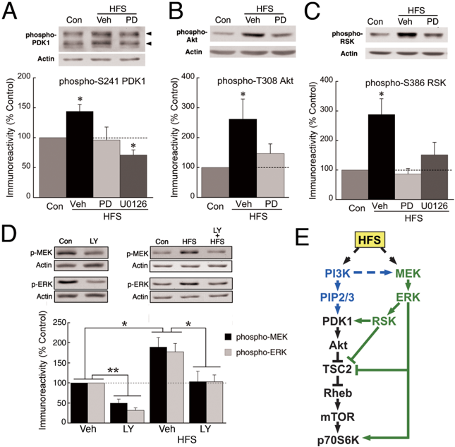
Protein synthesis is required for persistent forms of synaptic plasticity, including long-term potentiation (LTP). A key regulator of LTP-related protein synthesis is mammalian target of rapamycin (mTOR), which is thought to modulate translational capacity by facilitating the synthesis of particular components of the protein synthesis machinery. Recently, extracellularly regulated kinase (ERK) also was shown to mediate plasticity-related translation, an effect that may involve regulation of the mTOR pathway. We studied the interaction between the mTOR and ERK pathways in hippocampal LTP induced at CA3–CA1 synapses by high-frequency synaptic stimulation (HFS). Within minutes after HFS, the expression of multiple translational proteins, the synthesis of which is under the control of mTOR, increased in area CA1 stratum radiatum. This upregulation was detected in pyramidal cell dendrites and was blocked by inhibitors of the ERK pathway. In addition, ERK mediated the stimulation of mTOR by HFS. The possibility that ERK regulates mTOR by acting at a component further upstream in the phosphatidylinositide 3-kinase (PI3K)–mTOR pathway was tested by probing the phosphorylation of p90-S6 kinase, phosphoinositide-dependent kinase 1 (PDK1), and Akt. ERK inhibitors blocked HFS-induced phosphorylation of all three proteins at sites implicated in the regulation of mTOR. Moreover, a component of basal and HFS-induced ERK activity depended on PI3K, indicating that mTOR-mediated protein synthesis in LTP requires coincident and mutually dependent activity in the PI3K and ERK pathways. The role of ERK in regulating PDK1 and Akt, with their extensive effects on cellular function, has important implications for the coordinated response of the neuron to LTP-inducing stimulation.
Overview
The claims and statistics of this paper haven’t been checked yet.
Run the check — free, no account needed. It reads the open-access text and checks the sources it cites.
Integrity signals
Registry
Cites
Statistics
Figures
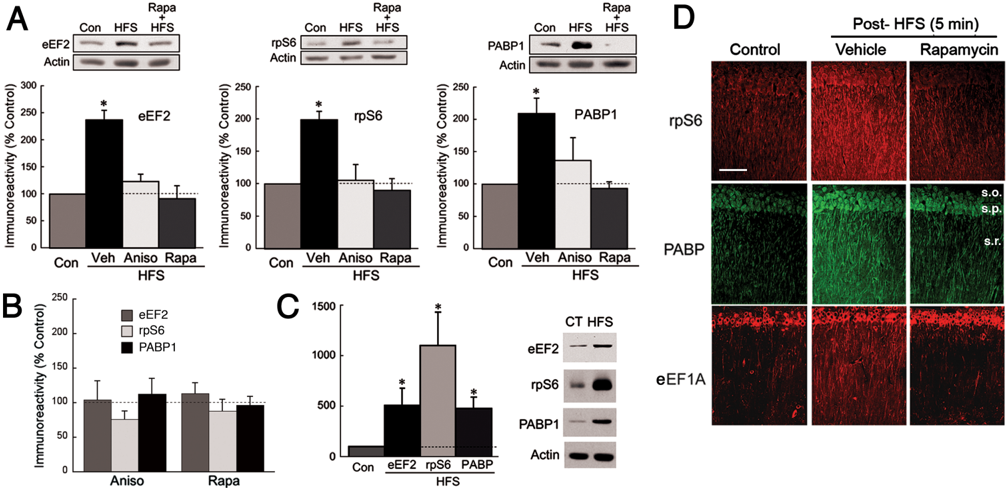
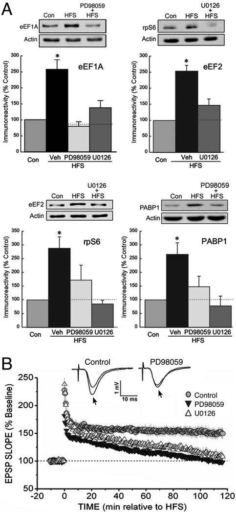
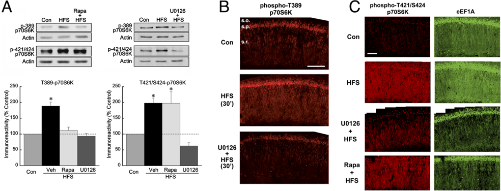
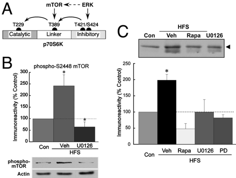
Verification
Claims the paper makes
The claims and statistics of this paper haven’t been checked yet.
Run the check — free, no account needed. It reads the open-access text and checks the sources it cites.
Claims other papers make about this one
Each is checked against this paper’s own text: does it say what the citing paper says it does?
In the citing paper
“Previous studies have demonstrated that the activation of PI3K and ERK pathways play essential roles in LTP induction and maintenance (Man et al., 2003;Kelleher et al., 2004;Tsokas et al., 2007).”
From BDNF-Dependent Recycling Facilitates TrkB Translocation to Postsynaptic Density during LTP via a Rab11-Dependent Pathway (2013) · cites this paper as Tsokas et al., 2007
Self-check
Percentages, sums and reported differences re-computed from the paper’s own numbers.
The self-check needs the paper’s full text.
Citations
Cites
Synaptic tagging and long-term potentiation
Frey, U., & Morris, R. G. M. (1997) · Nature · doi:10.1038/385533a0 · Cited by 1,736 · cited 3×
“Experiments that have addressed the issue of local protein synthesis in LTP have been inconclusive, with some supporting the tagging model (Frey and Morris, 1997, 1998) and others suggesting that LTP depends, at least in part, on local protein synthesis (Barco et al., 2002; Alarcon et al., 2006).”
BackgroundResultThe MAPK cascade is required for mammalian associative learning
Atkins, C. M., Selcher, J. C., Petraitis, J. J., Trzaskos, J. M., & Sweatt, J. D. (1998) · Nature Neuroscience · doi:10.1038/2836 · Cited by 1,083
Wu, L., Wells, D., Tay, J., Mendis, D., Abbott, M.-A., Barnitt, A., Quinlan, E., Heynen, A., Fallon, J. R., & Richter, J. D. (1998) · Neuron · doi:10.1016/s0896-6273(00)80630-3 · Cited by 488 · cited 1×
“…can supply the necessary PRPs. LTP-related translation is controlled by coordinated mechanisms that regulate mRNA availability and the activity of translation factors (Wu et al., 1998; Scheetz et al., 2000; Kelleher et al., 2004; Atkins et al., 2005; Banko et al., 2005; Sutton and Schuman, 2006).”
Background
Research works printed but not linked: 78
- 10.1523/JNEUROSCI.3196-05.2006 · DOI
- 10.1016/S0960-9822(06)00122-9 · DOI
- 10.1016/S0960-9822(06)00336-8 · DOI
- 10.1523/JNEUROSCI.5051-04.2005 · DOI
- 10.1111/j.1471-4159.2004.02734.x · DOI
- 10.1523/JNEUROSCI.2423-05.2005 · DOI · Paper page
- 10.1016/S0092-8674(02)00657-8 · DOI
- 10.1093/emboj/19.5.979 · DOI
- 10.1016/j.biopsych.2004.02.031 · DOI
- 10.1016/S0968-0004(01)01958-2 · DOI · Paper page
- 10.1073/pnas.2336098100 · DOI
- 10.1146/annurev.biochem.68.1.965 · DOI · Paper page
- 10.1073/pnas.95.17.9849 · DOI
- 10.1016/S0960-9822(98)70444-0 · DOI
- 10.1002/hipo.20078 · DOI
- 10.1042/0264-6021:3370575 · DOI
- Davis, 2000, The MAPK/ERK cascade targets both Elk-1 and cAMP response element-binding protein to control long-term potentiation-dependent gene expression in the dentate gyrus in vivo, J Neurosci, 20, 4563, 10.1523/JNEUROSCI.20-12-04563.2000 · DOI
- 10.1074/jbc.M408194200 · DOI
- 10.1016/S0960-9822(98)00017-7 · DOI
- 10.1074/jbc.272.31.19103 · DOI
- 10.1016/S0028-3908(98)00040-9 · DOI
- 10.1016/0006-8993(88)90008-X · DOI
- 10.1016/0304-3940(89)90152-3 · DOI
- 10.1093/emboj/19.12.2924 · DOI
- 10.1101/gad.13.11.1422 · DOI
- 10.1038/nrn1937 · DOI · Paper page
- 10.1074/jbc.M504045200 · DOI
- 10.1523/JNEUROSCI.0995-04.2004 · DOI · Paper page
- 10.1093/emboj/21.9.2139 · DOI
- 10.1101/lm.1.1.74 · DOI
- 10.1016/S0896-6273(00)80602-9 · DOI
- 10.1038/ncb839 · DOI · Paper page
- 10.1002/cne.903620103 · DOI · Paper page
- 10.1093/emboj/16.12.3693 · DOI · Paper page
- 10.1126/science.273.5280.1402 · DOI
- 10.1016/S0092-8674(04)00115-1 · DOI · Paper page
- 10.1016/S0896-6273(01)00508-6 · DOI
- 10.1074/jbc.M300376200 · DOI
- 10.1016/S0896-6273(01)00433-0 · DOI
- 10.1016/j.cell.2005.02.031 · DOI · Paper page
- 10.1016/S1097-2765(02)00568-3 · DOI · Paper page
- Marin, 1997, Glutamate-dependent phosphorylation of elongation factor-2 and inhibition of protein synthesis in neurons, J Neurosci, 17, 3445, 10.1523/JNEUROSCI.17-10-03445.1997 · DOI
- 10.1016/S0306-4522(00)00496-6 · DOI · Paper page
- 10.1016/j.semcdb.2003.12.022 · DOI
- 10.1074/jbc.273.30.19080 · DOI
- 10.1042/0264-6021:3440427 · DOI
- Opazo, 2003, Phosphatidylinositol 3-kinase regulates the induction of long-term potentiation through extracellular signal-related kinase-independent mechanisms, J Neurosci, 23, 3679, 10.1523/JNEUROSCI.23-09-03679.2003 · DOI · Paper page
- Osten, 1996, Protein synthesis-dependent formation of protein kinase Mζ in long-term potentiation, J Neurosci, 16, 2444, 10.1523/JNEUROSCI.16-08-02444.1996 · DOI
- Ouyang, 1999, Tetanic stimulation leads to increased accumulation of Ca2+/calmodulin-dependent protein kinase II via dendritic protein synthesis in hippocampal neurons, J Neurosci, 19, 7823, 10.1523/JNEUROSCI.19-18-07823.1999 · DOI
- 10.1074/jbc.M105916200 · DOI
- 10.1016/S0959-4388(00)00208-7 · DOI
- 10.1073/pnas.96.8.4438 · DOI
- 10.1038/ncb840 · DOI · Paper page
- 10.1016/S0014-5793(97)00323-2 · DOI
- 10.1073/pnas.0405659101 · DOI
- Sanna, 2002, Phosphatidylinositol 3-kinase is required for the expression but not for the induction or the maintenance of long-term potentiation in the hippocampal CA1 region, J Neurosci, 22, 3359, 10.1523/JNEUROSCI.22-09-03359.2002 · DOI
- 10.1074/jbc.M205141200 · DOI
- 10.1038/72915 · DOI
- 10.1523/JNEUROSCI.1739-04.2004 · DOI
- Sekulic, 2000, A direct linkage between the phosphoinositide 3-kinase-AKT signaling pathway and the mammalian target of rapamycin in mitogen-stimulated and transformed cells, Cancer Res, 60, 3504
- 10.1128/MCB.22.23.8101-8113.2002 · DOI
- 10.1016/j.cell.2006.09.014 · DOI
- 10.1074/jbc.M103237200 · DOI
- 10.1523/JNEUROSCI.1427-04.2004 · DOI · Paper page
- 10.1073/pnas.012605299 · DOI · Paper page
- 10.1523/JNEUROSCI.0599-05.2005 · DOI
- 10.1042/0264-6021:3460561 · DOI
- 10.1113/jphysiol.2005.092924 · DOI
- 10.1093/emboj/20.16.4349 · DOI
- 10.1016/S0959-4388(99)00050-1 · DOI
- Wells, 2001, A role for the cytoplasmic polyadenylation element in NMDA receptor-regulated mRNA translation in neurons, J Neurosci, 21, 9541, 10.1523/JNEUROSCI.21-24-09541.2001 · DOI
- 10.1074/jbc.M003937200 · DOI
- 10.1074/jbc.M304172200 · DOI
- 10.1016/S0896-6273(00)81124-1 · DOI
- 10.1126/science.279.5348.222 · DOI
- Yuan, 2002, Protein kinase modulation of dendritic K+ channels in hippocampus involves a mitogen-activated protein kinase pathway, J Neurosci, 22, 4860, 10.1523/JNEUROSCI.22-12-04860.2002 · DOI
- 10.1093/hmg/11.23.2929 · DOI
- 10.1074/jbc.M009047200 · DOI
Cited by
Sorted by citations
Translational control of long-lasting synaptic plasticity and memory
Costa-Mattioli, M., Sossin, W. S., Klann, E., & Sonenberg, N. (2009) · Neuron · doi:10.1016/j.neuron.2008.10.055 · Cited by 944
Translational control in synaptic plasticity and cognitive dysfunction
Buffington, S. A., Huang, W., & Costa-Mattioli, M. (2014) · Annual Review of Neuroscience · doi:10.1146/annurev-neuro-071013-014100 · Cited by 348
Translational Control in the Brain in Health and Disease
Sossin, W. S., & Costa-Mattioli, M. (2019) · Cold Spring Harbor Perspectives in Biology · doi:10.1101/cshperspect.a032912 · Cited by 138
Gafford, G. M., Parsons, R. G., & Helmstetter, F. J. (2011) · Neuroscience · doi:10.1016/j.neuroscience.2011.03.023 · Cited by 110
Chen, X., Lin, R., Chang, L., Xu, S., Wei, X., Zhang, J., Wang, C., Anwyl, R., & Wang, Q. (2013) · Neuroscience · doi:10.1016/j.neuroscience.2013.08.054 · Cited by 93
Metadata
- Authors
- Tsokas, P., Ma, T., Iyengar, R., Landau, E. M., & Blitzer, R. D.
- Venue
- The Journal of Neuroscience
- Publisher
- Society for Neuroscience
- Published
- 2007-05-30
- Volume
- 27
- Issue
- 22
- Pages
- 5885-5894
- ISSN
- 0270-6474
- 1529-2401
- Cited by
- 151
- References
- 3
- Language
- en
About these checks
Registry, retraction, Cites and statistics results are computed, not judged, with one exception: a printed reference that no index lists is read once by a model before it counts as a citation error. Claims are extracted by a model and kept only when their quote appears verbatim in the text; each cited source is then checked, with a model’s help, for whether it says what the claim relies on. Those verdicts can be wrong — read the claim’s quote and the cited source before relying on one.
Statistics and arithmetic are recomputed, not judged.
Confidence bands are shown only for fields calibrated on our annotated test set. None is calibrated yet, so no band is shown.
What counts as a claim ›How we measure confidence ›How we check this ›
Are you an author, or do you see a problem with this page? Request a review
How to cite
Cite CoChat for the checks on this page, and the paper for its own findings.
- This page
- CoChat. (2026). Citation check: Mitogen-Activated Protein Kinase Upregulates the Dendritic Translation Machinery in Long-Term Potentiation by Controlling the Mammalian Target of Rapamycin Pathway. https://app.cochat.ai/papers/10.1523/jneurosci.4548-06.2007
- The paper
- Tsokas, P., Ma, T., Iyengar, R., Landau, E. M., & Blitzer, R. D. (2007). Mitogen-Activated Protein Kinase Upregulates the Dendritic Translation Machinery in Long-Term Potentiation by Controlling the Mammalian Target of Rapamycin Pathway. The Journal of Neuroscience. https://doi.org/10.1523/jneurosci.4548-06.2007
