

The DOI matches its Crossref record; 72 of the 72 references it cites were found in a registry or an index.
Amygdalar Inactivation Blocks Stress-Induced Impairments in Hippocampal Long-Term Potentiation and Spatial Memory
Open accessAbstract
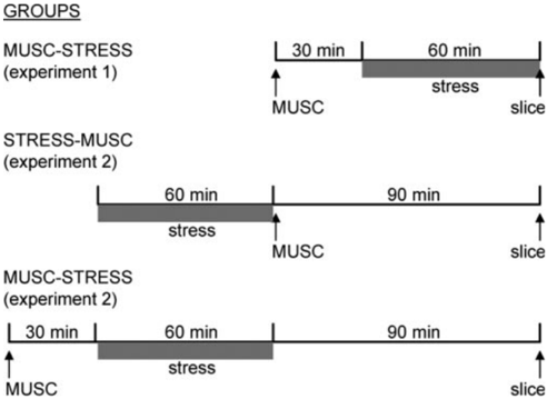
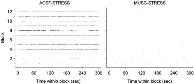
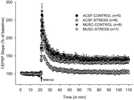
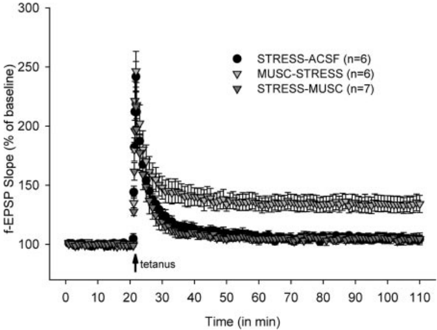
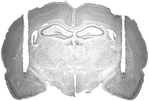
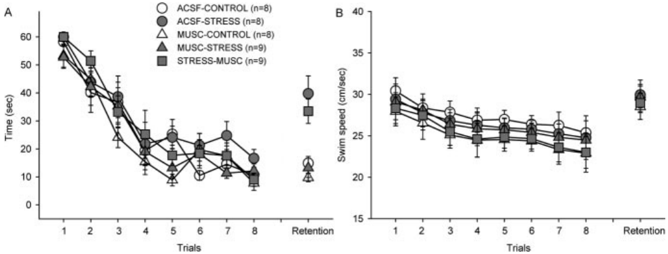
Electrolytic lesions to the amygdala, a limbic structure implicated in stress-related behaviors and memory modulation, have been shown to prevent stress-induced impairments of hippocampal long-term potentiation (LTP) and spatial memory in rats. The present study investigated the role of intrinsic amygdalar neurons in mediating stress effects on the hippocampus by microinfusing the GABA(A) receptor agonist muscimol into the amygdala and examining stress effects on Schaffer collateral/commissural-CA1 LTP and spatial memory. The critical period of the amygdalar contribution to stress effects on hippocampal functions was determined by applying muscimol either before stress or immediately after stress. Our results indicate that intra-amygdalar muscimol infusions before uncontrollable restraint-tailshock stress effectively blocked stress-induced physiological and behavioral effects. Specifically, hippocampal slices prepared from vehicle-infused stressed animals exhibited markedly impaired LTP, whereas slices obtained from muscimol-infused stressed animals demonstrated robust LTP comparable with that of unstressed animals. Correspondingly, vehicle-infused stressed animals displayed impaired spatial memory (on a hidden platform version of the Morris water maze task), whereas muscimol-infused stressed animals revealed unimpaired spatial memory. In contrast to prestress muscimol effects, however, immediate poststress infusions of muscimol into the amygdala failed to interfere with stress impairments of LTP and spatial memory. Together, these results suggest that the amygdalar neuronal activity during stress, but not shortly after stress, is essential for the emergence of stress-induced alterations in hippocampal LTP and memory.
Overview
The claims and statistics of this paper haven’t been checked yet.
Run the check — free, no account needed. It reads the open-access text and checks the sources it cites.
Integrity signals
Registry
Cites
Statistics
Figures
Verification
Claims the paper makes
The claims and statistics of this paper haven’t been checked yet.
Run the check — free, no account needed. It reads the open-access text and checks the sources it cites.
Claims other papers make about this one
Each is checked against this paper’s own text: does it say what the citing paper says it does?
In the citing paper
“damage to, or inactivation of, the amygdala blocks stress effects on hippocampal LTP and spatial memory (Almaguer-Melian et al. [214]; Kim et al. [154]; Korz and Frey [215]; Kim and Jung [21]).”
From The Temporal Dynamics Model of Emotional Memory Processing: A Synthesis on the Neurobiological Basis of Stress-Induced Amnesia, Flashbulb and Traumatic Memories, and the Yerkes-Dodson Law (2007) · cites this paper as Kim et al. [154]
In the citing paper
“Overall, these findings, in conjunction with related work by Kim et al. [154] support our hypothesis that a strong stressor generates a powerful inhibitory influence on hippocampal memory processing for events occurring 30–60 minutes after the onset of a stressful experience.”
From The Temporal Dynamics Model of Emotional Memory Processing: A Synthesis on the Neurobiological Basis of Stress-Induced Amnesia, Flashbulb and Traumatic Memories, and the Yerkes-Dodson Law (2007) · cites this paper as Kim et al. [154]
Self-check
Percentages, sums and reported differences re-computed from the paper’s own numbers.
The self-check needs the paper’s full text.
Citations
Cites
Bliss, T. V. P., & Gardner-Medwin, A. R. (1973) · The Journal of Physiology · doi:10.1113/jphysiol.1973.sp010274 · Cited by 7,424
LOSS OF RECENT MEMORY AFTER BILATERAL HIPPOCAMPAL LESIONS
Scoville, W. B., & Milner, B. (1957) · Journal of Neurology, Neurosurgery & Psychiatry · doi:10.1136/jnnp.20.1.11 · Cited by 7,322
The Organization of Behavior; A Neuropsychological Theory.
Attneave, F., B., M., & Hebb, D. O. (1950) · The American Journal of Psychology · doi:10.2307/1418888 · Cited by 5,146
Memory - A century of consolidation
McGaugh, J. L. (2000) · Science · doi:10.1126/science.287.5451.248 · Cited by 4,250
Morris, R. G. M., Anderson, E., Lynch, G. S., & Baudry, M. (1986) · Nature · doi:10.1038/319774a0 · Cited by 3,640
Long-Term Potentiation--A Decade of Progress?
Malenka, R. C., & Nicoll, A. R. A. (1999) · Science · doi:10.1126/science.285.5435.1870 · Cited by 2,729
Two receptor systems for corticosterone in rat brain: Microdistribution and differential occupation
REUL, J. M. H. M., & KLOET, E. R. D. (1985) · Endocrinology · doi:10.1210/endo-117-6-2505 · Cited by 2,580
Learned helplessness: Theory and evidence
Maier, S. F., & Seligman, M. E. (1976) · Journal of Experimental Psychology: General · doi:10.1037/0096-3445.105.1.3 · Cited by 2,494
Failure to escape traumatic shock.
Seligman, M. E., & Maier, S. F. (1967) · Journal of Experimental Psychology · doi:10.1037/h0024514 · Cited by 1,981
McEwen, B. S., & Sapolsky, R. M. (1995) · Current Opinion in Neurobiology · doi:10.1016/0959-4388(95)80028-x · Cited by 1,889
Research works printed but not linked: 6
- 1999, Physiol Behav, 65, 739
- 1973, J Physiol (Lond), 232, 331, 10.1113/jphysiol.1973.sp010273 · DOI · Paper page
- 1990, Psychol Sci, 3, 201
- 10.1037/0735-7044.106.3.518 · DOI
- 10.1037/0735-7044.108.5.1005 · DOI
- 10.1037//0096-3445.105.1.3 · DOI · Paper page
Cited by
Sorted by citations
Physiology and neurobiology of stress and adaptation: central role of the brain
McEwen, B. S. (2007) · Physiological Reviews · doi:10.1152/physrev.00041.2006 · Cited by 5,429
Stress, memory and the amygdala
Roozendaal, B., McEwen, B. S., & Chattarji, S. (2009) · Nature Reviews Neuroscience · doi:10.1038/nrn2651 · Cited by 1,760
The Brain on Stress: Vulnerability and Plasticity of the Prefrontal Cortex over the Life Course
McEwen, B. S., & Morrison, J. H. (2013) · Neuron · doi:10.1016/j.neuron.2013.06.028 · Cited by 1,079
Diamond, D. M., Campbell, A. M., Park, C. R., Halonen, J., & Zoladz, P. R. (2007) · Neural Plasticity · doi:10.1155/2007/60803 · Cited by 762
Stress effects on memory: An update and integration
Schwabe, L., Joëls, M., Roozendaal, B., Wolf, O. T., & Oitzl, M. S. (2012) · Neuroscience & Biobehavioral Reviews · doi:10.1016/j.neubiorev.2011.07.002 · Cited by 714
Stress and Memory: Behavioral Effects and Neurobiological Mechanisms
Sandi, C., & Pinelo-Nava, M. T. (2007) · Neural Plasticity · doi:10.1155/2007/78970 · Cited by 611
Stress and anxiety: Structural plasticity and epigenetic regulation as a consequence of stress
McEwen, B. S., Eiland, L., Hunter, R. G., & Miller, M. M. (2012) · Neuropharmacology · doi:10.1016/j.neuropharm.2011.07.014 · Cited by 596
Stress effects on the hippocampus: a critical review
Kim, E. J., Pellman, B., & Kim, J. J. (2015) · Learning & Memory · doi:10.1101/lm.037291.114 · Cited by 620
Self-citationSandi, C. (2013) · Wiley Interdisciplinary Reviews Cognitive Science · doi:10.1002/wcs.1222 · Cited by 580
Emotional Arousal and Memory Binding: An Object-Based Framework
Mather, M. (2007) · Perspectives on Psychological Science · doi:10.1111/j.1745-6916.2007.00028.x · Cited by 491
Acute corticosterone treatment is sufficient to induce anxiety and amygdaloid dendritic hypertrophy
Mitra, R., & Sapolsky, R. M. (2008) · Proceedings of the National Academy of Sciences · doi:10.1073/pnas.0705615105 · Cited by 409
Chronic stress: Implications for neuronal morphology, function and neurogenesis
Joëls, M., Karst, H., Krugers, H. J., & Lucassen, P. J. (2007) · Frontiers in Neuroendocrinology · doi:10.1016/j.yfrne.2007.04.001 · Cited by 381
Stress, sex, and neural adaptation to a changing environment: mechanisms of neuronal remodeling
McEwen, B. S. (2010) · Annals of the New York Academy of Sciences · doi:10.1111/j.1749-6632.2010.05568.x · Cited by 335
Synaptic plasticity in depression: Molecular, cellular and functional correlates
Marsden, W. N. (2012) · Progress in Neuro-Psychopharmacology and Biological Psychiatry · doi:10.1016/j.pnpbp.2012.12.012 · Cited by 323
Zoladz, P. R., & Diamond, D. M. (2013) · Neuroscience & Biobehavioral Reviews · doi:10.1016/j.neubiorev.2013.03.024 · Cited by 339
Stress, anxiety, and dendritic spines: What are the connections?
Leuner, B., & Shors, T. J. (2013) · Neuroscience · doi:10.1016/j.neuroscience.2012.04.021 · Cited by 284
McCutcheon, J. E., & Marinelli, M. (2009) · European Journal of Neuroscience · doi:10.1111/j.1460-9568.2009.06648.x · Cited by 299
Neighborhood matters: divergent patterns of stress-induced plasticity across the brain
Chattarji, S., Tomar, A., Suvrathan, A., Ghosh, S., & Rahman, M. M. (2015) · Nature Neuroscience · doi:10.1038/nn.4115 · Cited by 248
Stress, neurotransmitters, corticosterone and body–brain integration
Mora, F., Segovia, G., del Arco, A., de Blas, M., & Garrido, P. (2012) · Brain Research · doi:10.1016/j.brainres.2011.12.049 · Cited by 253
Stress effects in the hippocampus: Synaptic plasticity and memory
Kim, J. J., Song, E. Y., Kim, J. J., Song, E. Y., & Kosten, T. A. (2006) · Stress · doi:10.1080/10253890600678004 · Cited by 261
Self-citationPrefrontal cortex and hippocampus subserve different components of working memory in ratsPreprint
Yoon, T., Okada, J., Jung, M. W., & Kim, J. J. (2008) · Learning & Memory · doi:10.1101/lm.850808 · Cited by 267
Self-citationChapter 8 Synaptic plasticity in learning and memory: Stress effects in the hippocampus
Howland, J. G., & Wang, Y. T. (2008) · Progress in brain research · doi:10.1016/s0079-6123(07)00008-8 · Cited by 243
Stressful Experience and Learning Across the Lifespan
Shors, T. J. (2005) · Annual Review of Psychology · doi:10.1146/annurev.psych.57.102904.190205 · Cited by 234
Hippocampal long-term depression mediates spatial reversal learning in the Morris water maze
Dong, Z., Bai, Y., Wu, X., Li, H., Gong, B., Howland, J. G., Huang, Y., He, W., Li, T., & Wang, Y. T. (2013) · Neuropharmacology · doi:10.1016/j.neuropharm.2012.06.027 · Cited by 219
Hippocampal long-term depression mediates acute stress-induced spatial memory retrieval impairment
Wong, T. P., Howland, J. G., Robillard, J. M., Ge, Y., Yu, W. K., Titterness, A. K., Brebner, K., Liu, L., Weinberg, J., Christie, B. R., et al. (2007) · Proceedings of the National Academy of Sciences · doi:10.1073/pnas.0702308104 · Cited by 228
Social Transmission of Fear in Rats: The Role of 22-kHz Ultrasonic Distress Vocalization
Kim, E. J., Kim, E. S., Covey, E., & Kim, J. J. (2010) · PLoS ONE · doi:10.1371/journal.pone.0015077 · Cited by 216
Self-citationPark, C. R., Zoladz, P. R., Conrad, C. D., Fleshner, M., & Diamond, D. M. (2008) · Learning & Memory · doi:10.1101/lm.721108 · Cited by 170
Jiang, X., Xing, G., Yang, C., Verma, A. M., Zhang, L., & Li, H. (2008) · Neuropsychopharmacology · doi:10.1038/npp.2008.71 · Cited by 146
Joëls, M., & Krugers, H. J. (2007) · Neural Plasticity · doi:10.1155/2007/93202 · Cited by 141
Joca, S. R. L., Ferreira, F. R., & Guimarães, F. S. (2007) · Stress · doi:10.1080/10253890701223130 · Cited by 139
Stress-induced metaplasticity: From synapses to behavior
Schmidt, M. V., Abraham, W. C., Maroun, M., Stork, O., & Richter‐Levin, G. (2013) · Neuroscience · doi:10.1016/j.neuroscience.2013.06.059 · Cited by 137
Lissner, L. J., Wartchow, K. M., Toniazzo, A. P., Gonçalves, C., & Rodrigues, L. (2021) · Pharmacology Biochemistry and Behavior · doi:10.1016/j.pbb.2021.173273 · Cited by 141
Stamatakis, A. M., Sparta, D. R., Jennings, J. H., McElligott, Z. A., Decot, H., & Stuber, G. D. (2014) · Neuropharmacology · doi:10.1016/j.neuropharm.2013.05.046 · Cited by 132
Diamond, D. M., Park, C. R., Campbell, A. M., & Woodson, J. C. (2005) · Hippocampus · doi:10.1002/hipo.20107 · Cited by 128
Neurocognitive effects of stress: a metaparadigm perspective
Kim, E. J., & Kim, J. J. (2023) · Molecular Psychiatry · doi:10.1038/s41380-023-01986-4 · Cited by 154
Self-citationStress-induced alterations in hippocampal plasticity, place cells, and spatial memory
Kim, J. J., Lee, H. J., Welday, A. C., Song, E., Cho, J., Sharp, P. E., Jung, M. W., & Blair, H. T. (2007) · Proceedings of the National Academy of Sciences · doi:10.1073/pnas.0708644104 · Cited by 126
Self-citationAguilar‐Valles, A., Sánchez, E., de Gortari, P., Balderas, I., Ramı́rez-Amaya, V., Bermúdez‐Rattoni, F., & Joseph‐Bravo, P. (2005) · Neuroendocrinology · doi:10.1159/000093129 · Cited by 121
Oxytocin Protects Hippocampal Memory and Plasticity from Uncontrollable Stress
Lee, S.-Y., Park, S.-H., Chung, C., Kim, J. J., Choi, S.-Y., & Han, J.-S. (2015) · Scientific Reports · doi:10.1038/srep18540 · Cited by 119
Self-citationFunctional Connectivity from the Amygdala to the Hippocampus Grows Stronger after Stress
Ghosh, S., Laxmi, T. R., & Chattarji, S. (2013) · Journal of Neuroscience · doi:10.1523/jneurosci.0638-13.2013 · Cited by 115
Barzegar, M., Sajjadi, F. S., Talaei, S. A., Hamidi, G. A., & Salami, M. (2014) · Hippocampus · doi:10.1002/hipo.22363 · Cited by 108
Amygdala-hippocampal interactions in synaptic plasticity and memory formation
Roesler, R., Parent, M. B., LaLumiere, R. T., & McIntyre, C. K. (2021) · Neurobiology of Learning and Memory · doi:10.1016/j.nlm.2021.107490 · Cited by 121
Effects of stress and corticosterone on activity and plasticity in the amygdala
Kavushansky, A., & Richter‐Levin, G. (2006) · Journal of Neuroscience Research · doi:10.1002/jnr.21058 · Cited by 109
Establishing an Agenda for Translational Research on PTSD
MILLER, M. M., & McEWEN, B. S. (2006) · Annals of the New York Academy of Sciences · doi:10.1196/annals.1364.023 · Cited by 101
Emotional tagging—A simple hypothesis in a complex reality
Bergado, J. A., Lucas, M., & Richter‐Levin, G. (2011) · Progress in Neurobiology · doi:10.1016/j.pneurobio.2011.03.004 · Cited by 98
Zoladz, P. R., Clark, B., Warnecke, A., Smith, L., Tabar, J., & Talbot, J. N. (2011) · Physiology & Behavior · doi:10.1016/j.physbeh.2011.01.016 · Cited by 102
Cazakoff, B. N., Johnson, K. J., & Howland, J. G. (2010) · Progress in Neuro-Psychopharmacology and Biological Psychiatry · doi:10.1016/j.pnpbp.2010.04.002 · Cited by 96
Pochwat, B., Szewczyk, B., Sowa-Kućma, M., Siwek, A., Doboszewska, U., Piekoszewski, W., Gruca, P., Papp, M., & Nowak, G. (2013) · The International Journal of Neuropsychopharmacology · doi:10.1017/s1461145713001089 · Cited by 84
Effects of stressors in adolescence on learning and memory in rodent models
Green, M. R., & McCormick, C. M. (2013) · Hormones and Behavior · doi:10.1016/j.yhbeh.2012.09.012 · Cited by 86
Zoladz, P. R., Park, C. R., Halonen, J. D., Salim, S., Alzoubi, K. H., Srivareerat, M., Fleshner, M., Alkadhi, K. A., & Diamond, D. M. (2011) · Hippocampus · doi:10.1002/hipo.20922 · Cited by 83
Critical brain circuits at the intersection between stress and learning
Bangasser, D. A., & Shors, T. J. (2010) · Neuroscience & Biobehavioral Reviews · doi:10.1016/j.neubiorev.2010.02.002 · Cited by 83
Impact of glucocorticoids on brain function: Relevance for mood disorders
Joëls, M. (2010) · Psychoneuroendocrinology · doi:10.1016/j.psyneuen.2010.03.004 · Cited by 83
Murty, V. P., LaBar, K. S., Hamilton, D. A., & Adcock, R. A. (2011) · Learning & Memory · doi:10.1101/lm.023549.111 · Cited by 77
Amygdala inactivation reverses fear's ability to impair divided attention and make time stand still.
Meck, W. H., & MacDonald, C. J. (2007) · Behavioral Neuroscience · doi:10.1037/0735-7044.121.4.707 · Cited by 79
Vouimba, R., Yaniv, D., & Richter‐Levin, G. (2006) · Neuropharmacology · doi:10.1016/j.neuropharm.2006.07.007 · Cited by 71
Exposure to Stressors Facilitates Long-Term Synaptic Potentiation in the Lateral Habenula
Park, H.-Y., Rhee, J., Park, K., Han, J., Malinow, R., & Chung, C. (2017) · Journal of Neuroscience · doi:10.1523/jneurosci.2281-16.2017 · Cited by 71
Self-citationTrifilieff, P., Calandreau, L., Herry, C., Mons, N. E., & Micheau, J. (2007) · Neurobiology of Learning and Memory · doi:10.1016/j.nlm.2007.05.004 · Cited by 73
Glucocorticoid Hyper‐ and Hypofunction
Kim, J. J., & Haller, J. (2007) · Annals of the New York Academy of Sciences · doi:10.1196/annals.1391.014 · Cited by 66
Self-citationReznikov, L. R., Reagan, L. P., & Fadel, J. R. (2008) · The Journal of Comparative Neurology · doi:10.1002/cne.21687 · Cited by 65
Hoffman, A. N., Krigbaum, A. M., Ortiz, J. B., Mika, A., Hutchinson, K. M., Bimonte‐Nelson, H. A., & Conrad, C. D. (2011) · European Journal of Neuroscience · doi:10.1111/j.1460-9568.2011.07820.x · Cited by 67
Park, C. R., Campbell, A. M., Woodson, J. C., Smith, T. P., Fleshner, M., & Diamond, D. M. (2006) · Dose-Response · doi:10.2203/dose-response.004.01.005.park · Cited by 63
Nygard, S. K., Hourguettes, N. J., Sobczak, G. G., Carlezon, W. A., & Bruchas, M. R. (2016) · Journal of Neuroscience · doi:10.1523/jneurosci.0953-16.2016 · Cited by 60
Castro, J. E., Varea, E., Márquez, C., Cordero, M. I., Poirier, G. L., & Sandi, C. (2010) · PLoS ONE · doi:10.1371/journal.pone.0008618 · Cited by 63
Rei, D., Mason, X. L., Seo, J., Gräff, J., Rudenko, A., Wang, J., Rueda, R., Siegert, S., Cho, S., Canter, R. G., et al. (2015) · Proceedings of the National Academy of Sciences · doi:10.1073/pnas.1415845112 · Cited by 65
c-Jun N-terminal kinases in memory and synaptic plasticity
Sherrin, T., Blank, T., & Todorovic, C. (2011) · Reviews in the Neurosciences · doi:10.1515/rns.2011.032 · Cited by 56
Graham, L. K., Yoon, T., Lee, H. J., & Kim, J. J. (2009) · Psychology & Neuroscience · doi:10.3922/j.psns.2009.2.015 · Cited by 55
Self-citationThe adaptive and maladaptive continuum of stress responses – a hippocampal perspective
Suri, D., & Vaidya, V. A. (2015) · Reviews in the Neurosciences · doi:10.1515/revneuro-2014-0083 · Cited by 66
The Endocannabinoid System and Anxiety
Lisboa, S. F. D. S., Gomes, F. V., Terzian, A. L. B., de Aguiar, D. C., Moreira, F. A., Resstel, L. B. M., & Guimarães, F. S. (2016) · Vitamins and hormones · doi:10.1016/bs.vh.2016.09.006 · Cited by 56
Chewing ameliorates stress-induced suppression of hippocampal long-term potentiation
Ono, Y., Kataoka, T., Miyake, S., Cheng, S., Tachibana, A., Sasaguri, K., & Onozuka, M. (2008) · Neuroscience · doi:10.1016/j.neuroscience.2008.04.057 · Cited by 53
Sadowski, R. N., Jackson, G. R., Wieczorek, L., & Gold, P. E. (2009) · Behavioural Brain Research · doi:10.1016/j.bbr.2009.06.027 · Cited by 51
Acute stress throughout the memory cycle: Diverging effects on associative and item memory.
Goldfarb, E. V., Tompary, A., Davachi, L., & Phelps, E. A. (2018) · Journal of Experimental Psychology General · doi:10.1037/xge0000472 · Cited by 54
The dose makes the poison: From glutamate-mediated neurogenesis to neuronal atrophy and depression
Rubio-Casillas, A., & Fernández-Guasti, A. (2016) · Reviews in the Neurosciences · doi:10.1515/revneuro-2015-0066 · Cited by 49
Neuronal localization of the 5-HT2 receptor family in the amygdaloid complex
Bombardi, C. (2014) · Frontiers in Pharmacology · doi:10.3389/fphar.2014.00068 · Cited by 48
Cadle, C. E., & Zoladz, P. R. (2015) · Frontiers in Psychology · doi:10.3389/fpsyg.2015.00910 · Cited by 52
Setiawan, E., Jackson, M. F., MacDonald, J. F., & Matthews, S. G. (2007) · Journal of Physiology · doi:10.1113/jphysiol.2006.127381 · Cited by 48
Tripathi, S. J., Chakraborty, S., Srikumar, B. N., Raju, T. R., & Shankaranarayana Rao, B. S. (2019) · Progress in Neuro-Psychopharmacology and Biological Psychiatry · doi:10.1016/j.pnpbp.2018.07.016 · Cited by 43
Chronic Clomipramine Treatment Reverses Core Symptom of Depression in Subordinate Tree Shrews
Wang, J., Chai, A., Zhou, Q., Lv, L., Wang, L., Yang, Y., & Xu, L. (2013) · PLoS ONE · doi:10.1371/journal.pone.0080980 · Cited by 46
Hadad-Ophir, O., Albrecht, A., Stork, O., & Richter‐Levin, G. (2014) · Frontiers in Behavioral Neuroscience · doi:10.3389/fnbeh.2014.00003 · Cited by 45
Bredemann, T. M., & McMahon, L. L. (2014) · Psychoneuroendocrinology · doi:10.1016/j.psyneuen.2014.01.004 · Cited by 43
Do Stress and Long-Term Potentiation Share the Same Molecular Mechanisms?
Huang, C., Yang, C., & Hsu, K. (2005) · Molecular Neurobiology · doi:10.1385/mn:32:3:223 · Cited by 46
Stress impairs optimal behavior in a water foraging choice task in ratsPreprint
Graham, L. K., Yoon, T., & Kim, J. J. (2009) · Learning & Memory · doi:10.1101/lm.1605510 · Cited by 42
Self-citationBlaise, J. H., Koranda, J. L., Chow, U., Haines, K. E., & Dorward, E. C. (2007) · Brain Research · doi:10.1016/j.brainres.2007.11.049 · Cited by 41
Stress within a Restricted Time Window Selectively Affects the Persistence of Long-Term Memory
Yang, C., Liu, J., Chai, B.-S., Fang, Q., Chai, N., Zhao, L., Xue, Y.-X., Luo, Y., Jian, M., Han, Y., et al. (2013) · PLoS ONE · doi:10.1371/journal.pone.0059075 · Cited by 41
The dynamic impact of repeated stress on the hippocampal spatial map
Tomar, A., Polygalov, D., Chattarji, S., & McHugh, T. J. (2014) · Hippocampus · doi:10.1002/hipo.22348 · Cited by 38
Aguilar‐Valles, A., Sánchez, E., de Gortari, P., García-Vázquez, A., Ramı́rez-Amaya, V., Bermúdez‐Rattoni, F., & Joseph‐Bravo, P. (2006) · Neurochemistry International · doi:10.1016/j.neuint.2006.09.009 · Cited by 39
Giachero, M., Calfa, G. D., & Molina, V. A. (2013) · Learning & Memory · doi:10.1101/lm.031724.113 · Cited by 40
VanElzakker, M. B., Zoladz, P. R., Thompson, V. M., Park, C. R., Halonen, J. D., Spencer, R. L., & Diamond, D. M. (2011) · Frontiers in Behavioral Neuroscience · doi:10.3389/fnbeh.2011.00030 · Cited by 41
Zoladz, P. R., & Diamond, D. M. (2008) · Dose-Response · doi:10.2203/dose-response.08-015.zoladz · Cited by 37
Enhancement of spatial learning by predator odor in mice: Involvement of amygdala and hippocampus
Galliot, E., Vaillant, M. L., Béard, E., Millot, J., & Pourié, G. (2009) · Neurobiology of Learning and Memory · doi:10.1016/j.nlm.2009.09.011 · Cited by 36
Zoladz, P., Warnecke, A. J., Woelke, S., Burke, H. M., Frigo, R. M., Pisansky, J. M., Lyle, S. M., & Talbot, J. N. (2013) · Neurobiology of Learning and Memory · doi:10.1016/j.nlm.2012.12.012 · Cited by 35
Stressful stimuli modulate memory formation in Lymnaea stagnalis
Martens, K., Caigny, P. D., Parvez, K., Amarell, M., Wong, C., & Lukowiak, K. (2006) · Neurobiology of Learning and Memory · doi:10.1016/j.nlm.2006.10.005 · Cited by 36
Temporal modulation of hippocampal excitatory transmission by corticosteroids and stress
Chaouloff, F., & Groc, L. (2010) · Frontiers in Neuroendocrinology · doi:10.1016/j.yfrne.2010.07.004 · Cited by 50
Li, Z., & Richter‐Levin, G. (2012) · Frontiers in Cellular Neuroscience · doi:10.3389/fncel.2012.00021 · Cited by 35
Amygdalar Stimulation Produces Alterations on Firing Properties of Hippocampal Place Cells
Kim, E. J., Kim, E. S., Park, M.-J., Cho, J., & Kim, J. J. (2012) · Journal of Neuroscience · doi:10.1523/jneurosci.1108-12.2012 · Cited by 36
Self-citationStress and Glucocorticoid Contributions to Normal and Pathological Aging
Goosens, K. A., & Sapolsky, R. M. (2007) · CRC Press eBooks · doi:10.1201/9781420005523-13 · Cited by 41
Activation pattern of the limbic system following spatial learning under stress
Kogan, I., & Richter‐Levin, G. (2008) · European Journal of Neuroscience · doi:10.1111/j.1460-9568.2008.06034.x · Cited by 34
Miyake, S., Yoshikawa, G., Yamada, K., Sasaguri, K., Yamamoto, T., Onozuka, M., & Sato, S. (2012) · Brain Research · doi:10.1016/j.brainres.2012.01.011 · Cited by 34
Know thy SEFL: Fear sensitization and its relevance to stressor-related disorders
Nishimura, K., Poulos, A. M., Drew, M. R., & Rajbhandari, A. K. (2022) · Neuroscience & Biobehavioral Reviews · doi:10.1016/j.neubiorev.2022.104884 · Cited by 38
Tripathi, S. J., Chakraborty, S., Srikumar, B. N., Raju, T. R., & Rao, B. S. S. (2017) · Neurobiology of Learning and Memory · doi:10.1016/j.nlm.2017.05.005 · Cited by 35
Ishikawa, S., Saito, Y., Yanagawa, Y., Otani, S., Hiraide, S., Shimamura, K., Matsumoto, M., & Togashi, H. (2011) · European Journal of Neuroscience · doi:10.1111/j.1460-9568.2011.07921.x · Cited by 33
Canadian Association of Neurosciences Review: Learning at a Snail's Pace
Parvez, K., Rosenegger, D. G., Orr, M. V., Martens, K., & Lukowiak, K. (2006) · Canadian Journal of Neurological Sciences / Journal Canadien des Sciences Neurologiques · doi:10.1017/s0317167100005291 · Cited by 33
Metadata
- Authors
- Kim, J. J., Koo, J. W., Lee, H. J., & Han, J.-S.
- Affiliations
- University of Washington (US)
- Yale University (US)
- Johns Hopkins University (US)
- Venue
- The Journal of Neuroscience
- Publisher
- Society for Neuroscience
- Published
- 2005-02-09
- Volume
- 25
- Issue
- 6
- Pages
- 1532-1539
- ISSN
- 0270-6474 (ISSN-L)
- 1529-2401
- PMC
- PMC6725997
- OpenAlex
- W2053502537
- Cited by
- 184
- References
- 71
- Citations by year
- 2026: 2 · 2025: 2 · 2024: 1 · 2023: 2 · 2022: 4 · 2021: 8 · 2020: 3 · 2019: 4 · 2018: 6 · 2017: 6 · 2016: 12 · 2015: 6 · 2014: 14 · 2013: 19 · 2012: 16
- FWCI
- 7.12
- Type
- journal article
- Language
- en
- Open access
- hybrid
- doi.org
- Licence
- Locations
- Journal of Neuroscience (publishedVersion, cc-by-nc-sa)
- Europe PMC (PubMed Central) (publishedVersion)
- PubMed Central (publishedVersion)
- PubMed (publishedVersion)
- Funders
- Whitehall Foundation
- National Institute of Mental Health (MH64457, R01MH064457, 1R01 MH64457)
- Topics
- Life Sciences › Neuroscience › Cognitive Neuroscience › Memory and Neural Mechanisms
- Life Sciences › Neuroscience › Behavioral Neuroscience › Stress Responses and Cortisol
- Life Sciences › Neuroscience › Cellular and Molecular Neuroscience › Neuroscience and Neuropharmacology Research
- Keywords
- amygdala, stress, hippocampal long-term potentiation, spatial memory, muscimol, GABAA receptor, stress-induced memory impairment, limbic system
- MeSH terms
- Amygdala, Animals, Corticosterone, Drug Administration Schedule, Electroshock, Hippocampus, Male, Memory Disorders, Muscimol, Stress, Physiological, Vocalization, Animal, Rats, Sprague-Dawley, Long-Term Potentiation, GABA Agonists, Maze Learning, Rats, GABA-A Receptor Agonists
About these checks
Registry, retraction, Cites and statistics results are computed, not judged, with one exception: a printed reference that no index lists is read once by a model before it counts as a citation error. Claims are extracted by a model and kept only when their quote appears verbatim in the text; each cited source is then checked, with a model’s help, for whether it says what the claim relies on. Those verdicts can be wrong — read the claim’s quote and the cited source before relying on one.
Statistics and arithmetic are recomputed, not judged.
Confidence bands are shown only for fields calibrated on our annotated test set. None is calibrated yet, so no band is shown.
What counts as a claim ›How we measure confidence ›How we check this ›
Are you an author, or do you see a problem with this page? Request a review
