

The DOI matches its Crossref record; 43 of the 43 references it cites were found in a registry or an index.
Neurokinin B and Dynorphin A in Kisspeptin Neurons of the Arcuate Nucleus Participate in Generation of Periodic Oscillation of Neural Activity Driving Pulsatile Gonadotropin-Releasing Hormone Secretion in the Goat
Open accessAbstract
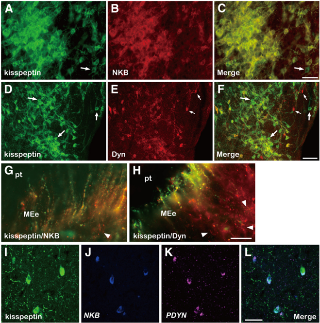
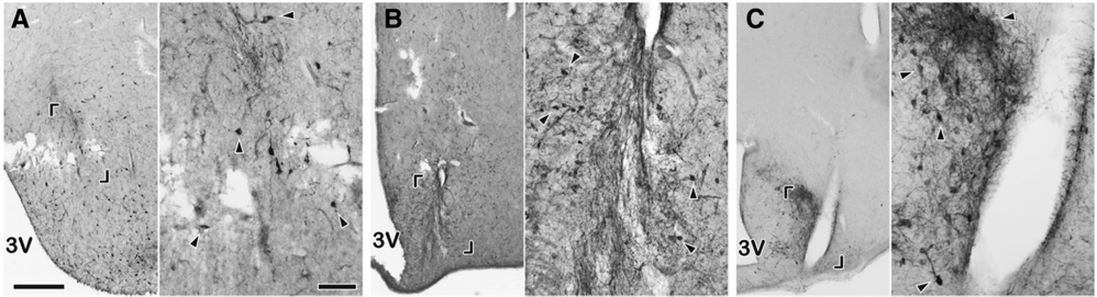
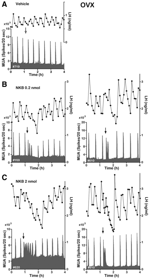
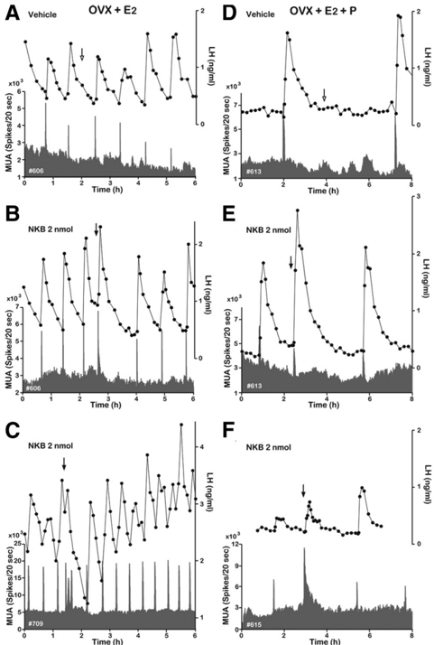
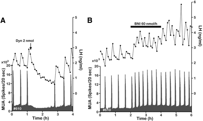
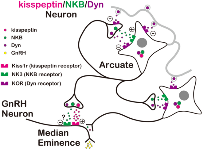
Gonadotropin-releasing hormone (GnRH) neurons in the basal forebrain are the final common pathway through which the brain regulates reproduction. GnRH secretion occurs in a pulsatile manner, and indirect evidence suggests the kisspeptin neurons in the arcuate nucleus (ARC) serve as the central pacemaker that drives pulsatile GnRH secretion. The purpose of this study was to investigate the possible coexpression of kisspeptin, neurokinin B (NKB), and dynorphin A (Dyn) in neurons of the ARC of the goat and evaluate their potential roles in generating GnRH pulses. Using double and triple labeling, we confirmed that all three neuropeptides are coexpressed in the same population of neurons. Using electrophysiological techniques to record multiple-unit activity (MUA) in the medial basal hypothalamus, we found that bursts of MUA occurred at regular intervals in ovariectomized animals and that these repetitive bursts (volleys) were invariably associated with discrete pulses of luteinizing hormone (LH) (and by inference GnRH). Moreover, the frequency of MUA volleys was reduced by gonadal steroids, suggesting that the volleys reflect the rhythmic discharge of steroid-sensitive neurons that regulate GnRH secretion. Finally, we observed that central administration of Dyn-inhibit MUA volleys and pulsatile LH secretion, whereas NKB induced MUA volleys. These observations are consistent with the hypothesis that kisspeptin neurons in the ARC drive pulsatile GnRH and LH secretion, and suggest that NKB and Dyn expressed in those neurons are involved in the process of generating the rhythmic discharge of kisspeptin.
Overview
The claims and statistics of this paper haven’t been checked yet.
Run the check — free, no account needed. It reads the open-access text and checks the sources it cites.
Integrity signals
Registry
Cites
Statistics
Figures
Verification
Claims the paper makes
The claims and statistics of this paper haven’t been checked yet.
Run the check — free, no account needed. It reads the open-access text and checks the sources it cites.
Claims other papers make about this one
Each is checked against this paper’s own text: does it say what the citing paper says it does?
The cited source says what the claim attributes to it.
In the citing paper
“administration of NKB or Dyn modulates MUA volley frequency in the ovariectomized goat (Wakabayashi et al., 2010)”
From The Origin of GnRH Pulse Generation: An Integrative Mathematical-Experimental Approach (2019) · cites this paper as Wakabayashi et al., 2010
In the citing paper
“Emerging evidence that KNDy neurons are an important part of the GnRH pulse generator (Lehman et al., 2010; Rance et al., 2010; Wakabayashi et al., 2010) strongly supports the hypothesis that they could also be involved in the generation of flushes.”
From Modulation of body temperature and LH secretion by hypothalamic KNDy (kisspeptin, neurokinin B and dynorphin) neurons: A novel hypothesis on the mechanism of hot flushes (2013) · cites this paper as Wakabayashi et al., 2010
In the citing paper
“Emerging evidence that KNDy neurons are an important part of the GnRH pulse generator (Lehman et al., 2010; Rance et al., 2010; Wakabayashi et al., 2010) strongly supports the hypothesis that they could also be involved in the generation of flushes.”
From Modulation of body temperature and LH secretion by hypothalamic KNDy (kisspeptin, neurokinin B and dynorphin) neurons: A novel hypothesis on the mechanism of hot flushes (2013) · cites this paper as Wakabayashi et al., 2010
Self-check
Percentages, sums and reported differences re-computed from the paper’s own numbers.
The self-check needs the paper’s full text.
Citations
Cites
Belchetz, P. E., Plant, T. M., Nakai, Y., Keogh, E. J., & Knobil, E. (1978) · Science · doi:10.1126/science.100883 · Cited by 1,385
The Neuroendocrine Control of the Menstrual Cycle
Knobil, E. (1980) · Elsevier eBooks · doi:10.1016/b978-0-12-571136-4.50008-5 · Cited by 1,285 · cited 1×
“The pulsatile release of gonadotropin-releasing hormone (GnRH) is a prerequisite for sustaining normal gonadotropin secretion in mammals (Knobil, 1980; Karsch, 1984); however, the cellular and molecular mechanisms that generate the rhythmic discharge of GnRH are unknown.”
BackgroundAlgorithms for the study of episodic hormone secretion
Merriam, G. R., & Wachter, K. W. (1982) · American Journal of Physiology-Endocrinology and Metabolism · doi:10.1152/ajpendo.1982.243.4.e310 · Cited by 1,221 · cited 1×
“LH pulses were identified by the PULSAR computer program (Merriam and Wachter, 1982).”
MethodAdvances in Male Contraception
Page, S. T., Amory, J. K., & Bremner, W. J. (2008) · Endocrine Reviews · doi:10.1210/er.2007-0041 · Cited by 1,138
Regulation of Kiss1 gene expression in the brain of the female mouse
Smith, J. T., Cunningham, M. J., Rissman, E. F., Clifton, D. K., & Steiner, R. A. (2005) · Endocrinology · doi:10.1210/en.2005-0488 · Cited by 1,102 · cited 1×
“Finally, kisspeptin/ NKB/Dyn neurons in the ARC have been proposed to be a primary site of negative feedback action of E2 and P (Smith et al., 2005a,b, 2007; Burke et al., 2006; Foradori et al., 2006; Goodman et al., 2007), and we demonstrated here that E2 and P suppress the occurrence of MUA…”
BackgroundSelf-citationHan, S.-K., Gottsch, M. L., Lee, K. J., Popa, S. M., Smith, J. T., Jakawich, S. K., Clifton, D. K., Steiner, R. A., & Herbison, A. E. (2005) · The Journal of Neuroscience · doi:10.1523/jneurosci.3328-05.2005 · Cited by 1,036 · cited 2×
“duces a prolonged activation of GnRH neurons in the mouse (Han et al., 2005; Pielecka-Fortuna et al., 2008).”
BackgroundResultTopaloglu, A. K., Reimann, F., Guclu, M., Yalin, A. S., Kotan, L. D., Porter, K. M., Serin, A., Mungan, N. O., Cook, J. R., Ozbek, M. N., et al. (2009) · Nature Genetics · doi:10.1038/ng.306 · Cited by 961 · cited 2×
“Indeed, Topaloglu et al. (2009) have recently shown that mutations of either Trc3 or Tacr3, which encode NKB and NK3, respectively, produce gonadotropin deficiency and pubertal failure in humans.”
BackgroundUse of cryoprotectant to maintain long-term peptide immunoreactivity and tissue morphology
Watson, R. E., Wiegand, S. J., Clough, R. W., & Hoffman, G. E. (1986) · Peptides · doi:10.1016/0196-9781(86)90076-8 · Cited by 933
Kisspeptin Signaling in the Brain
Oakley, A. E., Clifton, D. K., & Steiner, R. A. (2009) · Endocrine Reviews · doi:10.1210/er.2009-0005 · Cited by 842 · cited 1×
“Kisspeptin neurons in the hypothalamus play a key role in the regulation of GnRH neurons (Oakley et al., 2009), but the precise nature of the interaction between kisspeptin and GnRH neurons is just emerging.”
BackgroundNavarro, V. M., Gottsch, M. L., Chavkin, C., Okamura, H., Clifton, D. K., & Steiner, R. A. (2009) · The Journal of Neuroscience · doi:10.1523/jneurosci.1569-09.2009 · Cited by 779 · cited 19×
“Finally, we tested the hypothesis that NKB and Dyn play crucial roles in driving GnRH pulse generator activity by analyzing the effects of centrally administered NKB, Dyn, and a KOR antagonist on pulsatile MUA and LH secretion.”
BackgroundResultInfluential
Cited by
Sorted by citations
Items 201–300 of 599
García-Galiano, D., Pineda, R., Roa, J., Ruíz-Pino, F., Sanchez-Garrido, M. A., Castellano, J. M., Aguilar, E., Navarro, V. M., Pinilla, L., & Tena‐Sempere, M. (2012) · American Journal of Physiology-Endocrinology and Metabolism · doi:10.1152/ajpendo.00250.2012 · Cited by 39
Hawken, P., & Martin, G. (2012) · Domestic Animal Endocrinology · doi:10.1016/j.domaniend.2012.03.005 · Cited by 46
“[41] Wakabayashi Y, Nakada T, Murata K, et al.”
BackgroundNaufahu, J., Cunliffe, A., & Murray, J. F. (2013) · Reproduction · doi:10.1530/rep-12-0385 · Cited by 39
“In various mammalian species, NKB is co-expressed in the arcuate nucleus (ARC) with kisspeptin and dynorphin (DYN) (Goodman et al. 2007, Wakabayashi et al. 2010), an inhibitory opiod peptide.”
BackgroundKisspeptins and the neuroendocrine control of reproduction
Navarro, V. M. (2010) · Frontiers in Bioscience-Scholar · doi:10.2741/s150 · Cited by 42
“This co-expression has also been shown in the goat (55) and there are evidences to believe that the NKB/dyn neurons described in rats (33) are in fact Kiss1 neurons.”
Does Dynorphin Play a Role in the Onset of Puberty in Female Sheep?
Lopez, J. A., Bedenbaugh, M. N., McCosh, R. B., Weems, P. W., Meadows, L. J., Wisman, B. A., Coolen, L. M., Goodman, R. L., & Hileman, S. M. (2016) · Journal of Neuroendocrinology · doi:10.1111/jne.12445 · Cited by 41
Yamamura, T., Wakabayashi, Y., Ohkura, S., Navarro, V. M., & OKAMURA, H. (2014) · Journal of Reproduction and Development · doi:10.1262/jrd.2014-109 · Cited by 41
“In a variety of mammals including mice [4], rats [7], sheep [8], goats [9] and monkeys [10], KNDy neurons exist exclusively in the arcuate nucleus (ARC) of the hypothalamus.”
BackgroundMethodInfluentialHypothalamic Kisspeptin Neurons: Integral Elements of the GnRH System
Prashar, V., Arora, T., Singh, R., Sharma, A., & Parkash, J. (2022) · Reproductive Sciences · doi:10.1007/s43032-022-01027-5 · Cited by 46
“Pharmacological studies using NK3R antagonists and agonists [132] and KOR [25] also supported the stimulatory and inhibitory effect of NKB and dynorphin on the GnRH pulses generation in the ARC region of sheep, respectively [44].”
BackgroundInfluentialNarayanaswamy, S., Prague, J., Jayasena, C. N., Papadopoulou, D., Mizamtsidi, M., Shah, A., Bassett, P., Comninos, A. N., Abbara, A., Bloom, S. R., et al. (2016) · The Journal of Clinical Endocrinology & Metabolism · doi:10.1210/jc.2016-1911 · Cited by 39
“Multiunit activity (MUA) volleys near KNDy neurons in the ARC closely correspond with LH pulses (8).”
BackgroundIkegami, K., Minabe, S., Ieda, N., Goto, T., Sugimoto, A., Nakamura, S., Inoue, N., Oishi, S., Maturana, A. D., Sanbo, M., et al. (2017) · Journal of Neuroendocrinology · doi:10.1111/jne.12480 · Cited by 40
Borsay, B. Á., Skrapits, K., Herczeg, L., Ciofi, P., Bloom, S. R., Ghatei, M. A., Dhillo, W. S., Liposits, Z., & Hrabovszky, E. (2014) · Neuroendocrinology · doi:10.1159/000368362 · Cited by 39
Estradiol alters body temperature regulation in the female mouse
Krajewski, S. J., Blackmore, E. M., McMinn, J. R., & Rance, N. E. (2017) · Temperature · doi:10.1080/23328940.2017.1384090 · Cited by 39
Advances in clinical applications of kisspeptin-GnRH pathway in female reproduction
Hu, K.-L., Chen, Z., Li, X., Cai, E., Yang, H., Chen, Y., Wang, C., Ju, L., Deng, W., & Mu, L. (2022) · Reproductive Biology and Endocrinology · doi:10.1186/s12958-022-00953-y · Cited by 47
Puberty in Non-human Primates and Man
Plant, T. M., Terasawa, E., & Witchel, S. F. (2014) · Elsevier eBooks · doi:10.1016/b978-0-12-397175-3.00032-6 · Cited by 39
Overgaard, A., Ruíz-Pino, F., Castellano, J. M., Tena‐Sempere, M., & Mikkelsen, J. D. (2014) · Endocrinology · doi:10.1210/en.2014-1200 · Cited by 38
McCarthy, E. A., Dischino, D., Maguire, C., León, S., Talbi, R., Cheung, E., Schteingart, C. D., Rivière, P. J.-M., Reed, S. D., Steiner, R. A., et al. (2021) · The Journal of Clinical Endocrinology & Metabolism · doi:10.1210/clinem/dgab602 · Cited by 41
Arcuate Kisspeptin Neurons Coordinate Reproductive Activities with Metabolism
Rønnekleiv, O. K., Qiu, J., & Kelly, M. J. (2019) · Seminars in Reproductive Medicine · doi:10.1055/s-0039-3400251 · Cited by 38
Kunimura, Y., Iwata, K., Ishigami, A., & Ozawa, H. (2016) · Neurobiology of Aging · doi:10.1016/j.neurobiolaging.2016.10.018 · Cited by 37
Tacr3/NK3R: Beyond Their Roles in Reproduction
Zhang, W., Wang, Y.-Q., & Chu, Y. (2020) · ACS Chemical Neuroscience · doi:10.1021/acschemneuro.0c00421 · Cited by 38
Vanacker, C., Moya, M. R., DeFazio, R. A., Johnson, M. L., & Moenter, S. M. (2017) · Endocrinology · doi:10.1210/en.2017-00382 · Cited by 36
Wright, H., Aylwin, C. F., Toro, C. A., Ojeda, S. R., & Lomniczi, A. (2021) · Scientific Reports · doi:10.1038/s41598-021-81689-4 · Cited by 36
Two Types of Burst Firing in Gonadotrophin‐Releasing Hormone Neurones
Chu, Z., Tomaiuolo, M., Bertram, R., & Moenter, S. M. (2012) · Journal of Neuroendocrinology · doi:10.1111/j.1365-2826.2012.02313.x · Cited by 35
Estrogen receptor-α signaling in tanycytes lies at the crossroads of fertility and metabolism
Fernandois, D., Rusidzé, M., Müller‐Fielitz, H., Sauvé, F., Deligia, E., Silva, M. S. B., Evrard, F., Franco‐García, A., Mazur, D., Martínez‐Corral, I., et al. (2024) · Metabolism · doi:10.1016/j.metabol.2024.155976 · Cited by 35
Sakamoto, K., Wakabayashi, Y., Yamamura, T., TANAKA, T., Takeuchi, Y., Mori, Y., & Okamura, H. (2013) · PLoS ONE · doi:10.1371/journal.pone.0081017 · Cited by 36
“Five goats, ovariectomized at least 6 months prior to the experiment, were implanted with an array of bilateral recording electrodes consisting of six Teflon-insulated platinum-iridium wires (75 μm in diameter) [17,24,40].”
BackgroundMethodInfluentialTsuchida, H., Mostari, P., Yamada, K., Miyazaki, S., Enomoto, Y., Inoue, N., Uenoyama, Y., & Tsukamura, H. (2020) · Endocrinology · doi:10.1210/endocr/bqaa161 · Cited by 35
KISS1R: Hallmarks of an Effective Regulator of the Neuroendocrine Axis
Millar, R. P., & Babwah, A. V. (2015) · Neuroendocrinology · doi:10.1159/000381457 · Cited by 36
Pawsey, S., Mills, E. G., Ballantyne, E., Donaldson, K. M., Kerr, M., Trower, M., & Dhillo, W. S. (2021) · The Journal of Clinical Endocrinology & Metabolism · doi:10.1210/clinem/dgab108 · Cited by 34
Cross-talk between metabolism and reproduction: the role of POMC and SF1 neurons
Xu, Y. (2012) · Frontiers in Endocrinology · doi:10.3389/fendo.2011.00098 · Cited by 36
“…neurons co-express neurokinin B (NKB) and dynorphin (Dyn) in rat (Burke et al., 2006), mouse (Navarro et al., 2009), sheep (Goodman et al., 2007), goat (Wakabayashi et al., 2010), and possibly human (Rance, 2009) leading to these neurons acquiring the moniker of KNDy neurons (Cheng et al., 2010).”
BackgroundNeuroendocrine Control of Gonadotropin Secretion
Goodman, R. L. (2014) · Elsevier eBooks · doi:10.1016/b978-0-12-397175-3.00033-8 · Cited by 34
Goldfish neurokinin B: Cloning, tissue distribution, and potential role in regulating reproduction
Qi, X., Zhou, W., Li, S., Liu, Y., Ye, G., Liu, X., Peng, C., Zhang, Y., & Lin, H. (2014) · General and Comparative Endocrinology · doi:10.1016/j.ygcen.2014.10.017 · Cited by 33
Shirazi, M. R. J., Zamiri, M. J., Salehi, M. S., Moradi, S., Tamadon, A., Namavar, M. R., Akhlaghi, A., Tsutsui, K., & Caraty, A. (2014) · Journal of Neuroendocrinology · doi:10.1111/jne.12137 · Cited by 33
Weems, P. W., Smith, J., Clarke, I. J., Coolen, L. M., Goodman, R. L., & Lehman, M. N. (2017) · Endocrinology · doi:10.1210/en.2016-1830 · Cited by 33
Minabe, S., Uenoyama, Y., Tsukamura, H., & Maeda, K. (2011) · Journal of Reproduction and Development · doi:10.1262/jrd.11-078s · Cited by 33
“The interval seemed much shorter than in other animal species undergoing ovariectomy, such as rats (32 min) [17], rhesus monkeys (50 min) [7], goat (25 min) [18], ewes (62 min) [19], gilts (79 min) [20] and beef heifers (127 min) [21].”
BackgroundNuclear Receptor LRH-1 Induces the Reproductive Neuropeptide Kisspeptin in the Hypothalamus
Atkin, S. D., Owen, B. M., Bookout, A. L., Cravo, R. M., Lee, C., Elias, C. F., Elmquist, J. K., Kliewer, S. A., & Mangelsdorf, D. J. (2013) · Molecular Endocrinology · doi:10.1210/me.2012-1371 · Cited by 35
Stamou, M. I., Cox, K. H., & Crowley, W. F. (2015) · Endocrine Reviews · doi:10.1210/er.2015-1045.2016.1.test · Cited by 32
New advances in menopause symptom management
Koysombat, K., McGown, P., Nyunt, S., Abbara, A., & Dhillo, W. S. (2023) · Best Practice & Research Clinical Endocrinology & Metabolism · doi:10.1016/j.beem.2023.101774 · Cited by 36
Mechanism of pulsatile GnRH release in primates: Unresolved questions
Terasawa, E. (2019) · Molecular and Cellular Endocrinology · doi:10.1016/j.mce.2019.110578 · Cited by 32
“A quarter century later, another group reported that GnRH pulse-generating activity by MUA is readily recorded when electrodes are located in or near the arcuate nucleus (ARC), in close proximity to kisspeptin neurons in goats (Wakabayashi et al., 2010).”
BackgroundLehman, M. N., He, W., Coolen, L. M., Levine, J. E., & Goodman, R. L. (2019) · Seminars in Reproductive Medicine · doi:10.1055/s-0039-3400254 · Cited by 35
Kondo, M., Osuka, S., Iwase, A., Nakahara, T., Saito, A., Bayasula, Nakamura, T., Goto, M., Kotani, T., & Kikkawa, F. (2016) · Metabolic Brain Disease · doi:10.1007/s11011-016-9807-0 · Cited by 33
Lippincott, M. F., León, S., Chan, Y.-M., Fergani, C., Talbi, R., Farooqi, I. S., Jones, C. M., Arlt, W., Stewart, S. E., Cole, T. R. P., et al. (2019) · The Journal of Clinical Endocrinology & Metabolism · doi:10.1210/jc.2019-00146 · Cited by 32
Roseweir, A. K., & Millar, R. P. (2013) · Advances in experimental medicine and biology · doi:10.1007/978-1-4614-6199-9_8 · Cited by 32
Hypothalamic kisspeptin neurons as potential mediators of estradiol negative and positive feedback
Starrett, J. R., & Moenter, S. M. (2023) · Peptides · doi:10.1016/j.peptides.2023.170963 · Cited by 29
KNDy neurones and GnRH / LH pulse generation: Current understanding and future aspects
Uenoyama, Y., & Tsukamura, H. (2023) · Journal of Neuroendocrinology · doi:10.1111/jne.13285 · Cited by 32
“Furthermore, MUA volley frequency was decreased by LV administration of dynorphin A in female goats.(28,95) The stimulatory effects of NK3R agonists and inhibitory effects of KOR agonists were validated by previous studies on the firing of GFP-labelled ARC KNDy neurones in male mice.”
BackgroundInfluentialEffects of menopause on temperature regulation
Gombert, M., Vesterdorf, K., Fuller, A., Maloney, S. K., & Baker, F. C. (2025) · Temperature · doi:10.1080/23328940.2025.2484499 · Cited by 45
“…is thought to be controlled by recurrent glutamate, NKB, and DYN expression, and the release of kisspeptin from the KNDy neurons onto the GnRH somata or dendrons which consequently results in the release of GnRH [66] in several species including ruminants, humans and rodents [71,82,95,111,118,119].”
Matsui, H., & Asami, T. (2014) · Neuroendocrinology · doi:10.1159/000357809 · Cited by 32
Ikegami, K., Watanabe, Y., Nakamura, S., Goto, T., Inoue, N., Uenoyama, Y., & Tsukamura, H. (2021) · Frontiers in Neuroendocrinology · doi:10.1016/j.yfrne.2021.100968 · Cited by 32
Kisspeptin and neurokinin B: roles in reproductive health
Koysombat, K., Tsoutsouki, J., Patel, A., Comninos, A. N., Dhillo, W. S., & Abbara, A. (2025) · Physiological Reviews · doi:10.1152/physrev.00015.2024 · Cited by 39
Indirect Suppression of Pulsatile LH Secretion by CRH Neurons in the Female Mouse
Yip, S. H., Liu, X., Hessler, S., Cheong, I., Porteous, R. W., & Herbison, A. E. (2020) · Endocrinology · doi:10.1210/endocr/bqaa237 · Cited by 31
Ramaswamy, S., Dwarki, K., Ali, B. F., Gibbs, R. B., & Plant, T. M. (2013) · Endocrinology · doi:10.1210/en.2012-2154 · Cited by 30
Porter, K. L., Hileman, S. M., Hardy, S. L., Nestor, C. C., Lehman, M. N., & Goodman, R. L. (2014) · Journal of Neuroendocrinology · doi:10.1111/jne.12180 · Cited by 30
Control of the Menstrual Cycle
Zeleznik, A. J., & Plant, T. M. (2014) · Elsevier eBooks · doi:10.1016/b978-0-12-397175-3.00028-4 · Cited by 30
Neurokinin B regulates reproduction via inhibition of kisspeptin in a teleost, the striped bass
Zmora, N., Wong, T.-T., Stubblefield, J., Levavi‐Sivan, B., & Zohar, Y. (2017) · Journal of Endocrinology · doi:10.1530/joe-16-0575 · Cited by 30
KNDy neuron as a gatekeeper of puberty onset
Uenoyama, Y., Tsukamura, H., & Maeda, K. (2014) · Journal of obstetrics and gynaecology research · doi:10.1111/jog.12398 · Cited by 29
“examined the effect of neurokinin B and dynorphin A on GnRH pulse generation and demonstrated that neurokinin B facilitated and dynorphin A inhibited the MUA volley in goats.(69) These findings suggest that KNDy neurons are an intrinsic source of GnRH pulse generator, in which neurokinin B and dynorphin A function as…”
BackgroundRasri-Klosen, K., Simonneaux, V., & Klosen, P. (2017) · Journal of Neuroendocrinology · doi:10.1111/jne.12529 · Cited by 29
Characterization of Kisspeptin Neurons in the Human Rostral Hypothalamus
Rumpler, É., Skrapits, K., Takács, S., Göcz, B., Trinh, S. H., Rácz, G. A., Matolcsy, A., Kozma, Z., Ciofi, P., Dhillo, W. S., et al. (2020) · Neuroendocrinology · doi:10.1159/000507891 · Cited by 29
Opioidergic pathways and kisspeptin in the regulation of female reproduction in mammals
Uenoyama, Y., Tsuchida, H., Nagae, M., Inoue, N., & Tsukamura, H. (2022) · Frontiers in Neuroscience · doi:10.3389/fnins.2022.958377 · Cited by 30
“Using immunohistochemistry, Goodman and colleagues first demonstrated that dynorphin and NKB are largely coexpressed in a single population of ARC neurons in ewes (Foradori et al., 2006) and then revealed that kisspeptin is also expressed in the majority of the same neuronal population (Goodman et al., 2007).”
BackgroundInfluentialYamada, S., Uenoyama, Y., Deura, C., Minabe, S., Naniwa, Y., Iwata, K., Kawata, M., Maeda, K., & Tsukamura, H. (2012) · Journal of Neuroendocrinology · doi:10.1111/j.1365-2826.2012.02330.x · Cited by 29
Cernea, M., Phillips, R., Padmanabhan, V., Coolen, L. M., & Lehman, M. N. (2016) · European Journal of Neuroscience · doi:10.1111/ejn.13373 · Cited by 28
“secretion (Wakabayashi et al., 2010; Okamura et al., 2013) and, in sheep, also appear to be activated during the preovulatory GnRH surge (Merkley et al.”
BackgroundA "Timed" Kiss Is Essential for Reproduction: Lessons from Mammalian Studies
Putteeraj, M., Soga, T., Ubuka, T., & Parhar, I. S. (2016) · Frontiers in Endocrinology · doi:10.3389/fendo.2016.00121 · Cited by 28
“Central administration of Dyn inhibited multiple-unit activity (MUA) in the medial basal hypothalamus and pulsatile LH secretion, whereas NKB induced MUA and pulsatile LH secretion (135).”
BackgroundDo Substance P and Neurokinin A Play Important Roles in the Control of LH Secretion in Ewes?
Fergani, C., Mazzella, L. K., Coolen, L. M., McCosh, R. B., Hardy, S. L., Newcomb, N., Grachev, P., Lehman, M. N., & Goodman, R. L. (2016) · Endocrinology · doi:10.1210/en.2016-1565 · Cited by 28
“bursts of multiunit electrical activity in goats (44), but this”
BackgroundLi, S. Y., Li, X. F., Hu, M., Shao, B., Poston, L., Lightman, S. L., & O'Byrne, K. T. (2014) · Journal of Neuroendocrinology · doi:10.1111/jne.12167 · Cited by 28
Jacobs, D. C., Veitch, R. E., & Chappell, P. E. (2016) · Endocrinology · doi:10.1210/en.2016-1294 · Cited by 28
Campo, A., Lafont, A.-G., Lefranc, B., Leprince, J., Tostivint, H., Kamech, N., Dufour, S., & Rousseau, K. (2018) · Frontiers in Endocrinology · doi:10.3389/fendo.2018.00304 · Cited by 28
“However, evidence for stimulatory [mouse (17, 18); monkey (20, 26, 65); sheep (19, 26, 66–68); and human (21, 22)] or inhibitory [rat (69); mouse (70); and goat (71)] effects of NKB on LH have also been documented.”
BackgroundOride, A., Kanasaki, H., Mijiddorj, T., Sukhbaatar, U., Ishihara, T., & Kyo, S. (2015) · Reproductive Biology and Endocrinology · doi:10.1186/s12958-015-0083-3 · Cited by 27
“In this series of experiment, we also examined NKB expression, which is coexpressed in kisspeptin-producing neurons in the arcuate nucleus (ARC) of the rat hypothalamus and positively regulates their activity [18, 19].”
BackgroundKelly, M. J., Zhang, C., Qiu, J., & Rønnekleiv, O. K. (2013) · Experimental Physiology · doi:10.1113/expphysiol.2013.074559 · Cited by 25
“As such, Kiss1 neurons may themselves be an endogenous source of GABA, whose action could be autoinhibitory and perhaps be responsible for hyperpolarizing Kiss1 neurons via GABAB receptors in addition to the actions of dynorphin via κ-opioid receptors (Navarro et al. 2009; Wakabayashi et al. 2010).”
BackgroundTanco, V. M., Whitlock, B. K., Jones, M. A., Wilborn, R. R., Brandebourg, T. D., & Foradori, C. D. (2016) · PeerJ · doi:10.7717/peerj.1833 · Cited by 29
“Dynorphin inhibits episodic LH secretion in rats, goats, and sheep (Gallo, 1990; Goodman et al., 2004; Wakabayashi et al., 2010).”
BackgroundInfluentialBartzen-Sprauer, J., Klosen, P., Ciofi, P., Mikkelsen, J. D., & Simonneaux, V. (2014) · Journal of Neuroendocrinology · doi:10.1111/jne.12171 · Cited by 28
Electrophysiology of Arcuate Neurokinin B Neurons in Female Tac2-EGFP Transgenic Mice
Cholanian, M., Krajewski, S. J., Levine, R. B., McMullen, N. T., & Rance, N. E. (2014) · Endocrinology · doi:10.1210/en.2014-1065 · Cited by 23
“The GnRH pulse generator consists of bursts of multiunit activity in the arcuate nucleus that are timed with pulses of serum LH, and this phenomenon has been demonstrated in the monkey (56), goat (13, 55), and rat (57, 58).”
BackgroundMajarune, S., Nima, P., Sugimoto, A., Nagae, M., Inoue, N., Tsukamura, H., & Uenoyama, Y. (2019) · Journal of Reproduction and Development · doi:10.1262/jrd.2019-048 · Cited by 26
Molecular causes of hypogonadotropic hypogonadism
Topaloglu, A. K., & Kotan, L. D. (2010) · Current Opinion in Obstetrics & Gynecology · doi:10.1097/gco.0b013e32833bb425 · Cited by 27
Ahn, T., Fergani, C., Coolen, L. M., Padmanabhan, V., & Lehman, M. N. (2014) · Journal of Neuroendocrinology · doi:10.1111/jne.12244 · Cited by 27
κ Agonists as a novel therapy for menopausal hot flashes
Oakley, A. E., Steiner, R. A., Chavkin, C., Clifton, D. K., Ferrara, L. K., & Reed, S. D. (2015) · Menopause The Journal of The North American Menopause Society · doi:10.1097/gme.0000000000000476 · Cited by 26
“If kisspeptin neurons trigger hot flashes, it might be possible to inhibit hot flashes pharmacologically with k agonists by inhibiting the neuronal activity of KNDy neurons.(15) Unfortunately, pure k agonists—when administered systemically to humans—cause dysphoria and nausea,(25) making them suboptimal choices for th…”
BackgroundSaedi, S., Khoradmehr, A., Shirazi, M. R. J., & Tamadon, A. (2018) · Journal of Chemical Neuroanatomy · doi:10.1016/j.jchemneu.2018.07.001 · Cited by 26
Cowan, M. E., Davie, A., & Migaud, H. (2012) · Comparative Biochemistry and Physiology Part A Molecular & Integrative Physiology · doi:10.1016/j.cbpa.2012.05.191 · Cited by 26
Delgadillo, J. A., Hernández, H., Abecia, J. A., Keller, M., & Chemineau, P. (2020) · Domestic Animal Endocrinology · doi:10.1016/j.domaniend.2020.106468 · Cited by 25
Skrapits, K., Borsay, B. Á., Herczeg, L., Ciofi, P., Bloom, S. R., Ghatei, M. A., Dhillo, W. S., Liposits, Z., & Hrabovszky, E. (2014) · PLoS ONE · doi:10.1371/journal.pone.0103977 · Cited by 24
“The network connectivity and peptidergic communication of KNDy neurons with each other and with GnRH neurons have also been implicated in the regulation of the GnRH/LH pulse generator [2,3,8]; while NKB increases, dynorphin decreases the frequency of LH secretory episodes in gonadectomized goats [3].”
BackgroundRole of Kisspeptin and Neurokinin B in Puberty in Female Non-Human Primates
Terasawa, E., Garcia, J. P., Seminara, S. B., & Keen, K. L. (2018) · Frontiers in Endocrinology · doi:10.3389/fendo.2018.00148 · Cited by 24
Ieda, N., Assadullah, Minabe, S., Ikegami, K., Watanabe, Y., Sugimoto, Y., Sugimoto, A., Kawai, N., Ishii, H., Inoue, N., et al. (2020) · Endocrine Journal · doi:10.1507/endocrj.ej19-0444 · Cited by 25
“The coronal forebrain sections (50-μm thickness) including the AVPV and the ARC were made on a cryostat, with one section every 100 and 200 μm, respectively, and were subjected to the ISH as described previously [13].”
BackgroundMethodIdentification of Hypothalamic Arcuate Nucleus-Specific Enhancer Region of Kiss1 Gene in Mice
Goto, T., Tomikawa, J., Ikegami, K., Minabe, S., Abe, H., Fukanuma, T., Imamura, T., Takase, K., Sanbo, M., Tomita, K., et al. (2014) · Molecular Endocrinology · doi:10.1210/me.2014-1289 · Cited by 25
“Multiple-unit activities recorded near kisspeptin neurons in the ARC were associated with pulsatile LH secretion in rats (13) and goats (3, 4).”
BackgroundKNDy Hypothesis for Generation of GnRH Pulses: Evidence from Sheep and Goats
Goodman, R. L., Okhura, S., Okamura, H., Coolen, L. M., & Lehman, M. N. (2018) · Wiley eBooks · doi:10.1002/9781119233275.ch12 · Cited by 24
Ikegami, K., Goto, T., Nakamura, S., Watanabe, Y., Sugimoto, A., Majarune, S., Horihata, K., Nagae, M., Tomikawa, J., Imamura, T., et al. (2020) · Journal of Reproduction and Development · doi:10.1262/jrd.2020-026 · Cited by 24
Deficiency of arcuate nucleus kisspeptin results in postpubertal central hypogonadism
Nandankar, N., Negrón, A. L., Wolfe, A., Levine, J. E., & Radovick, S. (2021) · American Journal of Physiology-Endocrinology and Metabolism · doi:10.1152/ajpendo.00088.2021 · Cited by 24
Magata, F., Tsukamura, H., & Matsuda, F. (2023) · Peptides · doi:10.1016/j.peptides.2023.170958 · Cited by 27
Moenter, S. M. (2015) · Neuroendocrinology · doi:10.1159/000438790 · Cited by 24
Cholanian, M., Krajewski, S. J., McMullen, N. T., & Rance, N. E. (2015) · Journal of Neuroendocrinology · doi:10.1111/jne.12263 · Cited by 23
Neurokinin B Causes Acute GnRH Secretion and Repression of GnRH Transcription in GT1–7 GnRH Neurons
Glidewell-Kenney, C., Shao, P., Iyer, A. K., Grove, A., Meadows, J. D., & Mellon, P. L. (2013) · Molecular Endocrinology · doi:10.1210/me.2012-1271 · Cited by 24
True, C. A., Takahashi, D. L., Kirigiti, M. A., Lindsley, S. R., Moctezuma, C., Arik, A. J., Smith, M. S., Kievit, P., & Grove, K. L. (2017) · Journal of Neuroendocrinology · doi:10.1111/jne.12491 · Cited by 24
“Coexpression of other neuropeptides with kisspeptin in arcuate neurons is hypothesized to play an important role in the regulation of this critical reproductive output signal [27, 30, 67-70].”
Yin, W., Maguire, S. M., Pham, B., Garcia, A. N., Dang, N.-V., Liang, J., Wolfe, A., Hofmann, H. A., & Gore, A. C. (2015) · Endocrinology · doi:10.1210/en.2015-1032 · Cited by 23
Minabe, S., Nakamura, S., Fukushima, E., Sato, M., Ikegami, K., Goto, T., Sanbo, M., Hirabayashi, M., Tomikawa, J., Imamura, T., et al. (2020) · Journal of Reproduction and Development · doi:10.1262/jrd.2019-164 · Cited by 23
Tsuchida, H., Kawai, N., Yamada, K., Takizawa, M., Inoue, N., Uenoyama, Y., & Tsukamura, H. (2021) · Endocrinology · doi:10.1210/endocr/bqab140 · Cited by 23
Joseph, N. T., Tello, J. A., Bédécarrats, G. Y., & Millar, R. P. (2013) · General and Comparative Endocrinology · doi:10.1016/j.ygcen.2013.05.018 · Cited by 23
“The ARC subpopulation of Kiss neurones co-express neurokinin B (NKB) and dynorphin (Dyn) and have been subsequently termed ‘KNDy neurones’ (Burke et al., 2006; Goodman et al., 2007; Lehman et al., 2010a; Navarro et al., 2009; Wakabayashi et al., 2010).”
BackgroundElectrophysiology of Kisspeptin Neurons
Alreja, M. (2013) · Advances in experimental medicine and biology · doi:10.1007/978-1-4614-6199-9_16 · Cited by 21
“Moreover, the recorded burst activity synchronized with discrete pulses of LH and was reduced by gonadal steroid treatments [ 4 ].”
BackgroundLights on MsFLASH: a review of contributions
Reed, S. D., LaCroix, A. Z., Anderson, G. L., Ensrud, K. E., Caan, B. J., Carpenter, J. S., Cohen, L. S., Diem, S. J., Freeman, E. W., Joffe, H., et al. (2020) · Menopause The Journal of The North American Menopause Society · doi:10.1097/gme.0000000000001461 · Cited by 24
Dicer ablation in Kiss1 neurons impairs puberty and fertility preferentially in female mice
Roa, J., Ruiz-Cruz, M., Ruiz-Pino, F., Onieva, R., Vazquez, M. J., Sanchez-Tapia, M. J., Ruiz-Rodriguez, J. M., Sobrino, V., Barroso, A., Heras, V., et al. (2022) · Nature Communications · doi:10.1038/s41467-022-32347-4 · Cited by 22
Nakamura, S., Wakabayashi, Y., Yamamura, T., Ohkura, S., & Matsuyama, S. (2017) · Biology of Reproduction · doi:10.1093/biolre/iox068 · Cited by 23
“Previously, NKB or senktide elicited an obvious increase in LH secretion in seasonally anestrus ewe [19] and juvenile monkeys [20], whose pulsatile GnRH secretions are suppressed, while LH secretions are inhibited by NKB or senktide in the absence or very low levels of sex steroid condition [6, 9, 23, 40].”
BackgroundInfluentialFaykoo-Martinez, M., Monks, D. A., Zovkic, I. B., & Holmes, M. M. (2018) · PLoS ONE · doi:10.1371/journal.pone.0193417 · Cited by 21
Campo, A., Dufour, S., & Rousseau, K. (2022) · Frontiers in Endocrinology · doi:10.3389/fendo.2022.1056939 · Cited by 23
Sun, W., Yang, F., Zhang, H., Yuan, Q., Ling, S., Wang, Y., Lv, P., Li, Z., Luo, Y., Liu, D., et al. (2023) · Cell Discovery · doi:10.1038/s41421-023-00564-w · Cited by 22
“It was recently demonstrated that the NKB and NK3R systems are involved in the central control of reproduction 15,18,19 .”
Huang, Q., Sun, M., Chen, Y., Li, X., & Wang, Y. (2016) · Journal of Ethnopharmacology · doi:10.1016/j.jep.2016.12.027 · Cited by 22
“Its specificity was also validated by the antigen absorption test from other laboratories (Wakabayashi et al., 2010; Yamada et al., 2013).”
BackgroundMisu, R., Oishi, S., Yamada, A. Y., Yamamura, T., Matsuda, F., Yamamoto, K., Noguchi, T., Ohno, H., Okamura, H., Ohkura, S., et al. (2014) · Journal of Medicinal Chemistry · doi:10.1021/jm500771w · Cited by 22
Grachev, P., Porter, K. L., Coolen, L. M., McCosh, R. B., Connors, J. M., Hileman, S. M., Lehman, M. N., & Goodman, R. L. (2016) · Journal of Neuroendocrinology · doi:10.1111/jne.12393 · Cited by 23
Metadata
- Authors
- Wakabayashi, Y., Nakada, T., Murata, K., Ohkura, S., Mogi, K., Navarro, V. M., Clifton, D. K., Mori, Y., Tsukamura, H., Maeda, K.-I., Steiner, R. A., & Okamura, H.
- Affiliations
- Institute of Agrobiological Sciences (JP)
- Nagoya University (JP)
- University of Washington (US)
- The University of Tokyo (JP)
- ORCID
- Venue
- The Journal of Neuroscience
- Publisher
- Society for Neuroscience
- Published
- 2010-02-24
- Volume
- 30
- Issue
- 8
- Pages
- 3124-3132
- ISSN
- 0270-6474 (ISSN-L)
- 1529-2401
- PMC
- PMC6633939
- OpenAlex
- W2045555035
- Cited by
- 597
- References
- 42
- Citations by year
- 2026: 12 · 2025: 27 · 2024: 19 · 2023: 24 · 2022: 32 · 2021: 36 · 2020: 38 · 2019: 36 · 2018: 42 · 2017: 39 · 2016: 42 · 2015: 42 · 2014: 54 · 2013: 48 · 2012: 47
- FWCI
- 58.30
- Type
- journal article
- Language
- en
- Open access
- hybrid
- doi.org
- Licence
- Locations
- Journal of Neuroscience (publishedVersion, cc-by-nc-sa)
- PubMed Central (publishedVersion)
- PubMed (publishedVersion)
- Funders
- National Institutes of Health (5u54hd012629-31)
- National Institute of Child Health and Human Development
- Eunice Kennedy Shriver National Institute of Child Health and Human Development (R01HD049651, U54-HD-12629, U54HD-012629)
- Topics
- Health Sciences › Medicine › Reproductive Medicine › Hypothalamic control of reproductive hormones
- Life Sciences › Neuroscience › Cellular and Molecular Neuroscience › Neuropeptides and Animal Physiology
- Life Sciences › Neuroscience › Endocrine and Autonomic Systems › Neuroendocrine regulation and behavior
- Keywords
- kisspeptin neurons, arcuate nucleus, neurokinin B, dynorphin A, pulsatile GnRH secretion, luteinizing hormone, multi-unit activity, goats, neuroendocrine regulation of reproduction
- MeSH terms
- Action Potentials, Animals, Arcuate Nucleus of Hypothalamus, Biological Clocks, Dynorphins, Electrophysiology, Female, Goats, Luteinizing Hormone, Gonadotropin-Releasing Hormone, Neurons, Neurosecretory Systems, Ovariectomy, Periodicity, Proteins, Reproduction, Gonadal Steroid Hormones, Neurokinin B, Tumor Suppressor Proteins, Estrous Cycle, Kisspeptins
About these checks
Registry, retraction, Cites and statistics results are computed, not judged, with one exception: a printed reference that no index lists is read once by a model before it counts as a citation error. Claims are extracted by a model and kept only when their quote appears verbatim in the text; each cited source is then checked, with a model’s help, for whether it says what the claim relies on. Those verdicts can be wrong — read the claim’s quote and the cited source before relying on one.
Statistics and arithmetic are recomputed, not judged.
Confidence bands are shown only for fields calibrated on our annotated test set. None is calibrated yet, so no band is shown.
What counts as a claim ›How we measure confidence ›How we check this ›
Are you an author, or do you see a problem with this page? Request a review
