

The DOI matches its Crossref record; 9 of the 9 references it cites were found in a registry or an index.
Helminth Therapy for Immune-Mediated Inflammatory Diseases: Current and Future Perspectives
Overview
The claims and statistics of this paper haven’t been checked yet.
Run the check — free, no account needed. It reads the open-access text and checks the sources it cites.
Integrity signals
Registry
Cites
Statistics
Figures
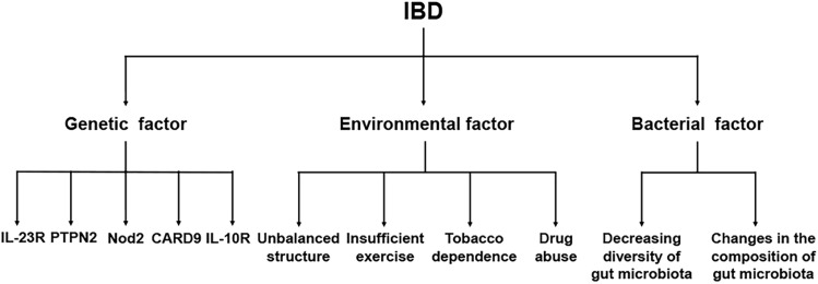
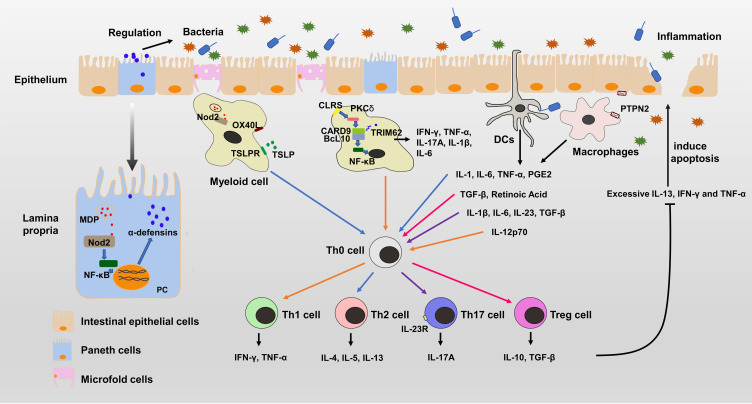
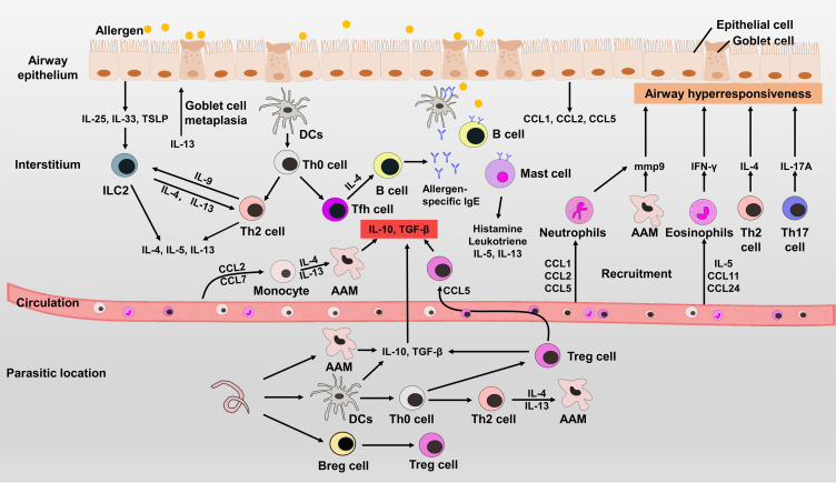
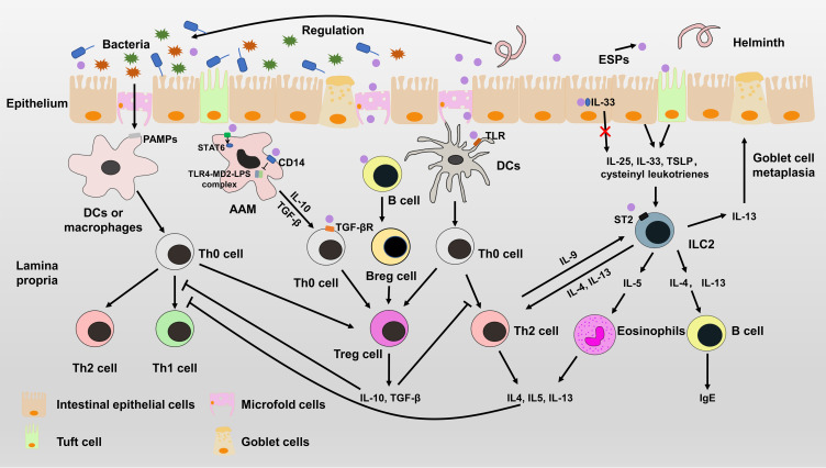
Verification
Claims the paper makes
The claims and statistics of this paper haven’t been checked yet.
Run the check — free, no account needed. It reads the open-access text and checks the sources it cites.
Claims other papers make about this one
Each is checked against this paper’s own text: does it say what the citing paper says it does?
In the citing paper
“Specifically, scientists are now focusing on identifying defined parasite-derived immunomodulatory molecules.”
From Parasites and allergies: a complex bidirectional relationship from evolutionary origins to modern therapeutics (2026) · cites this paper as Shi et al. 2022
In the citing paper
“Therefore, current research is shifting away from live organisms and toward a more targeted approach.”
From Parasites and allergies: a complex bidirectional relationship from evolutionary origins to modern therapeutics (2026) · cites this paper as Shi et al. 2022
In the citing paper
“These substances, which include various proteins and glycoproteins, could, in synthetic or recombinant form, exhibit anti-inflammatory and anti-allergic effects similar to those observed during natural helminth infection”
From Parasites and allergies: a complex bidirectional relationship from evolutionary origins to modern therapeutics (2026) · cites this paper as Shi et al. 2022
Self-check
Percentages, sums and reported differences re-computed from the paper’s own numbers.
The self-check needs the paper’s full text.
Citations
Cites
Epidemiology and risk factors for IBD
Ananthakrishnan, A. N. (2015) · Nature Reviews Gastroenterology & Hepatology · doi:10.1038/nrgastro.2015.34 · Cited by 1,741
Exposure to Environmental Microorganisms and Childhood Asthma
Ege, M. J., Mayer, M., Normand, A.-C., Genuneit, J., Cookson, W. O. C. M., Braun-Fahrländer, C., Heederik, D., Piarroux, R., & von Mutius, E. (2011) · New England Journal of Medicine · doi:10.1056/nejmoa1007302 · Cited by 1,529
Buck, A. H., Coakley, G., Simbari, F., McSorley, H. J., Quintana, J. F., Le Bihan, T., Kumar, S., Abreu-Goodger, C., Lear, M., Harcus, Y., et al. (2014) · Nature Communications · doi:10.1038/ncomms6488 · Cited by 770
Lupp, C., Robertson, M. L., Wickham, M. E., Sekirov, I., Champion, O. L., Gaynor, E. C., & Finlay, B. B. (2007) · Cell Host & Microbe · doi:10.1016/j.chom.2007.08.002 · Cited by 501
van Die, I. (2003) · Glycobiology · doi:10.1093/glycob/cwg052 · Cited by 316
HpARI protein secreted by a helminth parasite suppresses interleukin-33
Osbourn, M., Soares, D. C., Vacca, F., Cohen, E. S., Scott, I. C., Gregory, W. F., Smyth, D. J., Toivakka, M., Kemter, A. M., le Bihan, T., et al. (2017) · Immunity · doi:10.1016/j.immuni.2017.09.015 · Cited by 178
Hookworm recombinant protein promotes regulatory T cell responses that suppress experimental asthma
Navarro, S., Pickering, D. A., Ferreira, I. B., Jones, L., Ryan, S., Troy, S., Leech, A., Hotez, P. J., Zhan, B., Laha, T., et al. (2016) · Science Translational Medicine · doi:10.1126/scitranslmed.aaf8807 · Cited by 139
Haeberlein, S., Obieglo, K., Ozir-Fazalalikhan, A., Chayé, M. A. M., Veninga, H., van der Vlugt, L. E. P. M., Voskamp, A., Boon, L., den Haan, J. M. M., Westerhof, L. B., et al. (2017) · PLOS Pathogens · doi:10.1371/journal.ppat.1006539 · Cited by 96
Sulaiman, A. A., Zolnierczyk, K., Japa, O., Owen, J. P., Maddison, B. C., Emes, R. D., Hodgkinson, J. E., Gough, K. C., & Flynn, R. J. (2016) · PLOS Pathogens · doi:10.1371/journal.ppat.1005991 · Cited by 60
Research works printed but not linked: 138
- Ryan, 2020, PLoS Pathog, 16, e1008508, 10.1371/journal.ppat.1008508 · DOI
- Weinstock, 2014, J Immunol, 193, 3239, 10.4049/jimmunol.1400927 · DOI
- Weinstock, 2009, Inflamm Bowel Dis, 15, 128, 10.1002/ibd.20633 · DOI
- Rook, 2010, Clin Exp Immunol, 160, 70, 10.1111/j.1365-2249.2010.04133.x · DOI · Paper page
- Hirahara, 2016, Int Immunol, 28, 163, 10.1093/intimm/dxw006 · DOI · Paper page
- Metenou, 2010, J Immunol, 184, 5375, 10.4049/jimmunol.0904067 · DOI
- Heylen, 2014, Pharmacol Ther, 143, 153, 10.1016/j.pharmthera.2014.02.011 · DOI
- Summers, 2005, Gut, 54, 87, 10.1136/gut.2004.041749 · DOI · Paper page
- Bager, 2010, J Allergy Clin Immunol, 125, 10.1016/j.jaci.2009.08.006 · DOI
- Bourke, 2012, Clin Exp Allergy, 42, 1582, 10.1111/j.1365-2222.2012.04063.x · DOI · Paper page
- Croese, 2006, Gut, 55, 136, 10.1136/gut.2005.079129 · DOI · Paper page
- Kahl, 2018, Trends Parasitol, 34, 828, 10.1016/j.pt.2018.05.011 · DOI
- Maruszewska-Cheruiyot, 2021, Parasite Vector, 14, 10.1186/s13071-021-04613-9 · DOI
- Logan, 2020, PLoS Negl Trop Dis, 14, e0008237, 10.1371/journal.pntd.0008237 · DOI
- Namara, 2017, Pediat Allerg Imm, 28, 784, 10.1111/pai.12804 · DOI
- Park, 2017, Autoimmun Rev, 16, 416, 10.1016/j.autrev.2017.02.013 · DOI
- Na, 2019, Gut Liver, 13, 604, 10.5009/gnl19019 · DOI
- Joossens, 2007, Inflamm Bowel Dis, 13, 580, 10.1002/ibd.20086 · DOI
- Zhao, 2021, Aliment Pharmacol Ther, 10.1111/apt.16703 · DOI
- Cui, 2021, Front Med, 8, 2047
- Khor, 2011, Nature, 474, 307, 10.1038/nature10209 · DOI
- Glocker, 2009, N Engl J Med, 361, 2033, 10.1056/NEJMoa0907206 · DOI
- Liu, 2016, Annu Rev Pathol, 11, 127, 10.1146/annurev-pathol-012615-044152 · DOI · Paper page
- Lauro, 2016, Curr Opin Biotechnol, 40, 97, 10.1016/j.copbio.2016.02.028 · DOI
- Yang, 2021, Cell Prolif, 54, e12958, 10.1111/cpr.12958 · DOI
- Gubatan, 2021, World J Gastroenterol, 27, 7402, 10.3748/wjg.v27.i43.7402 · DOI
- Magalhaes, 2011, Proc Natl Acad Sci U S A, 108, 14896, 10.1073/pnas.1015063108 · DOI
- Glassner, 2020, J Allergy Clin Immunol, 145, 16, 10.1016/j.jaci.2019.11.003 · DOI
- Bouma, 2003, Nat Rev Immunol, 3, 521, 10.1038/nri1132 · DOI
- Qu, 2019, Int J Cancer, 145, 2225, 10.1002/ijc.32355 · DOI
- Strasser, 2012, Immunity, 36, 32, 10.1016/j.immuni.2011.11.015 · DOI
- Cao, 2015, Immunity, 43, 715, 10.1016/j.immuni.2015.10.005 · DOI
- Goel, 2021, J Clin Immunol, 10.1007/s10875-021-01173-6 · DOI
- Luo, 2020, J Cell Mol Med, 24, 34, 10.1111/jcmm.14770 · DOI
- Neurath, 2019, Cytokine Growth Factor Rev, 45, 1, 10.1016/j.cytogfr.2018.12.002 · DOI
- Spalinger, 2020, Gastroenterology, 159, 10.1053/j.gastro.2020.07.004 · DOI
- Garcia-Carbonell, 2018, Proc Natl Acad Sci U S A, 115, E9192, 10.1073/pnas.1810584115 · DOI
- Wallace, 2021, Front Immunol, 12, 767505, 10.3389/fimmu.2021.767505 · DOI
- Farache, 2013, Immunity, 38, 581, 10.1016/j.immuni.2013.01.009 · DOI
- Schirmer, 2018, Nat Microbiol, 3, 337, 10.1038/s41564-017-0089-z · DOI
- Imhann, 2018, Gut, 67, 108, 10.1136/gutjnl-2016-312135 · DOI · Paper page
- Furusawa, 2013, Nature, 504, 446, 10.1038/nature12721 · DOI · Paper page
- Levine, 2019, Gastroenterology, 157, 440, 10.1053/j.gastro.2019.04.021 · DOI
- Doherty, 2018, J Crohns Colitis, 12, 17, 10.1093/ecco-jcc/jjx101 · DOI
- Brown, 2016, Can J Hosp Pharm, 69, 466
- Bonovas, 2016, Clin Gastroenterol Hepatol, 14, 1385, 10.1016/j.cgh.2016.04.039 · DOI
- Papi, 2018, Lancet, 391, 783, 10.1016/S0140-6736(17)33311-1 · DOI
- Vasileiadou, 2021, PLoS One, 16, e0253322, 10.1371/journal.pone.0253322 · DOI
- Zhang, 2019, Allergy Asthma Immunol Res, 11, 156, 10.4168/aair.2019.11.2.156 · DOI
- Paaso, 2014, Respir Res, 15, 152, 10.1186/s12931-014-0152-8 · DOI
- Mukherjee, 2011, J Biol Chem, 286, 32883, 10.1074/jbc.R110.197046 · DOI
- Chan, 2019, Front Immunol, 10, 364, 10.3389/fimmu.2019.00364 · DOI
- Boberg, 2020, Int J Mol Sci, 21, 3751, 10.3390/ijms21113751 · DOI
- Kelsen, 2021, J Allergy Clin Immunol, 148, 790, 10.1016/j.jaci.2021.03.044 · DOI
- Murrison, 2019, J Clin Invest, 129, 1504, 10.1172/JCI124612 · DOI
- Hong, 2020, Allergy, 75, 2794, 10.1111/all.14526 · DOI
- Varricchi, 2016, Allergy, 71, 1086, 10.1111/all.12878 · DOI
- Nussbaum, 2013, Nature, 502, 245, 10.1038/nature12526 · DOI
- Chesne, 2014, Am J Respir Crit Care Med, 190, 1094, 10.1164/rccm.201405-0859PP · DOI
- Ventura, 2014, Allergy, 69, 898, 10.1111/all.12414 · DOI
- Giovannini-Chami, 2012, Eur Respir J, 39, 1197, 10.1183/09031936.00070511 · DOI
- Cho, 2005, Am J Respir Crit Care Med, 171, 224, 10.1164/rccm.200310-1416OC · DOI
- Magnan, 2000, Am J Respir Crit Care Med, 161, 1790, 10.1164/ajrccm.161.6.9906130 · DOI
- Tian, 2019, Nat Commun, 10, 4246, 10.1038/s41467-019-12243-0 · DOI
- Pfaar, 2021, Allergy, 10.1111/all.15023 · DOI
- Azam, 2021, Chest, 160, e633, 10.1016/j.chest.2021.07.052 · DOI
- McGregor, 2019, Am J Respir Crit Care Med, 199, 433, 10.1164/rccm.201810-1944CI · DOI
- Oray, 2016, Expert Opin Drug Saf, 15, 457, 10.1517/14740338.2016.1140743 · DOI
- Orons, 2000, J Vasc Interv Radiol, 11, 89, 10.1016/S1051-0443(07)61288-3 · DOI
- Gong, 2020, Nat Rev Immunol, 20, 95, 10.1038/s41577-019-0215-7 · DOI · Paper page
- Oh, 2014, Int J Mol Med, 33, 333, 10.3892/ijmm.2013.1569 · DOI
- Elliott, 2003, Am J Physiol Gastrointest Liver Physiol, 284, G385, 10.1152/ajpgi.00049.2002 · DOI
- Xu, 2019, Immunobiology, 224, 147, 10.1016/j.imbio.2018.09.005 · DOI
- Hasby, 2015, Cell Immunol, 295, 67, 10.1016/j.cellimm.2015.02.013 · DOI
- Ferreira, 2013, Infect Immun, 81, 2104, 10.1128/IAI.00563-12 · DOI
- Elliott, 2004, Eur J Immunol, 34, 2690, 10.1002/eji.200324833 · DOI
- Ramanan, 2016, Science, 352, 608, 10.1126/science.aaf3229 · DOI
- Shute, 2021, Microbiome, 9, 10.1186/s40168-021-01146-2 · DOI
- Hamid, 2015, Curr Top Microbiol Immunol, 388, 91, 10.1007/978-3-319-13725-4_5 · DOI
- Adjobimey, 2010, Ann Trop Med Parasitol, 104, 455, 10.1179/136485910X12786389891407 · DOI
- Kang, 2014, PLoS Negl Trop Dis, 8, e3410, 10.1371/journal.pntd.0003410 · DOI
- Layland, 2013, PLoS Negl Trop Dis, 7, e2379, 10.1371/journal.pntd.0002379 · DOI · Paper page
- Hartmann, 2009, Clin Exp Allergy, 39, 1585, 10.1111/j.1365-2222.2009.03290.x · DOI
- Liu, 2011, Parasite Immunol, 33, 525, 10.1111/j.1365-3024.2011.01308.x · DOI
- Kitagaki, 2006, J Immunol, 177, 1628, 10.4049/jimmunol.177.3.1628 · DOI
- Aranzamendi, 2013, Clin Exp Allergy, 43, 103, 10.1111/cea.12042 · DOI
- Mohammadzadeh, 2019, Infection, 47, 991, 10.1007/s15010-019-01343-0 · DOI
- Luo, 2019, Proc Natl Acad Sci U S A, 116, 5564, 10.1073/pnas.1812901116 · DOI
- von Moltke, 2016, Nature, 529, 221, 10.1038/nature16161 · DOI · Paper page
- McGinty, 2020, Immunity, 52, 528, 10.1016/j.immuni.2020.02.005 · DOI
- Luo, 2021, Biochem Soc Trans, 49, 2371, 10.1042/BST20210535 · DOI
- Nadjsombati, 2018, Immunity, 49, 33, 10.1016/j.immuni.2018.06.016 · DOI · Paper page
- Knuhr, 2018, Front Immunol, 9, 2293, 10.3389/fimmu.2018.02293 · DOI
- Jin, 2020, Front Immunol, 11, 2015, 10.3389/fimmu.2020.02015 · DOI
- Xu, 2019, Int Immunopharmacol, 66, 28, 10.1016/j.intimp.2018.10.043 · DOI
- Carson, 2021, Pathogens, 10, 863, 10.3390/pathogens10070863 · DOI
- Donnelly, 2008, FASEB J, 22, 4022, 10.1096/fj.08-106278 · DOI
- Pearce, 2004, Immunol Rev, 201, 117, 10.1111/j.0105-2896.2004.00187.x · DOI
- Everts, 2009, J Exp Med, 206, 1673, 10.1084/jem.20082460 · DOI
- Valverde, 2020, Chembiochem, 21, 2999, 10.1002/cbic.202000238 · DOI
- van Liempt, 2007, Mol Immunol, 44, 2605, 10.1016/j.molimm.2006.12.012 · DOI
- Figueroa-Santiago, 2014, Infect Immun, 82, 5005, 10.1128/IAI.02541-14 · DOI
- Martin, 2015, J Immunol, 194, 3924, 10.4049/jimmunol.1401182 · DOI · Paper page
- Alves, 2020, Mol Immunol, 124, 91, 10.1016/j.molimm.2020.05.011 · DOI
- Xu, 2021, J Immunol, 206, 963, 10.4049/jimmunol.2000290 · DOI
- Long, 2021, Front Immunol, 12, 664998, 10.3389/fimmu.2021.664998 · DOI
- Donnelly, 2010, J Biol Chem, 285, 3383, 10.1074/jbc.M109.060368 · DOI · Paper page
- Robinson, 2012, FASEB J, 26, 4614, 10.1096/fj.12-213876 · DOI
- Hartmann, 1997, Eur J Immunol, 27, 2253, 10.1002/eji.1830270920 · DOI
- Schnoeller, 2008, J Immunol, 180, 4265, 10.4049/jimmunol.180.6.4265 · DOI
- Danilowicz-Luebert, 2013, Int J Parasitol, 43, 201, 10.1016/j.ijpara.2012.10.014 · DOI · Paper page
- Bisht, 2019, Biomedicines, 7, 85, 10.3390/biomedicines7040085 · DOI
- Wang, 2016, Parasit Vectors, 9, 6, 10.1186/s13071-015-1288-1 · DOI · Paper page
- Khatri, 2020, Pathogens, 9, 431, 10.3390/pathogens9060431 · DOI
- Sun, 2019, Cells-Basel, 8, 1404, 10.3390/cells8111404 · DOI
- Cvetkovic, 2020, Sci Rep, 10, 20283, 10.1038/s41598-020-77497-x · DOI
- Rigano, 2007, Infect Immun, 75, 1667, 10.1128/IAI.01156-06 · DOI
- van der Kleij, 2002, J Biol Chem, 277, 48122, 10.1074/jbc.M206941200 · DOI · Paper page
- Saltykova, 2019, Front Vet Sci, 6, 332, 10.3389/fvets.2019.00332 · DOI
- Coronado, 2019, Front Immunol, 10, 2280, 10.3389/fimmu.2019.02280 · DOI · Paper page
- Bouchery, 2014, Front Immunol, 5, 487, 10.3389/fimmu.2014.00487 · DOI
- Johnston, 2017, Nat Commun, 8, 1741, 10.1038/s41467-017-01886-6 · DOI · Paper page
- Smyth, 2021, J Biotechnol, 340, 1, 10.1016/j.jbiotec.2021.08.006 · DOI
- Xie, 2021, Mol Immunol, 133, 86, 10.1016/j.molimm.2021.02.012 · DOI
- Li, 2021, Int J Parasitol, 10.1016/j.ijpara.2021.08.009 · DOI
- van der Vlugt, 2017, Int J Parasitol, 47, 545, 10.1016/j.ijpara.2017.02.002 · DOI
- Gao, 2019, Clin Exp Immunol, 198, 390, 10.1111/cei.13362 · DOI
- Vacca, 2020, Elife, 9, 10.7554/eLife.54017 · DOI
- Li, 2021, J Inflamm Res, 14, 5305, 10.2147/JIR.S334636 · DOI
- Shan, 2021, Parasit Vectors, 14, 455, 10.1186/s13071-021-04977-y · DOI
- Bancroft, 2019, Nat Commun, 10, 2344, 10.1038/s41467-019-09996-z · DOI
- Wang, 2017, Theranostics, 7, 3446, 10.7150/thno.20359 · DOI
- Ebner, 2021, J Immunol Res, 2021, 6234836, 10.1155/2021/6234836 · DOI
- Zhu, 2016, Sci Rep, 6, 25885, 10.1038/srep25885 · DOI
- Kuipers, 2020, J Extracell Vesicles, 9, 1753420, 10.1080/20013078.2020.1753420 · DOI
- Tanaka, 2018, J Allergy Clin Immunol, 141, 2316, 10.1016/j.jaci.2018.01.050 · DOI
- Coronado, 2017, Parasite Immunol, 39, e12425, 10.1111/pim.12425 · DOI · Paper page
- Aksoy, 2005, J Biol Chem, 280, 277, 10.1074/jbc.M411223200 · DOI
Cited by
Sorted by citations
Packi, K., Świerczewski, D., Klimczak, S., Ochab, D., Gałęziewska, J., Gromek, P., Kruczkowska, W., Płuciennik, E., Kałuzińska-Kołat, Ż., & Kołat, D. (2026) · Antonie van Leeuwenhoek · doi:10.1007/s10482-026-02425-4
Metadata
- Authors
- Shi, W., Xu, N., Wang, X., Vallée, I., Liu, M., & Liu, X.
- Venue
- Journal of Inflammation Research
- Publisher
- Informa UK Limited
- Published
- 2022-01
- Volume
- Volume 15
- Pages
- 475-491
- ISSN
- 1178-7031
- PMC
- PMC8789313
- Cited by
- 46
- References
- 9
- Language
- en
About these checks
Registry, retraction, Cites and statistics results are computed, not judged, with one exception: a printed reference that no index lists is read once by a model before it counts as a citation error. Claims are extracted by a model and kept only when their quote appears verbatim in the text; each cited source is then checked, with a model’s help, for whether it says what the claim relies on. Those verdicts can be wrong — read the claim’s quote and the cited source before relying on one.
Statistics and arithmetic are recomputed, not judged.
Confidence bands are shown only for fields calibrated on our annotated test set. None is calibrated yet, so no band is shown.
What counts as a claim ›How we measure confidence ›How we check this ›
Are you an author, or do you see a problem with this page? Request a review
