

The DOI matches its Crossref record; 47 of the 47 references it cites were found in a registry or an index.
Increased Secretion and Expression of Myostatin in Skeletal Muscle From Extremely Obese Women
Open accessAbstract
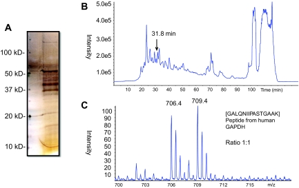
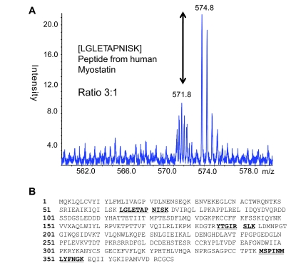
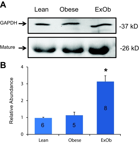
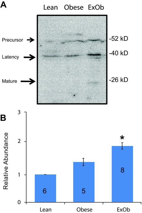
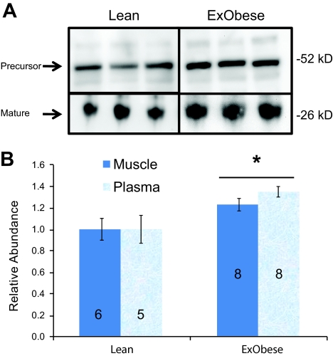
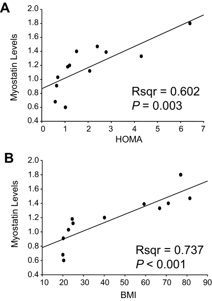
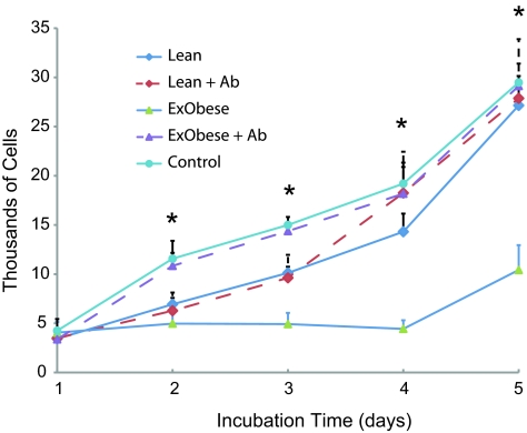
OBJECTIVE: Obesity is associated with endocrine abnormalities that predict the progression of insulin resistance to type 2 diabetes. Because skeletal muscle has been shown to secrete proteins that could be used as biomarkers, we characterized the secreted protein profile of muscle cells derived from extremely obese (BMI 48.8 +/- 14.8 kg/m(2); homeostasis model assessment [HOMA] 3.6 +/- 1.0) relative to lean healthy subjects (BMI 25.7 +/- 3.2 kg/m(2); HOMA 0.8 +/- 0.2). RESEARCH DESIGN AND METHODS: We hypothesized that skeletal muscle would secrete proteins that predict the severity of obesity. To test this hypothesis, we used a "bottom-up" experimental design using stable isotope labeling by amino acids in culture (SILAC) and liquid chromatography/mass spectometry/mass spectometry (LC-MS/MS) to both identify and quantify proteins secreted from cultured myotubes derived from extremely obese compared with healthy nonobese women. RESULTS: Using SILAC, we discovered a 2.9-fold increase in the secretion of myostatin from extremely obese human myotubes. The increased secretion and biological activity of myostatin were validated by immunoblot (3.16 +/- 0.18, P < 0.01) and a myoblast proliferation assay using conditioned growth medium. Myostatin was subsequently shown to increase in skeletal muscle (23%, P < 0.05) and plasma (35%, P < 0.05) and to correlate (r(2) = 0.6, P < 0.05) with the severity of insulin resistance. CONCLUSIONS: Myostatin is a potent antianabolic regulator of muscle mass that may also play a role in energy metabolism. These findings show that increased expression of myostatin in skeletal muscle with obesity and insulin resistance results in elevated circulating myostatin. This may contribute to systemic metabolic deterioration of skeletal muscle with the progression of insulin resistance to type 2 diabetes.
Overview
The claims and statistics of this paper haven’t been checked yet.
Run the check — free, no account needed. It reads the open-access text and checks the sources it cites.
Integrity signals
Registry
Cites
Statistics
Figures
Verification
Claims the paper makes
The claims and statistics of this paper haven’t been checked yet.
Run the check — free, no account needed. It reads the open-access text and checks the sources it cites.
Claims other papers make about this one
Each is checked against this paper’s own text: does it say what the citing paper says it does?
The source backs part of the claim, not all of it — or backs it for a different population, condition or comparison.
In the citing paper
“Myostatin mRNA levels increase substantially in genetically-modified obese mice (leptin-deficient ob/ob mice) and also in those receiving a high fat diet (HFD)”
From Physical Exercise-Induced Myokines and Muscle-Adipose Tissue Crosstalk: A Review of Current Knowledge and the Implications for Health and Metabolic Diseases (2018) · cites this paper as Hittel et al., 2009
Experimental — accuracy not yet measuredThe cited source says what the claim attributes to it.
In the citing paper
“so both circulating and SkM myostatin levels are present in obese subjects when compared to non-obese subjects (Allen et al., 2008; Hittel et al., 2009).”
From Physical Exercise-Induced Myokines and Muscle-Adipose Tissue Crosstalk: A Review of Current Knowledge and the Implications for Health and Metabolic Diseases (2018) · cites this paper as Hittel et al., 2009
Self-check
Percentages, sums and reported differences re-computed from the paper’s own numbers.
The self-check needs the paper’s full text.
Citations
Cites
Koves, T. R., Ussher, J. R., Noland, R. C., Slentz, D., Mosedale, M., Ilkayeva, O., Bain, J., Stevens, R., Dyck, J. R. B., Newgard, C. B., et al. (2008) · Cell Metabolism · doi:10.1016/j.cmet.2007.10.013 · Cited by 1,990
Inflammation and insulin resistance
de Luca, C., & Olefsky, J. M. (2008) · FEBS Letters · doi:10.1016/j.febslet.2007.11.057 · Cited by 1,161
Myostatin, a Negative Regulator of Muscle Growth, Functions by Inhibiting Myoblast Proliferation
Thomas, M., Langley, B., Berry, C., Sharma, M., Kirk, S., Bass, J., & Kambadur, R. (2000) · Journal of Biological Chemistry · doi:10.1074/jbc.m004356200 · Cited by 1,014
Cachexia: pathophysiology and clinical relevance1,2
Morley, J. E., Thomas, D. R., & Wilson, M.-M. G. (2006) · The American Journal of Clinical Nutrition · doi:10.1093/ajcn/83.4.735 · Cited by 786
Gonzalez-Cadavid, N. F., Taylor, W. E., Yarasheski, K., Sinha-Hikim, I., Ma, K., Ezzat, S., Shen, R., Lalani, R., Asa, S., Mamita, M., et al. (1998) · Proceedings of the National Academy of Sciences · doi:10.1073/pnas.95.25.14938 · Cited by 571
Muscle fiber type is associated with obesity and weight loss
Tanner, C. J., Barakat, H. A., Dohm, G. L., Pories, W. J., MacDonald, K. G., Cunningham, P. R. G., Swanson, M. S., & Houmard, J. A. (2002) · American Journal of Physiology-Endocrinology and Metabolism · doi:10.1152/ajpendo.00416.2001 · Cited by 493
Self-citationLouis, E., Raue, U., Yang, Y., Jemiolo, B., & Trappe, S. (2007) · Journal of Applied Physiology · doi:10.1152/japplphysiol.00679.2007 · Cited by 478
Hulver, M. W., Berggren, J. R., Carper, M. J., Miyazaki, M., Ntambi, J. M., Hoffman, E. P., Thyfault, J. P., Stevens, R., Dohm, G. L., Houmard, J. A., et al. (2005) · Cell Metabolism · doi:10.1016/j.cmet.2005.09.002 · Cited by 384
Lower skeletal muscle mass in male transgenic mice with muscle-specific overexpression of myostatin
Reisz‐Porszasz, S., Bhasin, S., Artaza, J. N., Shen, R., Sinha‐Hikim, I., Hogue, A., Fielder, T. J., & González-Cadavid, N. F. (2003) · American Journal of Physiology-Endocrinology and Metabolism · doi:10.1152/ajpendo.00107.2003 · Cited by 333
Hickey, M. S., Carey, J. O., Azevedo, J. L., Houmard, J. A., Pories, W. J., Israel, R. G., & Dohm, G. L. (1995) · American Journal of Physiology-Endocrinology and Metabolism · doi:10.1152/ajpendo.1995.268.3.e453 · Cited by 305
Research works printed but not linked: 1
- Allen DL, Cleary AS, Speaker KJ, Lindsay SF, Uyenishi J, Reed JM, Madden MC, Mehan RS: Myostatin, activin receptor IIb, and follistatin-like-3 gene expression are altered in adipose tissue and skeletal muscle of obese mice. Am J Physiol Endocrinol Metab 294:E918–E927,2008 · DOI · Paper page
Cited by
Sorted by citations
Muscles, exercise and obesity: skeletal muscle as a secretory organ
Pedersen, B. K., & Febbraio, M. A. (2012) · Nature Reviews Endocrinology · doi:10.1038/nrendo.2012.49 · Cited by 2,909
Interleukin-1 in the pathogenesis and treatment of inflammatory diseases
Dinarello, C. A. (2011) · Blood · doi:10.1182/blood-2010-07-273417 · Cited by 2,248
Exercise is the real polypill.
Fiuza-Luces, C., Garatachea, N., Berger, N. A., & Lucia, A. (2013) · Physiology · doi:10.1152/physiol.00019.2013 · Cited by 791
A clinical perspective of IL‐1β as the gatekeeper of inflammation
Dinarello, C. A. (2011) · European Journal of Immunology · doi:10.1002/eji.201141550 · Cited by 792
Skeletal muscle inflammation and insulin resistance in obesity
Wu, H., & Ballantyne, C. M. (2017) · Journal of Clinical Investigation · doi:10.1172/jci88880 · Cited by 695
Metadata
- Authors
- Hittel, D. S., Berggren, J. R., Shearer, J., Boyle, K., & Houmard, J. A.
- Affiliations
- University of Calgary (CA)
- East Carolina University (US)
- Venue
- Diabetes
- Publisher
- American Diabetes Association
- Published
- 2009-01-01
- Volume
- 58
- Issue
- 1
- Pages
- 30-38
- ISSN
- 0012-1797 (ISSN-L)
- 1939-327X
- PMC
- PMC2606890
- OpenAlex
- W2132813206
- Cited by
- 324
- References
- 47
- Citations by year
- 2026: 10 · 2025: 19 · 2024: 12 · 2023: 18 · 2022: 19 · 2021: 21 · 2020: 20 · 2019: 14 · 2018: 26 · 2017: 13 · 2016: 11 · 2015: 25 · 2014: 31 · 2013: 24 · 2012: 22
- FWCI
- 5.35
- Type
- journal article
- Language
- en
- Open access
- hybrid
- doi.org
- Licence
- cc-by-nc-nd
- Locations
- Diabetes (publishedVersion, cc-by-nc-nd)
- PubMed Central (publishedVersion, cc-by-nc-nd)
- PubMed (publishedVersion)
- The Scholarship East Carolina University's Institutional Repository (East Carolina University) (submittedVersion)
- Funders
- Heart and Stroke Foundation of Canada
- Canadian Diabetes Association
- Fondation pour la Recherche Médicale
- National Institutes of Health
- National Institute of Diabetes and Digestive and Kidney Diseases (R01DK056112)
- Topics
- Life Sciences › Biochemistry, Genetics and Molecular Biology › Molecular Biology › Muscle Physiology and Disorders
- Health Sciences › Medicine › Endocrinology, Diabetes and Metabolism › Adipokines, Inflammation, and Metabolic Diseases
- Health Sciences › Medicine › Endocrinology, Diabetes and Metabolism › Diabetes, Cardiovascular Risks, and Lipoproteins
- Keywords
- myostatin, obesity, insulin resistance, skeletal muscle, myotubes, secreted proteins, type 2 diabetes, myoblast proliferation, energy metabolism, women
- MeSH terms
- Adult, Chromatography, Liquid, Female, Humans, Insulin Resistance, Middle Aged, Obesity, Immunoblotting, Muscle, Skeletal, Muscle Fibers, Skeletal, Tandem Mass Spectrometry, Myostatin
About these checks
Registry, retraction, Cites and statistics results are computed, not judged, with one exception: a printed reference that no index lists is read once by a model before it counts as a citation error. Claims are extracted by a model and kept only when their quote appears verbatim in the text; each cited source is then checked, with a model’s help, for whether it says what the claim relies on. Those verdicts can be wrong — read the claim’s quote and the cited source before relying on one.
Statistics and arithmetic are recomputed, not judged.
Confidence bands are shown only for fields calibrated on our annotated test set. None is calibrated yet, so no band is shown.
What counts as a claim ›How we measure confidence ›How we check this ›
Are you an author, or do you see a problem with this page? Request a review
