

Adipose Tissue Macrophages Function As Antigen-Presenting Cells and Regulate Adipose Tissue CD4+ T Cells in Mice
Open accessThe DOI matches its Crossref record; 48 of the 48 references it cites were found in a registry or an index.
Abstract
The proinflammatory activation of leukocytes in adipose tissue contributes to metabolic disease. How crosstalk between immune cells initiates and sustains adipose tissue inflammation remains an unresolved question. We have examined the hypothesis that adipose tissue macrophages (ATMs) interact with and regulate the function of T cells. Dietary obesity was shown to activate the proliferation of effector memory CD4+ T cells in adipose tissue. Our studies further demonstrate that ATMs are functional antigen-presenting cells that promote the proliferation of interferon-γ–producing CD4+ T cells in adipose tissue. ATMs from lean and obese visceral fat process and present major histocompatibility complex (MHC) class II–restricted antigens. ATMs were sufficient to promote proliferation and interferon-γ production from antigen-specific CD4+ T cells in vitro and in vivo. Diet-induced obesity increased the expression of MHC II and T-cell costimulatory molecules on ATMs in visceral fat, which correlated with an induction of T-cell proliferation in that depot. Collectively, these data indicate that ATMs provide a functional link between the innate and adaptive immune systems within visceral fat in mice.
Overview
The claims and statistics of this paper haven’t been checked yet.
Free: it reads the open-access text and checks the first 5 claims that cite a source we can check. Create a free account to check every claim.
Integrity signals
Registry
Cites
Statistics
Figures
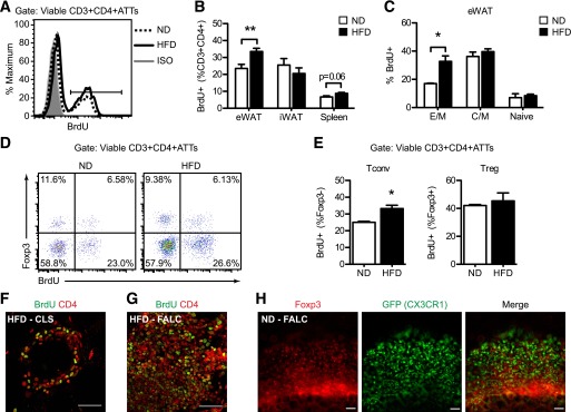

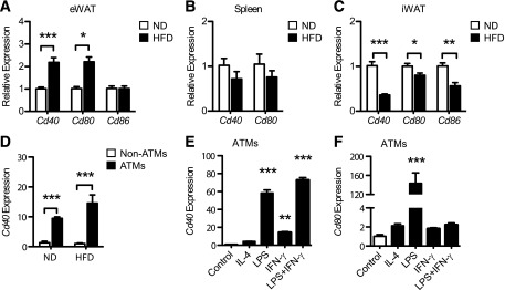

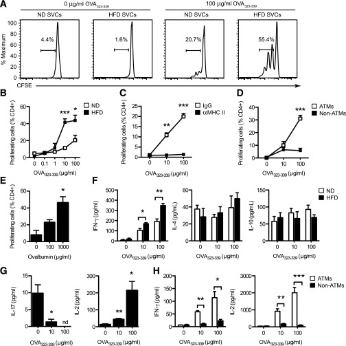

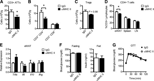
Verification
Claims the paper makes
The claims and statistics of this paper haven’t been checked yet.
Free: it reads the open-access text and checks the first 5 claims that cite a source we can check. Create a free account to check every claim.
Claims other papers make about this one
Each is checked against this paper’s own text: does it say what the citing paper says it does?
In the citing paper
“The infiltrated macrophages in muscle tissue present adipocyte-derived antigens and activate antigen-specific CD4+ T cells [39].”
From Sarcopenic obesity, insulin resistance, and their implications in cardiovascular and metabolic consequences (2020) · cites this paper as [39]
Self-check
Percentages, sums and reported differences re-computed from the paper’s own numbers.
The self-check needs the paper’s full text.
Citations
Cites
Obesity is associated with macrophage accumulation in adipose tissue
Weisberg, S. P., McCann, D., Desai, M., Rosenbaum, M., Leibel, R. L., & Ferrante, A. W. (2003) · Journal of Clinical Investigation · doi:10.1172/jci200319246 · Cited by 10,041 · cited 1×
“ATMs that co-express the dendritic cell (DC) marker CD11c and pro-inflammatory genes typical of ‘classically activated’ or M1 polarized macrophages (11-13).”
Xu, H., Barnes, G. T., Yang, Q., Tan, G., Yang, D., Chou, C. J., Sole, J., Nichols, A., Ross, J. S., Tartaglia, L. A., et al. (2003) · Journal of Clinical Investigation · doi:10.1172/jci200319451 · Cited by 6,760
Obesity induces a phenotypic switch in adipose tissue macrophage polarization
Lumeng, C. N., Bodzin, J. L., & Saltiel, A. R. (2007) · Journal of Clinical Investigation · doi:10.1172/jci29881 · Cited by 4,664 · cited 1×
“Current models suggest that obesity promotes metainflammation in part by altering the balance between type 2 and type 1 ATMs in visceral fat (13,18).”
BackgroundSelf-citationAdipokines in inflammation and metabolic disease
Ouchi, N., Parker, J. L., Lugus, J. J., & Walsh, K. (2011) · Nature Reviews Immunology · doi:10.1038/nri2921 · Cited by 4,625 · cited 2×
“Classically, APCs (specifically macrophages and dendritic cells (DCs)) shape CD4 + T cell activation by three signals: (1) presentation of peptide antigens on Major histocompatibility complex (MHC) Class II molecules (signal 1), (2) expression of T cell costimulatory molecules (e.”
Inflammation, stress, and diabetes
Wellen, K. E., & Hotamisligil, G. S. (2005) · Journal of Clinical Investigation · doi:10.1172/jci25102 · Cited by 4,143
Cinti, S., Mitchell, G., Barbatelli, G., Murano, I., Ceresi, E., Faloia, E., Wang, S., Fortier, M., Greenberg, A. S., & Obin, M. S. (2005) · Journal of Lipid Research · doi:10.1194/jlr.m500294-jlr200 · Cited by 2,529 · cited 1×
“M1 ATMs form multicellular lipidladen clusters, known as crown-like structures (CLS), around dead adipocytes in obese fat (6,14,15) and produce inflammatory cytokines (e.”
BackgroundInflammatory links between obesity and metabolic disease
Lumeng, C. N., & Saltiel, A. R. (2011) · Journal of Clinical Investigation · doi:10.1172/jci57132 · Cited by 2,319 · cited 1×
“Accumulation of activated leukocytes in metabolic tissues is a driving force for obesity-associated metabolic inflammation (metainflammation) and insulin resistance (3,4).”
BackgroundSelf-citationNishimura, S., Manabe, I., Nagasaki, M., Eto, K., Yamashita, H., Ohsugi, M., Otsu, M., Hara, K., Ueki, K., Sugiura, S., et al. (2009) · Nature Medicine · doi:10.1038/nm.1964 · Cited by 2,230 · cited 1×
“In contrast, T helper 1 (Th1) CD4 T cells and CD8 adipose tissue T cells (ATTs) accumulate in fat during obesity, promoting IFN-g and TNF-a production and insulin resistance (20,21,27).”
Feuerer, M., Herrero, L., Cipolletta, D., Naaz, A., Wong, J., Nayer, A., Lee, J., Goldfine, A. B., Benoist, C., Shoelson, S., et al. (2009) · Nature Medicine · doi:10.1038/nm.2002 · Cited by 2,145
Macrophage-specific PPARγ controls alternative activation and improves insulin resistance
Odegaard, J. I., Ricardo-Gonzalez, R. R., Goforth, M. H., Morel, C. R., Subramanian, V., Mukundan, L., Eagle, A. R., Vats, D., Brombacher, F., Ferrante, A. W., et al. (2007) · Nature · doi:10.1038/nature05894 · Cited by 2,107 · cited 1×
“Disruption of macrophage M2 polarization increases the susceptibility to high fat diet (HFD)-induced insulin resistance (8-10).”
Cited by
Items 101–200 of 217
A method for long-term live imaging of tissue macrophages in adipose tissue explants
Gericke, M. T., Weyer, U., Braune, J., Bechmann, I., & Eilers, J. (2015) · American Journal of Physiology-Endocrinology and Metabolism · doi:10.1152/ajpendo.00075.2015 · Cited by 41
Brooks-Worrell, B. M., & Palmer, J. P. (2019) · Diabetes Care · doi:10.2337/dc19-0475 · Cited by 38
“Adipose tissue macrophages (ATMs) and adipocytes have been shown to act as antigen-presenting cells, promoting the activation and proliferation of effector T cells (36).”
BackgroundKouidhi, S., Noman, M. Z., Kieda, C., Elgaaied, A. B., & Chouaib, S. (2016) · Frontiers in Immunology · doi:10.3389/fimmu.2016.00114 · Cited by 36
“How WAT inflammation is triggered is not completely understood, but it is suggested that lipotoxicity (106), endoplasmic reticulum (ER) stress, and Toll-like receptor activation (107) are involved.”
BackgroundInfluentialA vicious circle in chronic lymphoedema pathophysiology? An adipocentric view
Cucchi, F., Rossmeislová, L., Simonsen, L., Jensen, M. R., & Bülow, J. (2017) · Obesity Reviews · doi:10.1111/obr.12565 · Cited by 38
Kim, B., Tilstam, P. V., Arnke, K., Leng, L., Ruhl, T., Piecychna, M., Schulte, W., Sauler, M., Frueh, F. S., Storti, G., et al. (2020) · The FASEB Journal · doi:10.1096/fj.201901511r · Cited by 35
Zhang, S.-Y., Lv, Y., Zhang, H., Gao, S., Wang, T., Feng, J., Wang, Y., Liu, G., Xu, M.-J., Wang, X., et al. (2016) · Diabetes · doi:10.2337/db15-1626 · Cited by 36
“However, other evidence revealed that adipose macrophages also present antigen and play a key role in the late stage of obesity-induced adipose T-cell activation and insulin resistance (28,42).”
BackgroundGauci, M.-L., Boudou, P., Baroudjian, B., Vidal-Trécan, T., Meda, L. D., Madelaine, I., Basset‐Séguin, N., Bagot, M., Pagès, C., Mourah, S., et al. (2018) · Cancer Immunology Immunotherapy · doi:10.1007/s00262-018-2178-0 · Cited by 39
Kumar, R. K., Jin, Y., Watts, S. W., & Rockwell, C. E. (2020) · Frontiers in Physiology · doi:10.3389/fphys.2020.00058 · Cited by 35
“By contrast, our study has identified ∼80–95% macrophages that are MHCII+ but other studies in mice that Frontiers in Physiology | www.frontiersin.org 8 February 2020 | Volume 11 | Article 58 have reported 55% of MHCII+macrophages in epididymal WAT in mice (Morris et al., 2013).”
ResultTraining of Primary Chicken Monocytes Results in Enhanced Pro-Inflammatory Responses
Verwoolde, M. B., van den Biggelaar, R. H. G. A., van Baal, J., Jansen, C. A., & Lammers, A. (2020) · Veterinary Sciences · doi:10.3390/vetsci7030115 · Cited by 39
Roselli, M., Finamore, A., Brasili, E., Rami, R., Nobili, F., Orsi, C., Zambrini, A. V., & Mengheri, E. (2018) · Journal of Functional Foods · doi:10.1016/j.jff.2018.03.039 · Cited by 39
Drastic transformation of visceral adipose tissue and peripheral CD4 T cells in obesity
Shirakawa, K., & Sano, M. (2023) · Frontiers in Immunology · doi:10.3389/fimmu.2022.1044737 · Cited by 38
“, dendritic cells (DCs) (28), macrophages (27, 29), and B cells (30), adipocytes (26) also play an important role in the formation of T-cell responses as antigen-presenting cells (31).”
BackgroundCaloric restriction for the immunometabolic control of human health
Procaccini, C., de Candia, P., Russo, C., Rosa, G. D., Lepore, M. T., Colamatteo, A., & Matarese, G. (2023) · Cardiovascular Research · doi:10.1093/cvr/cvad035 · Cited by 37
EMT reversal in human cancer cells after IR knockdown in hyperinsulinemic mice
Zelenko, Z., Gallagher, E. J., Antoniou, I. M., Sachdev, D., Nayak, A., Yee, D., & LeRoith, D. (2016) · Endocrine Related Cancer · doi:10.1530/erc-16-0142 · Cited by 30
OX40 promotes obesity-induced adipose inflammation and insulin resistance
Liu, B., Yu, H., Sun, G., Sun, X., Jin, H., Zhang, C., Shi, W., Tian, D., Liu, K., Xu, H., et al. (2017) · Cellular and Molecular Life Sciences · doi:10.1007/s00018-017-2552-7 · Cited by 32
Laparra, A., Tricot, S., Le Van, M., Damouche, A., Gorwood, J., Vaslin, B., Favier, B., Benoist, S., Ho Tsong Fang, R., Bosquet, N., et al. (2019) · Frontiers in Immunology · doi:10.3389/fimmu.2019.00117 · Cited by 29
“Lastly, we evaluated the proportion of macrophages, which can potentially be modulated by Tregs (65–68) but may also favor Treg accumulation (69, 70) The proportion of macrophages in the SVF fraction was not defined in the same way in all the three species: F4/80 expression in mice, and CD14 expression in cynomolgus m…”
BackgroundNicholas, D., Andrieu, G. P., Strissel, K. J., Nikolajczyk, B. S., & Denis, G. V. (2016) · Cellular and Molecular Life Sciences · doi:10.1007/s00018-016-2320-0 · Cited by 35
López‐Ortega, O., Moreno-Corona, N. C., Cruz-Holguín, V. J., Garcia-Gonzalez, L. D., Helguera‐Repetto, A. C., Romero‐Valdovinos, M., Arévalo-Romero, H., Cedillo‐Barrón, L., & León‐Juárez, M. (2022) · International Journal of Molecular Sciences · doi:10.3390/ijms23116154 · Cited by 33
“The immune cells present in AT can also be affected by obesity; for example, adipose tissue macrophages from obese animals have more cells expressing I-Ab (mouse class II MHC molecule) that are implicated in antigen presentation as well as in the induction of T-cell proliferation [43], which is indicative of their ess…”
BackgroundKumar, D., Pandya, S. M., Varshney, S., Shankar, K., Rajan, S., Srivastava, A., Gupta, A., Gupta, S., Vishwakarma, A. L., Misra, A., et al. (2018) · International Journal of Obesity · doi:10.1038/s41366-018-0228-5 · Cited by 33
mTORC2/Akt activation in adipocytes is required for adipose tissue inflammation in tuberculosis
Martínez, N., Cheng, C. Y., Ketheesan, N., Cullen, A. M., Tang, Y., Lum, J., West, K., Poidinger, M., Guertin, D. A., Singhal, A., et al. (2019) · EBioMedicine · doi:10.1016/j.ebiom.2019.06.052 · Cited by 28
“In mouse models of obesity (in the absence of infection) antigen-specific adipose-resident T cell become activated and this dependsmainly onMHC class II expression by resident and recruited MΦ [28] and by other non-immune (CD45) cells [29].”
BackgroundKyung, D. S., Sung, H., Kim, Y. J., Kim, K., Cho, S. Y., Choi, J., Lee, Y.-H., Kim, I. Y., & Seong, J. K. (2017) · International Journal of Obesity · doi:10.1038/ijo.2017.297 · Cited by 26
Interaction between human mature adipocytes and lymphocytes induces T-cell proliferation
Poloni, A., Maurizi, G., Ciarlantini, M., Medici, M., Mattiucci, D., Mancini, S., Maurizi, A., Falconi, M., Olivieri, A., & Leoni, P. (2015) · Cytotherapy · doi:10.1016/j.jcyt.2015.06.007 · Cited by 32
“[12] examined the hypothesis that adipose tissue macrophages interact with and regulate the function of T cells.”
BackgroundJorgačević, B. Z., Vučević, D. B., Samardžić, J., Mladenović, D. R., Vesković, M. N., Vukićević, D., Ješić, R., & Radosavljević, T. S. (2020) · Current Medicinal Chemistry · doi:10.2174/0929867327666200303122734 · Cited by 25
Peripheral and central macrophages in obesity
Mukherjee, S., Skrede, S., Haugstøyl, M. E., López, M., & Fernø, J. (2023) · Frontiers in Endocrinology · doi:10.3389/fendo.2023.1232171 · Cited by 30
Translating MSC Therapy in the Age of Obesity
Boland, L. K., Bitterlich, L., Hogan, A. E., Ankrum, J. A., & English, K. (2022) · Frontiers in Immunology · doi:10.3389/fimmu.2022.943333 · Cited by 31
Liu, M., Li, Y., Han, L., Zhang, Y.-Y., Wang, D., Wang, Z., Zhou, H., Song, M., Li, Y., Tang, M., et al. (2017) · Molecular Immunology · doi:10.1016/j.molimm.2017.03.020 · Cited by 23
Immunopathology of adipose tissue during metabolic syndrome
Ghazarian, M., Luck, H., Revelo, X. S., Winer, S., & Winer, D. A. (2015) · Turkish Journal of Pathology · doi:10.5146/tjpath.2015.01323 · Cited by 30
“Interestingly, IFNγ can also act on adipose tissue macrophages to enhance expression of MHC II, which promotes antigen presentation to Th1 cells resulting in their activation, proliferation and cytokine production (29).”
Adipose tissue lymphocytes and obesity
Gao, F., Litchfield, B. A., & Wu, H.-Z. (2023) · The Journal of Cardiovascular Aging · doi:10.20517/jca.2023.38 · Cited by 24
“MHC II on either macrophages or adipocytes is sufficient to promote Th1 cell polarization and IFN-γ production [35,36] .”
BackgroundJang, J. H., Sung, J. H., & Huh, J. Y. (2025) · Immune Network · doi:10.4110/in.2025.25.e12 · Cited by 32
Vpr Is a VIP: HIV Vpr and Infected Macrophages Promote Viral Pathogenesis
Lubow, J., & Collins, K. L. (2020) · Viruses · doi:10.3390/v12080809 · Cited by 30
Quante, M., Iske, J., Uehara, H., Minami, K., Nian, Y., Maenosono, R., Matsunaga, T., Liu, Y., Azuma, H., Perkins, D. L., et al. (2021) · American Journal of Transplantation · doi:10.1111/ajt.16856 · Cited by 21
Immune Microenvironment Dysregulation: A Contributing Factor to Obesity-Associated Male Infertility
Feng, R., Cheng, D., Zhang, W., Zhang, J., Chen, S., & Xia, Y. (2025) · Biomedicines · doi:10.3390/biomedicines13061314 · Cited by 25
“…into pro-inflammatory subtypes (such as Th1 and Th17) [92], which alters the function of APCs and adipose tissue macrophages, enhancing the inflammatory response and inhibiting the function of Tregs, thus leading to immune dysregulation and the suppression of immune defense against infections [93].”
Naruse, K., Akasaka, J., 重光, 愛., Tsunemi, T., Koike, N., Yoshimoto, C., & Kobayashi, H. (2015) · Mediators of Inflammation · doi:10.1155/2015/325932 · Cited by 20
Innate Lymphoid Cell Immunometabolism
O’Sullivan, T. E., & Sun, J. C.-Y. (2017) · Journal of Molecular Biology · doi:10.1016/j.jmb.2017.08.014 · Cited by 23
Teixeira, L., Moreira, J., Melo, J., Bezerra, F., Marques, R. M., Ferreirinha, P., Correia, A., Monteiro, M. P., Ferreira, P. G., & Vilanova, M. (2015) · Immunology · doi:10.1111/imm.12440 · Cited by 26
Stephens, J. M., Bailey, J. L., Hang, H., Rittell, V., Dietrich, M. A., Mynatt, R. L., & Elks, C. M. (2018) · Obesity · doi:10.1002/oby.22254 · Cited by 19
Subramanian, M., Özcan, L., Ghorpade, D. S., Ferrante, A. W., & Tabas, I. A. (2015) · PLoS ONE · doi:10.1371/journal.pone.0135842 · Cited by 17
Teixeira, L., Marques, R. M., Ferreirinha, P., Bezerra, F., Melo, J., Moreira, J., Pinto, A., Correia, A., Ferreira, P. G., & Vilanova, M. (2016) · Scientific Reports · doi:10.1038/srep23475 · Cited by 20
Antony, A. K., Lian, Z., Perrard, X. D., Perrard, J. L., Liu, H., Cox, A. R., Saha, P. K., Hennighausen, L. G., Hartig, S. M., Ballantyne, C. M., et al. (2020) · Diabetes · doi:10.2337/db20-0634 · Cited by 19
Mogilenko, D. A., Caiazzo, R. J., L’homme, L., Pineau, L., Raverdy, V., Noulette, J., Derudas, B., Pattou, F. N., Staels, B., & Dombrowicz, D. (2021) · International Journal of Obesity · doi:10.1038/s41366-021-00826-1 · Cited by 19
Filgueiras, L. R., Koga, M. M., Quaresma, P. G. F., Ishizuka, E. K., Montes, M. B. A., Prada, P. O., Saad, M. J. A., Jancar, S., & Rios, F. J. O. (2016) · Clinical Science · doi:10.1042/cs20150538 · Cited by 17
“ATMs are also efficient antigen-presenting cells that are able to induce effector/memory CD4+ T cell proliferation (Th1) in visceral fat, potentiating adipose tissue inflammation (11).”
BackgroundThe Interactions Between Kynurenine, Folate, Methionine and Pteridine Pathways in Obesity
Engin, A. B., & Engin, A. (2017) · Advances in experimental medicine and biology · doi:10.1007/978-3-319-48382-5_22 · Cited by 18
Adipose tissue: a new target for electroporation-enhanced DNA vaccines
Fisher, P. D., Brambila, C. J., McCoy, J. R., Kiosses, W. B., Mendoza, J. M., Oh, J., Yung, B. S., Schultheis, K., Smith, T. R. F., & Broderick, K. E. (2017) · Gene Therapy · doi:10.1038/gt.2017.96 · Cited by 17
Capturing the multifaceted function of adipose tissue macrophages
Matz, A. J., Qu, L., Karlinsey, K., Vella, A. T., & Zhou, B. (2023) · Frontiers in Immunology · doi:10.3389/fimmu.2023.1148188 · Cited by 16
“Frontiers in Immunology 04 (MHCII) and co-stimulatory molecules to activate CD4+ T cells (67).”
BackgroundRETRACTED: Living with Yourself: Innate Lymphoid Cell Immunometabolism
Rolot, M., & O’Sullivan, T. E. (2020) · Cells · doi:10.3390/cells9020334 · Cited by 16
RetractedNutrients and Immunometabolism: Role of Macrophage NLRP3
Larson, K., Alvine, T., Wu, D., Kalupahana, N. S., Moustaïd‐Moussa, N., & Roemmich, J. N. (2020) · Journal of Nutrition · doi:10.1093/jn/nxaa085 · Cited by 17
Differential Gene Expression Associated with Soybean Oil Level in the Diet of Pigs
Fanalli, S. L., da Silva, B. P. M., Gomes, J. D., de Almeida, V. V., Freitas, F. A. O., Moreira, G. C. M., Silva‐Vignato, B., Afonso, J., Reecy, J. M., Koltes, J. E., et al. (2022) · Animals · doi:10.3390/ani12131632 · Cited by 18
“Studies have shown that diet-induced obesity increases the expression of T-cell and MHC II molecules in adipose tissue and fat spots in this tissue [65].”
BackgroundBoer, A. A. D., Monk, J. M., Liddle, D. M., Power, K. A., David, W. L., & Robinson, L. E. (2015) · Frontiers in Nutrition · doi:10.3389/fnut.2015.00031 · Cited by 19
“Overall, this data provide preliminary evidence that FO may mitigate antigen presentation in macrophages [key antigen presenting cells in obese AT (9, 62, 63)] by decreasing co-stimulatory molecule expression; however, this relationship requires further study.”
BackgroundAdipose tissue dendritic cells in steady‐state
Macdougall, C. E., & Longhi, M. P. (2018) · Immunology · doi:10.1111/imm.13034 · Cited by 15
Blaszczak, A. M., Wright, V. P., Anandani, K., Liu, J., Jalilvand, A., Bergin, S. M., Nicoloro, S. M. C., Czech, M., Lafuse, W. P., Deng, T., et al. (2019) · The Journal of Immunology · doi:10.4049/jimmunol.1801470 · Cited by 18
“The macrophage is one of the primary immune cells within SVF capable of presenting Ag (5), and macrophage accumulation in AT is positively correlated with adipocyte size, body mass, and insulin resistance in both humans and mice (6–8).”
BackgroundTryptophan Metabolism in Obesity: The Indoleamine 2,3-Dioxygenase-1 Activity and Therapeutic Options
Engin, A. B., & Engin, A. (2024) · Advances in experimental medicine and biology · doi:10.1007/978-3-031-63657-8_21 · Cited by 18
Immune Regulatory Crosstalk in Adipose Tissue Thermogenesis
Yildiz, R., Ganbold, K., Sparman, N. Z. R., & Rajbhandari, P. (2025) · Comprehensive physiology · doi:10.1002/cph4.70001 · Cited by 17
“Depending on the specific niche, these macrophages may exhibit a diversity of functional characteristics that are modulated by nutrients and interaction with other immune cells (Cho et al. 2016; Morris et al. 2013; Vandanmagsar et al. 2011) (Figure 1).”
Obese visceral adipose dendritic cells downregulate regulatory T cell development through IL-33
Soedono, S., Sharlene, S., Vo, D. H. N., Averia, M., Rosalie, E. E., Lee, Y. K., & Cho, K. W. (2024) · Frontiers in Immunology · doi:10.3389/fimmu.2024.1335651 · Cited by 21
“However, our studies emphasize APCs as the major MHC-II-expressing cells in VAT that professionally acquire, digest, and present antigens to T cells (22, 49).”
BackgroundSelf-citationPeterson, T. C., Lechtenberg, K. J., Piening, B., Lucas, T. A., Wei, E., Chaïb, H., Dowdell, A. K., Snyder, M. P., & Buckwalter, M. S. (2020) · Translational Stroke Research · doi:10.1007/s12975-020-00826-9 · Cited by 15
“Increased adaptive inflammation in obese animals happens in adipose tissue [8, 9, 84, 87, 96].”
BackgroundDynamic Roles and Expanding Diversity of Adipose Tissue Macrophages in Obesity
Soedono, S., Julietta, V., Nawaz, H., & Cho, K. W. (2024) · Journal of Obesity & Metabolic Syndrome · doi:10.7570/jomes24030 · Cited by 17
“70,71 Obese ATMs have been demonstrated to skew naïve CD4 + T-cell polarization toward pro-inflammatory T helper (Th)-1 and Th17 CD4 + T-cell subsets, aligning with the obese AT T-cell (ATT) profile.”
BackgroundSelf-citationFulham, M. A., Ratna, A., Gerstein, R. M., Kurt‐Jones, E. A., & Mandrekar, P. (2019) · American Journal of Physiology-Cell Physiology · doi:10.1152/ajpcell.00276.2017 · Cited by 14
Caslin, H. L., & Hasty, A. H. (2019) · Current Obesity Reports · doi:10.1007/s13679-019-00344-2 · Cited by 14
Anti‐obesity effects exerted by Dioscorea opposita Thunb. polysaccharides in diet‐induced obese mice
Li, S., Zhang, D., Wang, Z., Song, W., Chen, W., Hu, G., Han, L., & Zhou, J. (2023) · Food Science & Nutrition · doi:10.1002/fsn3.3588 · Cited by 11
Impact of microRNA Regulated Macrophage Actions on Adipose Tissue Function in Obesity
Matz, A., Qu, L., Karlinsey, K. S., & Zhou, B. (2022) · Cells · doi:10.3390/cells11081336 · Cited by 15
“During obesity, ATMs display increased major histocompatibility complex II (MHCII) and co-stimulatory molecules to activate CD4 + T cell proliferation and pro-inflammatory interferon-gamma (IFN-γ) production [84].”
BackgroundAnderson‐Baucum, E. K., Major, A. S., & Hasty, A. H. (2014) · Adipocyte · doi:10.4161/adip.27556 · Cited by 12
Zimmerer, J. M., Liu, X. L., Blaszczak, A. M., Avila, C. L., Pham, T., Warren, R. T., & Bumgardner, G. L. (2018) · The Journal of Immunology · doi:10.4049/jimmunol.1800333 · Cited by 10
“Macrophages are innate immune cells that have multiple functions, including phagocytosis, Ag presentation, and cytotoxic effector functions (27, 28).”
BackgroundObesity Heats Up Adipose Tissue Lymphocytes
O’Rourke, R. W., & Lumeng, C. N.-K. (2013) · Gastroenterology · doi:10.1053/j.gastro.2013.06.026 · Cited by 13
Self-citationUnraveling the complex roles of macrophages in obese adipose tissue: an overview
Peng, C. S., Chen, J., Wu, R., Jiang, H., & Li, J. X. (2024) · Frontiers of Medicine · doi:10.1007/s11684-023-1033-7 · Cited by 10
Klein, M., & Varga, I. (2018) · Inflammation · doi:10.1007/s10753-018-0798-3 · Cited by 13
Jacques, C., Marchand, F., Chatelais, M., & Floris, I. (2024) · Journal of Inflammation Research · doi:10.2147/jir.s445053 · Cited by 10
“44,45 In such inflammatory environments, it is thus possible that the actives from MIM could help in reducing both Th1 and Th2 responses, through its mediating action towards a reduction in the expression of HLA-II molecules, like HLA-DR and HLA-DP.”
BackgroundRuiz-Pablos, M., Paiva, B., & Zabaleta, A. (2025) · Frontiers in Immunology · doi:10.3389/fimmu.2025.1710571 · Cited by 13
Interrupting T-Cell Memory Ameliorates Exaggerated Metabolic Response to Weight Cycling
Garcia, J. N., Cottam, M. A., Rodriguez, A. S., Agha, A. F. H., Caslin, H. L., Winn, N. C., & Hasty, A. H. (2026) · Diabetes · doi:10.2337/db25-0605 · Cited by 12
Metabolic syndrome – Removing roadblocks to therapy: Antigenic immunotherapies
Coppieters, K. T., & von Herrath, M. G. (2014) · Molecular Metabolism · doi:10.1016/j.molmet.2013.12.005 · Cited by 10
“Especially in atherosclerosis, antigen-specific T cell responses have been identified, enabling pre-clinical proof-ofconcept studies using antigen-specific approaches.”
BackgroundCrohn’s Disease Increases the Mesothelial Properties of Adipocyte Progenitors in the Creeping Fat
Madeira, A., Serena, C., Ejarque, M., Maymó‐Masip, E., Millán, M., Navarro‐Ruiz, M. C., Guzmán‐Ruiz, R., Malagón, M. M., Espín, E., Martí, M., et al. (2021) · International Journal of Molecular Sciences · doi:10.3390/ijms22084292 · Cited by 8
Wilson, A., Garmo, L. C., & Podgorski, I. (2022) · Pharmacology & Therapeutics · doi:10.1016/j.pharmthera.2022.108274 · Cited by 8
Buryk, M. A., Dosch, H. ‐., Libman, I. M., Arena, V. C., Huang, Y., Cheung, R. K., Trucco, M., Pietropaolo, M., & BECKER, D. J. (2014) · Diabetes Care · doi:10.2337/dc14-1861 · Cited by 8
“This finding of amplified T-cell reactivities in the most obese patients is in keeping with the recent understanding of obesity-related inflammation (33), with adiposity creating a highly proinflammatory milieu (21).”
Adipose Tissue Macrophages Are Innate to the Immunological Awareness of Adipose Tissue
Dixit, V. D. (2013) · Diabetes · doi:10.2337/db13-0608 · Cited by 6
“Furthermore, HFD feeding increases the expression of costimulatory molecules on the ATMs (7).”
BackgroundInfluentialM6A modification and T cells in adipose tissue inflammation
Yang, X., Tang, H., Sun, X., & Gui, Q. (2024) · Cell Biochemistry and Function · doi:10.1002/cbf.4089 · Cited by 5
“98,99 M6A plays a regulatory role in modulating crosstalk between tissue, these aberrant T cells may further exacerbate the inflammatory response within the adipose tissue, creating a vicious circle.”
Noninvasive Gene Electrotransfer in Skin
Pasquet, L., Chabot, S., Bellard, E., Rols, M., Teissié, J., & Golzio, M. (2019) · Human Gene Therapy Methods · doi:10.1089/hgtb.2018.051 · Cited by 5
Burwitz, B. J., Yusova, S., Robino, J. J., Takahashi, D. L., Luo, A., Slayden, O. D., Bishop, C. V., Hennebold, J. D., Roberts, C. T., & Varlamov, O. (2023) · American Journal of Reproductive Immunology · doi:10.1111/aji.13773 · Cited by 4
CD11c−MHC2low Macrophages Are a New Inflammatory and Dynamic Subset in Murine Adipose Tissue
Wetzels, S., Bijnen, M., Wijnands, E., van de Gaar, J., Tan, A., Coort, S. L., Biessen, E. A. L., Schalkwijk, C. G., & Wouters, K. (2020) · Immunometabolism · doi:10.20900/immunometab20200015 · Cited by 3
Ariyasu, Y., Sato, Y., Isobe, Y., Taniguchi, K., Yanagita, M., & Arita, M. (2022) · International Journal of Molecular Sciences · doi:10.3390/ijms232415465 · Cited by 3
“Interestingly, TLTs also develop within adipose tissue, termed fat-associated lymphoid clusters, which also promotes T cell proliferation under HFD feeding [14].”
BackgroundGastric Bypass Regulates Early Inflammatory Responses in High-Fat Diet-Induced Obese Mice
Xu, R., Zhu, C., Li, Y., Andrade, M. S., & Yin, D. (2022) · Journal of Surgical Research · doi:10.1016/j.jss.2021.12.027 · Cited by 3
High-Fat Diet: A Paradoxical Factor in the Setting of Type 1 Diabetes
Lu, W.-Y., Rong, S.-J., Liu, S., Yang, C., Liu, Y.-C., Al-Siddiqi, H., Sun, F., & Wang, C.-Y. (2025) · Nutrition Reviews · doi:10.1093/nutrit/nuaf184 · Cited by 2
Blaszczak, A. M., Quiroga, D. M., Jalilvand, A., Matias, G. S. T., Wright, V. P., Liu, J., Yu, L., Bradley, D. P., Hsueh, W. A., & Carson, W. E. (2021) · Surgical Oncology · doi:10.1016/j.suronc.2021.101659 · Cited by 2
Araújo, M., Moreira, D., Mesquita, I., Ferreira, C., Mendes‐Frias, A., Barros‐Carvalho, S., Dinis‐Oliveira, R. J., Duarte‐Oliveira, C., Cunha, C., Carvalho, A., et al. (2023) · Immunology · doi:10.1111/imm.13686 · Cited by 3
“Previous studies have demonstrated that ATMs are functional antigen-presenting cells that promote the proliferation of interferon-γ -producing CD4 + T cells in adipose tissue [62, 63] yet none was performed in an infection context.”
ResultCaslin, H. L., Bolus, W. R., Thomas, C., Toki, S., Norlander, A. E., Peebles, R. S., & Hasty, A. H. (2023) · ImmunoHorizons · doi:10.4049/immunohorizons.2300061 · Cited by 2
The Immunology of Adipose Tissue
Revelo, X. S., Luck, H., Winer, S., & Winer, D. A. (2016) · Elsevier eBooks · doi:10.1016/b978-0-12-374279-7.19008-3 · Cited by 2
AGING INDUCES NLRP3 INFLAMMASOME DEPENDENT ADIPOSE B CELL EXPANSION TO IMPAIR METABOLIC HOMEOSTASISPreprint
Camell, C. D., Lee, A. H., Günther, P., Goldberg, E. L., Spadaro, O., Youm, Y., Bartke, A., Hubbard, G., Ikeno, Y., Ruddle, N., et al. (2019) · bioRxiv · doi:10.1101/607192 · Cited by 4
Bailin, S. S., Kropski, J. A., Gangula, R., Hannah, L., Simmons, J. D., Mashayekhi, M., Ye, F., Fan, R., Chopra, A., Ram, R., et al. (2022) · SSRN Electronic Journal · doi:10.2139/ssrn.4097122 · Cited by 2
“In the context of metabolic disease, CD11c+ macrophages can act as antigen-presenting cells and promote CD4+ T cell activation through major histocompatibility complex class II (Cho et al., 2014; Morris et al., 2013).”
BackgroundAnalysis of immune cell remodeling and functional alterations induced by aging and obesity in mice
Sim, H. B., Choi, Y.-J., Mun, S., Ramos, S., Park, D.-H., Lee, J.-B., Jeong, S., & Kim, J.-J. (2025) · International Immunopharmacology · doi:10.1016/j.intimp.2025.115735 · Cited by 2
Banxia Baizhu Tianma Decoction attenuates obesity via regulating adipocyte-immune cell communication
Niu, W., Zhang, D., Zhang, H., Qi, L., Wang, D., Gong, L., & Jiang, Y. (2026) · Journal of Ethnopharmacology · doi:10.1016/j.jep.2026.121246 · Cited by 1
Blaszczak, A. M., Shantaram, D., Rima, X. Y., Wright, V. P., Liu, J., Smith, A. J., Jalilvand, A., AMARI, A., Bender, C., Middendorf, D., et al. (2026) · Arteriosclerosis Thrombosis and Vascular Biology · doi:10.1161/atvbaha.125.324204 · Cited by 1
Zamarron, B. F. (2017) · Deep Blue (University of Michigan) · Cited by 1
TYPE 2 DIABETES MELLITUS – AN AUTOIMMUNE DISEASE?
Asfandiyarova, N. S. (2020) · Russian Journal of Immunology · doi:10.46235/1028-7221-001-tdm · Cited by 1
“Дисбаланс в системе Тh оказывает мощное влияние как на развитие воспаления, так и развитие аутоиммунных реакций [16, 26, 27, 32, 37, 43, 51, 53].”
Guo, C. (2013) · The Journal of the American Medical Association (JAMA) Network (American Medical Association) · doi:10.13028/m2vw2m · Cited by 1
“One study indicated that ATMs can function as antigen presenting cells (APCs) ([153]), whereas the other study suggested that adipocyte MHC II is functional, therefore, adipocytes themselves can perform APC functions and induce antigen-specific CD4 T cell activation via MHC II pathways ([154]).”
Linking Inflammation, Obesity, and Diabetes
McArdle, M. A., Kennedy, E. B., & Roche, H. M. (2023) · Springer eBooks · doi:10.1007/978-3-031-40116-9_29 · Cited by 1
Immunosenescence of Lymphocytes T and Implications for Age-Related DisordersPreprint
Tylutka, A., Morawin, B., Gramacki, A., & Zembroń-Łacny, A. (2020) · doi:10.21203/rs.3.rs-24968/v1 · Cited by 1
“Both CD4 + and CD8 + T cells have been shown to increase with obesity and preceded macrophage in�ltration [30,31].”
BackgroundJacks, R. D., Wu, P., DelProposto, J. L., & Lumeng, C. N.-K. (2025) · bioRxiv (Cold Spring Harbor Laboratory) · doi:10.1101/2025.10.22.683711 · Cited by 1
“MHC-II signals from ATMs and adipocytes have been implicated in ATT cell activation[10-12] Clonal TCR rearrangements have been described in ATT cells suggesting antigen dependent processes shape the ATT cell pool[13].”
Self-citationRole of immune mediators in metabolic syndrome and atherosclerosis
Strodthoff, D. (2013) · Figshare
O eixo LTB4/MYD88 na inflamação estéril e na sepse em modelos experimentais de diabetes.
Filgueiras, L. R. (2014) · Digital Library of Theses and Dissertations (Universidade de São Paulo) · doi:10.11606/t.42.2014.tde-25112014-165209
“A segunda via se vale das moléculas adaptadoras TRIF (do inglês “TIR-domain-containing adapterinducing IFN-β”) e TRAM (“TRIF-related adapter molecule”), que além da sinalização via NF-κB, é também responsável pela ativação do fator IRF-3, culminando na síntese de IFN-α e β (36).”
InfluentialMyD88: um modulador do perfil inflamatório e metabólico na obesidade experimental.
Castoldi, Â. (2015) · Digital Library of Theses and Dissertations (Universidade de São Paulo) · doi:10.11606/t.42.2015.tde-10122015-070834
Boer, A. D. (2015) · The Atrium (University of Guelph)
MorphologicalandInflammatoryChangesinVisceralAdipose Tissue During Obesity
Revelo, X. S., Luck, H., Winer, S., & Winer, D. A. (2014)
Obesity induces metabolic alterations of T lymphocytes in influenza-vaccinated adults
Green, W. (2017) · Carolina Digital Repository (University of North Carolina at Chapel Hill) · doi:10.17615/yh9d-8106
Metadata
- Authors
- Morris, D. L., Cho, K. W., DelProposto, J. L., Oatmen, K. E., Geletka, L. M., Martinez-Santibanez, G., Singer, K., & Lumeng, C. N.
- Affiliations
- Michigan Medicine (US)
- University of Michigan (US)
- Cellular and Molecular Biology Graduate Program, University of Michigan Medical School, Ann Arbor, Michigan
- Corresponding author
- Lumeng, Carey N.
- Venue
- Diabetes
- Publisher
- American Diabetes Association
- Published
- 2013-07-17
- Volume
- 62
- Issue
- 8
- Pages
- 2762-2772
- ISSN
- 0012-1797 (ISSN-L)
- 1939-327X
- PMC
- PMC3717880
- OpenAlex
- W2130657795
- Cited by
- 217
- References
- 48
- Citations by year
- 2026: 6 · 2025: 8 · 2024: 6 · 2023: 13 · 2022: 17 · 2021: 11 · 2020: 23 · 2019: 17 · 2018: 20 · 2017: 18 · 2016: 20 · 2015: 24 · 2014: 21 · 2013: 13
- FWCI
- 18.98
- Type
- journal article
- Language
- en
- Open access
- hybrid
- doi.org
- Licence
- Locations
- Diabetes (publishedVersion, cc-by-nc-nd)
- PubMed Central (publishedVersion, cc-by-nc-nd)
- PubMed (publishedVersion)
- Funders
- National Institute of General Medical Sciences (T32GM007315, T32GM008322)
- National Institute of Diabetes and Digestive and Kidney Diseases (DK-078851, DK-087999, P60DK020572, R21DK092873, F32DK091976, DK089503, R01DK090262, DK-091976, DK092873, DK090262, K08DK078851, R03DK087999, P30DK020572, P30DK089503)
- Eunice Kennedy Shriver National Institute of Child Health and Human Development (F32HD008513, T32-HD-008513, K12HD028820)
- Topics
- Health Sciences › Medicine › Endocrinology, Diabetes and Metabolism › Adipokines, Inflammation, and Metabolic Diseases
- Health Sciences › Medicine › Physiology (medical) › Adipose Tissue and Metabolism
- Health Sciences › Medicine › Cardiology and Cardiovascular Medicine › Cardiovascular Disease and Adiposity
- Keywords
- adipose tissue macrophages, antigen presentation, CD4+ T cells, diet-induced obesity, visceral fat, interferon-gamma, MHC class II, T cell costimulatory molecules, T cell proliferation, adipose tissue inflammation, innate-adaptive immune crosstalk, metabolic disorders
- MeSH terms
- Adipose Tissue, Animals, Antigen-Presenting Cells, Diet, Glucose Tolerance Test, Inflammation, Insulin, Insulin Resistance, Lymphocyte Activation, Macrophages, Obesity, Phagocytosis, CD4-Positive T-Lymphocytes, Mice
About these checks
Registry, retraction, Cites and statistics results are computed, not judged, with one exception: a printed reference that no index lists is read once by a model before it counts as a citation error. Claims are extracted by a model and kept only when their quote appears verbatim in the text; each cited source is then checked, with a model’s help, for whether it says what the claim relies on. Those verdicts can be wrong — read the claim’s quote and the cited source before relying on one.
Statistics and arithmetic are recomputed, not judged.
Confidence bands are shown only for fields calibrated on our annotated test set. None is calibrated yet, so no band is shown.
What counts as a claim ›How we measure confidence ›How we check this ›
Are you an author, or do you see a problem with this page? Request a review
Data sources
Bibliographic data: Semantic Scholar, OpenAlex, Crossref.
Contains information from the Semantic Scholar Academic Graph Datasets, made available under the ODC Attribution License (ODC-BY 1.0).
Semantic Scholar’s data is described in: Kinney, R., et al. (2023). The Semantic Scholar Open Data Platform. arXiv:2301.10140.
