

Adipose Tissue Macrophages Function As Antigen-Presenting Cells and Regulate Adipose Tissue CD4+ T Cells in Mice
Open accessThe DOI matches its Crossref record; 48 of the 48 references it cites were found in a registry or an index.
Abstract
The proinflammatory activation of leukocytes in adipose tissue contributes to metabolic disease. How crosstalk between immune cells initiates and sustains adipose tissue inflammation remains an unresolved question. We have examined the hypothesis that adipose tissue macrophages (ATMs) interact with and regulate the function of T cells. Dietary obesity was shown to activate the proliferation of effector memory CD4+ T cells in adipose tissue. Our studies further demonstrate that ATMs are functional antigen-presenting cells that promote the proliferation of interferon-γ–producing CD4+ T cells in adipose tissue. ATMs from lean and obese visceral fat process and present major histocompatibility complex (MHC) class II–restricted antigens. ATMs were sufficient to promote proliferation and interferon-γ production from antigen-specific CD4+ T cells in vitro and in vivo. Diet-induced obesity increased the expression of MHC II and T-cell costimulatory molecules on ATMs in visceral fat, which correlated with an induction of T-cell proliferation in that depot. Collectively, these data indicate that ATMs provide a functional link between the innate and adaptive immune systems within visceral fat in mice.
Overview
The claims and statistics of this paper haven’t been checked yet.
Free: it reads the open-access text and checks the first 5 claims that cite a source we can check. Create a free account to check every claim.
Integrity signals
Registry
Cites
Statistics
Figures
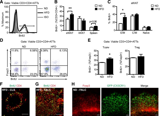

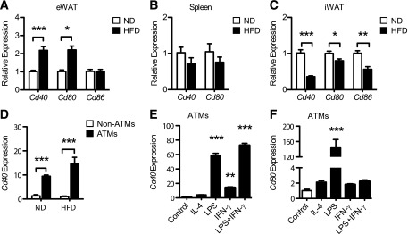

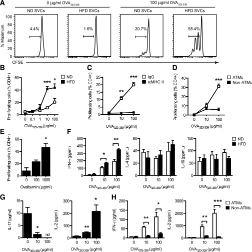

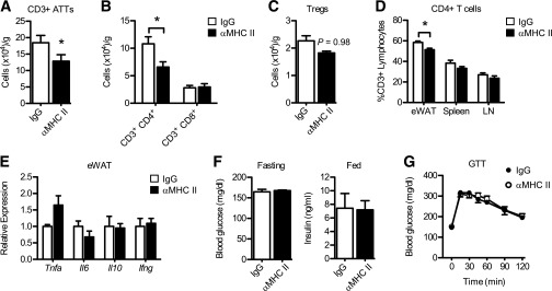
Verification
Claims the paper makes
The claims and statistics of this paper haven’t been checked yet.
Free: it reads the open-access text and checks the first 5 claims that cite a source we can check. Create a free account to check every claim.
Claims other papers make about this one
Each is checked against this paper’s own text: does it say what the citing paper says it does?
In the citing paper
“The infiltrated macrophages in muscle tissue present adipocyte-derived antigens and activate antigen-specific CD4+ T cells [39].”
From Sarcopenic obesity, insulin resistance, and their implications in cardiovascular and metabolic consequences (2020) · cites this paper as [39]
Self-check
Percentages, sums and reported differences re-computed from the paper’s own numbers.
The self-check needs the paper’s full text.
Citations
Cites
Obesity is associated with macrophage accumulation in adipose tissue
Weisberg, S. P., McCann, D., Desai, M., Rosenbaum, M., Leibel, R. L., & Ferrante, A. W. (2003) · Journal of Clinical Investigation · doi:10.1172/jci200319246 · Cited by 10,041 · cited 1×
“ATMs that co-express the dendritic cell (DC) marker CD11c and pro-inflammatory genes typical of ‘classically activated’ or M1 polarized macrophages (11-13).”
Xu, H., Barnes, G. T., Yang, Q., Tan, G., Yang, D., Chou, C. J., Sole, J., Nichols, A., Ross, J. S., Tartaglia, L. A., et al. (2003) · Journal of Clinical Investigation · doi:10.1172/jci200319451 · Cited by 6,760
Obesity induces a phenotypic switch in adipose tissue macrophage polarization
Lumeng, C. N., Bodzin, J. L., & Saltiel, A. R. (2007) · Journal of Clinical Investigation · doi:10.1172/jci29881 · Cited by 4,664 · cited 1×
“Current models suggest that obesity promotes metainflammation in part by altering the balance between type 2 and type 1 ATMs in visceral fat (13,18).”
BackgroundSelf-citationAdipokines in inflammation and metabolic disease
Ouchi, N., Parker, J. L., Lugus, J. J., & Walsh, K. (2011) · Nature Reviews Immunology · doi:10.1038/nri2921 · Cited by 4,625 · cited 2×
“Classically, APCs (specifically macrophages and dendritic cells (DCs)) shape CD4 + T cell activation by three signals: (1) presentation of peptide antigens on Major histocompatibility complex (MHC) Class II molecules (signal 1), (2) expression of T cell costimulatory molecules (e.”
Inflammation, stress, and diabetes
Wellen, K. E., & Hotamisligil, G. S. (2005) · Journal of Clinical Investigation · doi:10.1172/jci25102 · Cited by 4,143
Cinti, S., Mitchell, G., Barbatelli, G., Murano, I., Ceresi, E., Faloia, E., Wang, S., Fortier, M., Greenberg, A. S., & Obin, M. S. (2005) · Journal of Lipid Research · doi:10.1194/jlr.m500294-jlr200 · Cited by 2,529 · cited 1×
“M1 ATMs form multicellular lipidladen clusters, known as crown-like structures (CLS), around dead adipocytes in obese fat (6,14,15) and produce inflammatory cytokines (e.”
BackgroundInflammatory links between obesity and metabolic disease
Lumeng, C. N., & Saltiel, A. R. (2011) · Journal of Clinical Investigation · doi:10.1172/jci57132 · Cited by 2,319 · cited 1×
“Accumulation of activated leukocytes in metabolic tissues is a driving force for obesity-associated metabolic inflammation (metainflammation) and insulin resistance (3,4).”
BackgroundSelf-citationNishimura, S., Manabe, I., Nagasaki, M., Eto, K., Yamashita, H., Ohsugi, M., Otsu, M., Hara, K., Ueki, K., Sugiura, S., et al. (2009) · Nature Medicine · doi:10.1038/nm.1964 · Cited by 2,230 · cited 1×
“In contrast, T helper 1 (Th1) CD4 T cells and CD8 adipose tissue T cells (ATTs) accumulate in fat during obesity, promoting IFN-g and TNF-a production and insulin resistance (20,21,27).”
Feuerer, M., Herrero, L., Cipolletta, D., Naaz, A., Wong, J., Nayer, A., Lee, J., Goldfine, A. B., Benoist, C., Shoelson, S., et al. (2009) · Nature Medicine · doi:10.1038/nm.2002 · Cited by 2,145
Macrophage-specific PPARγ controls alternative activation and improves insulin resistance
Odegaard, J. I., Ricardo-Gonzalez, R. R., Goforth, M. H., Morel, C. R., Subramanian, V., Mukundan, L., Eagle, A. R., Vats, D., Brombacher, F., Ferrante, A. W., et al. (2007) · Nature · doi:10.1038/nature05894 · Cited by 2,107 · cited 1×
“Disruption of macrophage M2 polarization increases the susceptibility to high fat diet (HFD)-induced insulin resistance (8-10).”
Cited by
What we talk about when we talk about fat
Rosen, E. D., & Spiegelman, B. M. (2014) · Cell · doi:10.1016/j.cell.2013.12.012 · Cited by 2,373
“, 2013), though others suggest that adipose tissuemacrophages are the relevant APCs (Morris et al., 2013).”
Metabolic Inflammation and Insulin Resistance in Obesity
Wu, H., & Ballantyne, C. M. (2020) · Circulation Research · doi:10.1161/circresaha.119.315896 · Cited by 1,059
Kalinkovich, A., & Livshits, G. (2017) · Ageing Research Reviews · doi:10.1016/j.arr.2016.09.008 · Cited by 882
Wu, T.-R., Lin, C.-S., Chang, C.-J., Lin, T.-L., Martel, J., Ko, Y.-F., Ojcius, D. M., Lu, C.-C., Young, J. D., & Lai, H.-C. (2019) · Gut · doi:10.1136/gutjnl-2017-315458 · Cited by 758
Macrophages, Immunity, and Metabolic Disease
McNelis, J. C., & Olefsky, J. M. (2014) · Immunity · doi:10.1016/j.immuni.2014.05.010 · Cited by 720
“stimulate T cells was greater when theywere isolated fromobese compared to lean mice, possibly because of the higher amount of MHCII expression by these cells (Deng et al., 2013; Morris et al., 2013).”
BackgroundInfluentialSkeletal muscle inflammation and insulin resistance in obesity
Wu, H., & Ballantyne, C. M. (2017) · Journal of Clinical Investigation · doi:10.1172/jci88880 · Cited by 695
“MHC-II and CD11c, which are mainly expressed on M1-like macrophages/dendritic cells, play important roles in macrophage/dendritic cell–induced T cell activation in obese AT (46, 84).”
BackgroundMacrophage polarization in pathology
Sica, A., Erreni, M., Allavena, P., & Porta, C. (2015) · Cellular and Molecular Life Sciences · doi:10.1007/s00018-015-1995-y · Cited by 673
“AT macrophages also act as antigen-presenting cells promoting T cell proliferation and Th1 skewing of CD4 ? T cells with increased IFNc production, eventually supporting M1-like functions of AT macrophages [183].”
BackgroundLocal Proliferation of Macrophages Contributes to Obesity-Associated Adipose Tissue Inflammation
Amano, S. U., Cohen, J. L., Vangala, P., Tencerova, M., Nicoloro, S. M., Yawe, J. C., Shen, Y., Czech, M. P., & Aouadi, M. (2014) · Cell Metabolism · doi:10.1016/j.cmet.2013.11.017 · Cited by 613
“It is interesting to note that fasting had no effect on the proliferation of F4/80 /CD11b cells, which could include cell types that have been shown to proliferate in situ in the AT, including T cells (Morris et al., 2013a) and adipocyte progenitors (Lee et al., 2012) (Figure 1O).”
Properties and functions of adipose tissue macrophages in obesity
Russo, L., & Lumeng, C. N. (2018) · Immunology · doi:10.1111/imm.13002 · Cited by 598
Self-citationFoundations of Immunometabolism and Implications for Metabolic Health and Disease
Hotamisligil, G. S. (2017) · Immunity · doi:10.1016/j.immuni.2017.08.009 · Cited by 505
Immune Regulation of Metabolic Homeostasis in Health and Disease
Brestoff, J. R., & Artis, D. (2015) · Cell · doi:10.1016/j.cell.2015.02.022 · Cited by 497
“T-bet-dependent CD4 Th1 cells produce TNF-a and IFN-g to further potentiate insulin resistance in WAT (Cho et al., 2014; Morris et al., 2013; Stolarczyk et al., 2013).”
Obesity, Inflammation, and Immune System in Osteoarthritis
Nedunchezhiyan, U., Varughese, I., Sun, A. R., Wu, X., Crawford, R., & Prasadam, I. (2022) · Frontiers in Immunology · doi:10.3389/fimmu.2022.907750 · Cited by 466
“demonstrated that ATM in obese models had increased expression of MHC II and T cell co-stimulatory molecules, thus processing antigens and inducing antigen specific CD4 T cell population (36).”
BackgroundAdipose tissue as an immunological organ
Grant, R. W., & Dixit, V. D. (2015) · Obesity · doi:10.1002/oby.21003 · Cited by 453
“In addition to macrophages (58), B cell (15), and adipocytes (59) have also been demonstrated to present antigens.”
BackgroundImmunological Goings-on in Visceral Adipose Tissue
Mathis, D. (2013) · Cell Metabolism · doi:10.1016/j.cmet.2013.05.008 · Cited by 437
Huh, J. Y., Park, Y. J., Ham, M., & Kim, J. B. (2014) · Molecules and Cells · doi:10.14348/molcells.2014.0074 · Cited by 418
“Morris et al. (2013) have suggested that MHC class II molecules on macrophages, but not on adipocytes, could play critical roles in CD4 T cell activation in adipose tissue.”
BackgroundHong, S.-H., & Choi, K. M. (2020) · International Journal of Molecular Sciences · doi:10.3390/ijms21020494 · Cited by 390
“The infiltrated macrophages in muscle tissue present adipocyte-derived antigens and activate antigen-specific CD4+ T cells [39].”
BackgroundKolodin, D., van Panhuys, N., Li, C., Magnuson, A. M., Cipolletta, D., Miller, C. M., Wagers, A., Germain, R. N., Benoist, C., & Mathis, D. (2015) · Cell Metabolism · doi:10.1016/j.cmet.2015.03.005 · Cited by 365
“Our findings are consistent with a report highlighting the importance of MHCII+ MFs in the stimulation of VAT Tconv cells (Morris et al., 2013), but do not support previous contentions that MHCII molecule expression on adipocytes is key to VAT Tconv cell activation (Deng et al., 2013).”
ResultMacrophage functions in lean and obese adipose tissue
Thomas, D., & Apovian, C. (2017) · Metabolism · doi:10.1016/j.metabol.2017.04.005 · Cited by 316
The immune cells in adipose tissue
Ferrante, A. W. (2013) · Diabetes, Obesity and Metabolism · doi:10.1111/dom.12154 · Cited by 305
Adipose-resident group 1 innate lymphoid cells promote obesity-associated insulin resistance
O’Sullivan, T. E., Rapp, M., Fan, X., Weizman, O.-E., Bhardwaj, P., Adams, N. M., Walzer, T., Dannenberg, A. J., & Sun, J. C. (2016) · Immunity · doi:10.1016/j.immuni.2016.06.016 · Cited by 293
“…type 1 immune cells such as CD8+ T cells, Th1 cells, and NK cells, with subsequent accumulation of proinflammatory classically activated macrophage type 1 (M1) macrophages in the adipose tissue (Cho et al., 2014; Lee et al., 2016; Morris et al., 2013; Nishimura et al., 2009; Wensveen et al., 2015).”
BackgroundMoraes-Vieira, P. M., Yore, M. M., Dwyer, P. M., Syed, I., Aryal, P., & Kahn, B. B. (2014) · Cell Metabolism · doi:10.1016/j.cmet.2014.01.018 · Cited by 286
“Macrophages and dendritic cells (DC), which are both APCs (Steinman et al., 2005), are present in healthy AT throughout the lifespan (Bertola et al., 2012; Morris et al., 2013).”
BackgroundInfluentialMatt, S. M., & Gaskill, P. J. (2020) · Journal of Neuroimmune Pharmacology · doi:10.1007/s11481-019-09851-4 · Cited by 287
“Some studies suggest that adipocytes act as antigen presenting cells during inflammation (Deng et al. 2013; Morris et al. 2013), meaning that T-cells in adipose make direct contact with dopamine-producing adipocytes.”
Obesity accelerates T cell senescence in murine visceral adipose tissue
Shirakawa, K., Yan, X., Shinmura, K., Endo, J., Kataoka, M., Katsumata, Y., Yamamoto, T., Anzai, A., Isobe, S., Yoshida, N., et al. (2016) · Journal of Clinical Investigation · doi:10.1172/jci88606 · Cited by 271
Glutamine Metabolism in Macrophages: A Novel Target for Obesity/Type 2 Diabetes
Ren, W., Xia, Y., Chen, S., Wu, G., Bazer, F. W., Zhou, B., Tan, B., Zhu, G., Deng, J., & Yin, Y. (2019) · Advances in Nutrition · doi:10.1093/advances/nmy084 · Cited by 252
Dalmas, E., Venteclef, N., Caer, C., Poitou, C., Cremer, I., Aron-Wisnewsky, J., Lacroix-Desmazes, S., Bayry, J., Kaveri, S. V., Clément, K., et al. (2014) · Diabetes · doi:10.2337/db13-1511 · Cited by 240
“expansion of antigen-specific Th1 skewed CD4 T cells in diet-induced obese mice (12).”
BackgroundLocal proliferation of macrophages in adipose tissue during obesity-induced inflammation
Haase, J., Weyer, U., Immig, K., Klöting, N., Blüher, M., Eilers, J., Bechmann, I., & Gericke, M. (2014) · Diabetologia · doi:10.1007/s00125-013-3139-y · Cited by 236
A decade of progress in adipose tissue macrophage biology
Hill, A. A., Reid Bolus, W., & Hasty, A. H. (2014) · Immunological Reviews · doi:10.1111/imr.12216 · Cited by 222
Camell, C. D., Günther, P., Lee, A., Goldberg, E. L., Spadaro, O., Youm, Y.-H., Bartke, A., Hubbard, G. B., Ikeno, Y., Ruddle, N. H., et al. (2019) · Cell Metabolism · doi:10.1016/j.cmet.2019.10.006 · Cited by 214
“Fat-associated lymphoid clusters (FALCs), predominantly composed of B1innate B cells, serve as unique immunological sites that are acutely responsive to pathogens and increase with chronic inflammation (Bénézech et al., 2015; Jackson-Jones et al., 2016; Lumeng et al., 2011; Morris et al., 2013).”
BackgroundThe initiation of metabolic inflammation in childhood obesity
Singer, K., & Lumeng, C. N. (2017) · Journal of Clinical Investigation · doi:10.1172/jci88882 · Cited by 194
“Adaptive immunity is also triggered by chronic inflammation via the function of ATMs and adipose tissue dendritic cells as antigen-presenting cells (28, 29).”
BackgroundSelf-citationCho, K. W., Morris, D. L., DelProposto, J. L., Geletka, L., Zamarron, B., Martinez-Santibanez, G., Meyer, K. A., Singer, K., O’Rourke, R. W., & Lumeng, C. N. (2014) · Cell Reports · doi:10.1016/j.celrep.2014.09.004 · Cited by 172
“We have previously identified high concentrations of CD4+ ATTs in FALC structures in visceral adipose tissue that are also enriched for MHC II+ CX3CR1+ ATMs (Morris et al., 2013).”
BackgroundMethodResultInfluentialSelf-citationCho, K. W., Zamarron, B. F., Muir, L. A., Singer, K., Porsche, C. E., DelProposto, J. B., Geletka, L., Meyer, K. A., O’Rourke, R. W., & Lumeng, C. N. (2016) · The Journal of Immunology · doi:10.4049/jimmunol.1600820 · Cited by 149
“Ag-specific T cell activation assay was performed as previously described (18).”
MethodSelf-citationBiological plausibility linking sleep apnoea and metabolic dysfunction
Gileles‐Hillel, A., Gozal, D., & Gozal, D. (2016) · Nature Reviews Endocrinology · doi:10.1038/nrendo.2016.22 · Cited by 154
T Cells in Adipose Tissue: Critical Players in Immunometabolism
Wang, Q., & Wu, H. (2018) · Frontiers in Immunology · doi:10.3389/fimmu.2018.02509 · Cited by 145
“These adipocytes and macrophages/DCs can function as antigen-presenting cells (APCs) to promote adipose tissue CD4 T cell proliferation and production of IFN-γ, which further activate adipocytes and macrophages/DCs with elevated MHC II, thereby forming a positive loop to amplify Th1 cell-mediated inflammation in adipo…”
BackgroundRole of T Cells in Malnutrition and Obesity
Gerriets, V. A., & MacIver, N. J. (2014) · Frontiers in Immunology · doi:10.3389/fimmu.2014.00379 · Cited by 145
“There is evidence for both macrophages and B cells (51, 59), as well as adipocytes (60), in acting as antigen-presenting cells to stimulate T cell activation and inflammation of VAT in obesity.”
BackgroundB Lymphocytes in obesity-related adipose tissue inflammation and insulin resistance
Winer, D. A., Winer, S., Chng, M. H. Y., Shen, L., & Engleman, E. G. (2014) · Cellular and Molecular Life Sciences · doi:10.1007/s00018-013-1486-y · Cited by 142
“It is also interesting to speculate that priming of the adaptive immune response from potential VAT antigens may occur at least partially in FALCs [29].”
InfluentialDysregulation of immune response in PCOS organ system
Wang, J., Yin, T., & Liu, S. (2023) · Frontiers in Immunology · doi:10.3389/fimmu.2023.1169232 · Cited by 183
“, CD80 and CD86) in adipose tissue act as antigenpresenting cells (APCs) to promote CD4 T cell proliferation and Th1 differentiation and produce excessive IFN-g in adipose tissue (35).”
BackgroundZhou, Y., Yu, X., Chen, H., Sjöberg, S., Roux, J., Zhang, L., Ivoulsou, A.-H., Bensaid, F., Liu, C.-L., Liu, J., et al. (2015) · Cell Metabolism · doi:10.1016/j.cmet.2015.09.013 · Cited by 137
“Compared to lean mice, WAT from obese mice (Morris et al., 2013; Winer et al., 2009) contain increased numbers of CD4+ and CD8+ T cells.”
Adipose tissue B2 cells promote insulin resistance through leukotriene LTB4/LTB4R1 signaling
Ying, W., Wollam, J., Ofrecio, J. M., Bandyopadhyay, G., El Ouarrat, D., Lee, Y. S., Oh, D. Y., Li, P., Osborn, O., & Olefsky, J. M. (2017) · Journal of Clinical Investigation · doi:10.1172/jci90350 · Cited by 128
The Immune Landscape of Visceral Adipose Tissue During Obesity and Aging
Khan, S., Chan, Y. T., Revelo, X. S., & Winer, D. A. (2020) · Frontiers in Endocrinology · doi:10.3389/fendo.2020.00267 · Cited by 125
Dietary Regulation of Immunity
Lee, A. H., & Dixit, V. D. (2020) · Immunity · doi:10.1016/j.immuni.2020.08.013 · Cited by 123
Macrophage-targeted nanomedicine for chronic diseases immunotherapy
Song, Y., Huang, Y., Zhou, F., Ding, J., & Zhou, W. (2022) · Chinese Chemical Letters · doi:10.1016/j.cclet.2021.08.090 · Cited by 120
Flow Cytometry Analyses of Adipose Tissue Macrophages
Cho, K. W., Morris, D. L., & Lumeng, C. N. (2014) · Methods in Enzymology · doi:10.1016/b978-0-12-411619-1.00016-1 · Cited by 117
“This FACS strategy has also been successfully used to purify ATMs for gene expression analysis and functional assays (Morris et al., 2013; Singer et al., 2013).”
BackgroundMethodInfluentialSelf-citationStroke sensitivity in the aged: sex chromosome complement vs. gonadal hormones
McCullough, L. D., Mirza, M. A., Xu, Y., Bentivegna, K., Steffens, E. B., Ritzel, R., & Liu, F. (2016) · Aging · doi:10.18632/aging.100997 · Cited by 113
Li, C.-R., Wang, G., Sivasami, P., Ramírez, R. N., Zhang, Y.-B., Benoist, C., & Mathis, D. (2021) · Cell Metabolism · doi:10.1016/j.cmet.2021.06.007 · Cited by 107
Immune Cell Crosstalk in Obesity: A Key Role for Costimulation?
Seijkens, T., Kusters, P., Chatzigeorgiou, A., Chavakis, T., & Lutgens, E. (2014) · Diabetes · doi:10.2337/db14-0272 · Cited by 108
“AT macrophages also process and present antigen on MHCII molecules and express costimulatory molecules (34,35).”
BackgroundB Lymphocytes and Adipose Tissue Inflammation
Srikakulapu, P., & McNamara, C. A. (2020) · Arteriosclerosis Thrombosis and Vascular Biology · doi:10.1161/atvbaha.119.312467 · Cited by 117
Metabolic Inflammation in Inflammatory Bowel Disease: Crosstalk Between Adipose Tissue and Bowel
Gonçalves, P., Magro, F., & Martel, F. (2014) · Inflammatory Bowel Diseases · doi:10.1097/mib.0000000000000209 · Cited by 121
“antigen presentation function in this organ is mainly controlled by adipose tissue macrophages.(186) The percentage of macrophages resident in adipose tissue increases in obese mice(187) and humans.”
BackgroundObesity altered T cell metabolism and the response to infection
Green, W. D., & Beck, M. A. (2017) · Current Opinion in Immunology · doi:10.1016/j.coi.2017.03.008 · Cited by 104
“Recent findings by Morris et al. identified a mechanism by which ATMs function as antigen presenting cells (APCs) to regulate the activation of CD4 + T cells in mice [47].”
BackgroundAre Obesity-Related Insulin Resistance and Type 2 Diabetes Autoimmune Diseases?
Tsai, S., Clemente-Casares, X., Revelo, X. S., Winer, S., & Winer, D. A. (2015) · Diabetes · doi:10.2337/db14-1488 · Cited by 103
“The VAT environment in HFD-fed animals can support the activation and proliferation of CD4 and CD8 T cells (26,32), where macrophages (32,33), B cells, dendritic cells (DCs) (34), and potentially the adipocytes themselves (35,36) are all candidate professional APC types that can process and present antigens for T-cell…”
BackgroundAdipose tissue inflammation in glucose metabolism
Kammoun, H. L., Kraakman, M. J., & Febbraio, M. A. (2013) · Reviews in Endocrine and Metabolic Disorders · doi:10.1007/s11154-013-9274-4 · Cited by 111
Khan, I. M., Dai Perrard, X.-Y., Perrard, J. L., Mansoori, A., Wayne Smith, C., Wu, H., & Ballantyne, C. M. (2014) · Atherosclerosis · doi:10.1016/j.atherosclerosis.2014.01.011 · Cited by 97
“We found increased expression of the transcriptional regulator of the MHC II pathway (CIITA) and MHC II invariant chain (CD74) in skeletal muscle of obese WT mice, which is important for differentiation of naïve CD4þ T cells into TH1 cells [37,38].”
An update on immune dysregulation in obesity‐related insulin resistance
Daryabor, G., Kabelitz, D., & Kalantar, K. (2019) · Scandinavian Journal of Immunology · doi:10.1111/sji.12747 · Cited by 96
Myeloid cell dysfunction and the pathogenesis of the diabetic chronic wound
Wicks, K., Torbica, T., & Mace, K. A. (2014) · Seminars in Immunology · doi:10.1016/j.smim.2014.04.006 · Cited by 93
Morphological and Inflammatory Changes in Visceral Adipose Tissue During Obesity
Revelo, X. S., Luck, H., Winer, S., & Winer, D. A. (2013) · Endocrine Pathology · doi:10.1007/s12022-013-9288-1 · Cited by 115
“Adaptive Immunity During obesity, T lymphocytes expressing CD4 and CD8 produce high amounts of inflammatory cytokines such as interferon gamma (IFN-γ) that contribute to the proinflammatorymicroenvironment of adipose tissue [37].”
BackgroundInfluentialWatanabe, Y., Nagai, Y., & Takatsu, K. (2013) · Nutrients · doi:10.3390/nu5093757 · Cited by 102
“Intriguingly, CD4+ Th1 cells in visceral adipose tissue interact with antigen-presenting cells, such as adipose tissue macrophages (ATMs), and proliferate and produce IFN-γ in vitro and in vivo [23].”
BackgroundXu, E., Pereira, M. M. A., Karakasilioti, I., Theurich, S., Al-Maarri, M., Rappl, G., Waisman, A., Wunderlich, F. T., & Brüning, J. C. (2017) · Nature Communications · doi:10.1038/ncomms14803 · Cited by 89
Specific Biological Features of Adipose Tissue, and Their Impact on HIV Persistence
Bourgeois, C., Gorwood, J., Barrail-tran, A., Lagathu, C., Capeau, J., Desjardins, D., Grand, R. L., Damouche, A., Béréziat, V., & Lambotte, O. (2019) · Frontiers in Microbiology · doi:10.3389/fmicb.2019.02837 · Cited by 81
“…M1: TNF-α, MIP-1α producer Weisberg et al., 2003; Lumeng et al., 2007; Catrysse and van Loo, 2018; Ivanov et al., 2018; Lumeng et al., 2007; Morris et al., 2013; Suganami et al., 2007; Pang et al., 2008 CD4 T cells Shift from Th2 to Th1 Subset dependent Th2: maintaining glucose…”
BackgroundT Cell Immunosenescence in Aging, Obesity, and Cardiovascular Disease
Shirakawa, K., & Sano, M. (2021) · Cells · doi:10.3390/cells10092435 · Cited by 97
“MHC class II deficiency ameliorated HFD-induced VAT inflammation as well as insulin resistance [83,86,94].”
BackgroundIron homeostasis: a new job for macrophages in adipose tissue?
Hubler, M. J., Peterson, K. R., & Hasty, A. H. (2015) · Trends in Endocrinology & Metabolism · doi:10.1016/j.tem.2014.12.005 · Cited by 77
Fat-Associated Lymphoid Clusters in Inflammation and Immunity
Migoni, S. C., & Caamaño, J. (2016) · Frontiers in Immunology · doi:10.3389/fimmu.2016.00612 · Cited by 87
“org December 2016 | Volume 7 | Article 612 context of MHC II complexes, indicating that they can function as antigen-presenting cells to induce T cell proliferation (47).”
BackgroundChang, E., Hafner, H., Varghese, M., Griffin, C., Clemente, J., Islam, M., Carlson, Z., Zhu, A., Hak, L., Abrishami, S., et al. (2019) · Scientific Reports · doi:10.1038/s41598-019-52583-x · Cited by 75
Self-citationAlZaim, I., Hammoud, S. H., Al-Koussa, H., Ghazi, A., Eid, A. H., & El-Yazbi, A. F. (2020) · Frontiers in Cardiovascular Medicine · doi:10.3389/fcvm.2020.602088 · Cited by 73
Zhang, X., Hu, P., Zhang, X., & Li, X. (2020) · Carbohydrate Polymers · doi:10.1016/j.carbpol.2020.116357 · Cited by 77
Macrophage and T cell networks in adipose tissue
Jacks, R. D., & Lumeng, C. N.-K. (2023) · Nature Reviews Endocrinology · doi:10.1038/s41574-023-00908-2 · Cited by 86
Self-citationEssential Role of CD11a in CD8 + T-Cell Accumulation and Activation in Adipose Tissue
Jiang, E., Perrard, X. D., Yang, D., Khan, I. M., Perrard, J. L., Smith, C. W., Ballantyne, C. M., & Wu, H. (2014) · Arteriosclerosis, Thrombosis, and Vascular Biology · doi:10.1161/atvbaha.113.302077 · Cited by 69
“T-cell proliferation has recently been reported in peripheral blood of obese humans,(30) and CD4 T-cell proliferation has just been reported in mouse AT.(16) Here, we report CD8 T-cell proliferation in AT in obesity.”
BackgroundResultInfluentialAntigen Presentation and T-Cell Activation Are Critical for RBP4-Induced Insulin Resistance
Moraes-Vieira, P. M., Castoldi, A., Aryal, P., Wellenstein, K., Peroni, O. D., & Kahn, B. B. (2016) · Diabetes · doi:10.2337/db15-1696 · Cited by 67
“AT antigen presentation integrates the AT inflammatory responses (9,22,24,34,35) and regulates AT T-cell activation and polarization (24).”
BackgroundThe Role of T Cells in Obesity-Associated Inflammation and Metabolic Disease
Park, C., & Shastri, N. (2022) · Immune Network · doi:10.4110/in.2022.22.e13 · Cited by 88
Leukocyte Heterogeneity in Adipose Tissue, Including in Obesity
Weinstock, A., Silva, H. M., Moore, K. J., Schmidt, A. M., & Fisher, E. A. A. (2020) · Circulation Research · doi:10.1161/circresaha.120.316203 · Cited by 69
Autoimmune responses and inflammation in type 2 diabetes
Prasad, M., Chen, E. W., Toh, S.-A. E. S., & Gascoigne, N. R. J. (2020) · Journal of Leukocyte Biology · doi:10.1002/jlb.3mr0220-243r · Cited by 78
Catestatin as a Target for Treatment of Inflammatory Diseases
Muntjewerff, E. M., Dunkel, G., Nicolasen, M. J. T., Mahata, S. K., & van den Bogaart, G. (2018) · Frontiers in Immunology · doi:10.3389/fimmu.2018.02199 · Cited by 67
“Since adipose tissue macrophages (ATMs) have antigen-presenting capacities this shift could influence the adaptive immune response (123).”
Imaging White Adipose Tissue with Confocal Microscopy
Martinez-Santibañez, G., Cho, K. W., & Lumeng, C. N. (2014) · Methods in Enzymology · doi:10.1016/b978-0-12-411619-1.00002-1 · Cited by 64
Self-citationRusso, L., Muir, L., Geletka, L., Delproposto, J., Baker, N., Flesher, C., O'Rourke, R., & Lumeng, C. N. (2020) · Molecular Metabolism · doi:10.1016/j.molmet.2020.100983 · Cited by 58
Self-citationMacrophage-specific MHCII expression is regulated by a remote Ciita enhancer controlled by NFAT5
Buxadé, M., Encabo, H. H., Riera-Borrull, M., Quintana‐Gallardo, L., López-Cotarelo, P., Tellechea, M., Martínez-Martínez, S., Redondo, J. M., Martín‐Caballero, J., Flores, J. M., et al. (2018) · The Journal of Experimental Medicine · doi:10.1084/jem.20180314 · Cited by 80
“Finally, we analyzed the effect of NFAT5 in the expression of the different isoforms of Ciita (Muhlethaler-Mottet et al., 1997; Morris et al., 2013) in untreated and IFNγ-treated macrophages.”
BackgroundHIV Persistence in Adipose Tissue Reservoirs
Couturier, J. P., & Lewis, D. E. (2018) · Current HIV/AIDS Reports · doi:10.1007/s11904-018-0378-z · Cited by 67
Adipose‐tissue regulatory T cells: Critical players in adipose‐immune crosstalk
Becker, M., Levings, M. K., & Daniel, C. (2017) · European Journal of Immunology · doi:10.1002/eji.201646739 · Cited by 59
Interferon Family Cytokines in Obesity and Insulin Sensitivity
Huang, L.-Y., Chiu, C.-J., Hsing, C., & Hsu, Y. (2022) · Cells · doi:10.3390/cells11244041 · Cited by 64
The weight of nutrients: kynurenine metabolites in obesity and exercise
Dadvar, S., Ferreira, D. M. S., Červenka, I., & Ruas, J. L. (2018) · Journal of Internal Medicine · doi:10.1111/joim.12830 · Cited by 61
T Cells in Adipose Tissue in Aging
Kalathookunnel Antony, A., Lian, Z., & Wu, H. (2018) · Frontiers in Immunology · doi:10.3389/fimmu.2018.02945 · Cited by 56
“indicated that ATMs colocalized with T cells in lymphoid clusters within adipose tissue and may act as APCs, which express high levels of MHCII and also costimulatory molecules and process and present antigens to induce CD4+ Tcell proliferation and activation in adipose tissue of obese mice (29, 68, 105).”
BackgroundMinireview: 12-Lipoxygenase and Islet β-Cell Dysfunction in Diabetes
Tersey, S. A., Bolanis, E., Holman, T. R., Maloney, D. J., Nadler, J. L., & Mirmira, R. G. (2015) · Molecular Endocrinology · doi:10.1210/me.2015-1041 · Cited by 61
Tang, P., Virtue, S., Goie, J. Y. G., Png, C. W., Guo, J., Li, Y., Jiao, H., Chua, Y. L., Campbell, M., Moreno‐Navarrete, J. M., et al. (2021) · Cell Death and Differentiation · doi:10.1038/s41418-021-00798-9 · Cited by 55
CCR7 Maintains Nonresolving Lymph Node and Adipose Inflammation in Obesity
Hellmann, J. L., Sansbury, B. E., Holden, C. R., Tang, Y., Wong, B. C. K., Wysoczynski, M., Rodríguez, J., Bhatnagar, A., Hill, B. G., & Spite, M. R. (2016) · Diabetes · doi:10.2337/db15-1689 · Cited by 58
“Because previous studies demonstrated that both ATMs and DCs can activate adaptive immune cells (13,33), we hypothesized that by blocking CCR7-dependent recruitment of these cells to LNs, the subsequent recirculation of activated T cells and B cells to adipose tissue should be blunted.”
BackgroundResultSoedono, S., & Cho, K. W. (2021) · International Journal of Molecular Sciences · doi:10.3390/ijms22168666 · Cited by 60
“Furthermore, the IFNγ produced by Th1 will further activate DCs’ AP function to increase its MHC-II, in a feed-back loop, which would cause AT inflammation [58,65,68,70].”
BackgroundSelf-citationInnate PD-L1 limits T cell–mediated adipose tissue inflammation and ameliorates diet-induced obesity
Schwartz, C., Schmidt, V., Deinzer, A., Hawerkamp, H. C., Hams, E., Bayerlein, J., Röger, O., Bailer, M., Krautz, C., Gendy, A. E., et al. (2022) · Science Translational Medicine · doi:10.1126/scitranslmed.abj6879 · Cited by 54
Miranda, K., Mehrpouya-Bahrami, P., Nagarkatti, P., & Nagarkatti, M. (2019) · Frontiers in Immunology · doi:10.3389/fimmu.2019.01049 · Cited by 44
Martinez-Santibañez, G., Singer, K., Cho, K. W., DelProposto, J. L., Mergian, T. A., & Lumeng, C. N.-K. (2015) · Adipocyte · doi:10.1080/21623945.2015.1027848 · Cited by 57
“Metabolic studies Glucose and Insulin tolerance tests were performed as described.(36) Plasma insulin levels were measured using a mouse insulin ELISA kit (Crystal Chem, Downers Grove, IL).”
MethodSelf-citationHuman CD206+ macrophages associate with diabetes and adipose tissue lymphoid clusters
Muir, L. A., Cho, K. W., Geletka, L. M., Baker, N. A., Flesher, C. G., Ehlers, A. P., Kaciroti, N., Lindsly, S., Ronquist, S., Rajapakse, I., et al. (2022) · JCI Insight · doi:10.1172/jci.insight.146563 · Cited by 56
“Insulin resistance has been associated with ATM MHC II antigen presentation in murine obesity (25, 26) and in human CD11c+ ATMs (9), so we examined antigen presentation–related genes HLA-D, CD40, CD80, CD86, and ICAM1 in ATM subtypes.”
Self-citationLi, S., Kievit, P., Robertson, A.-K., Kolumam, G. A., Li, X.-M., von Wachenfeldt, K. A., Valfridsson, C., Bullens, S., Messaoudi, I., Bader, L., et al. (2013) · Molecular Metabolism · doi:10.1016/j.molmet.2013.06.001 · Cited by 52
Lymphocyte roles in metabolic dysfunction: of men and mice
Ip, B. C., Hogan, A. E., & Nikolajczyk, B. S. (2015) · Trends in Endocrinology & Metabolism · doi:10.1016/j.tem.2014.12.001 · Cited by 49
Zhou, H., & Liu, F. (2018) · Frontiers in Immunology · doi:10.3389/fimmu.2018.01961 · Cited by 50
“ATMs are thought to be the predominant MHCII-expressing cells in VAT under both ND and HFD feeding conditions (51).”
BackgroundObesity Programs Macrophages to Support Cancer Progression
Singh, A., Mayengbam, S. S., Yaduvanshi, H., Wani, M. R., & Bhat, M. K. (2022) · Cancer Research · doi:10.1158/0008-5472.can-22-1257 · Cited by 51
Chan, P., Wu, T., Chen, Y., Lu, C., Wabitsch, M., Tian, Y., & Hsieh, P. (2018) · Clinical Science · doi:10.1042/cs20180041 · Cited by 48
Félix, I., Jokela, H., Karhula, J., Kotaja, N., Savontaus, E., Salmi, M., & Rantakari, P. (2021) · Frontiers in Immunology · doi:10.3389/fimmu.2021.719979 · Cited by 54
“In contrast to previously published papers (48, 78), our studies revealed that although diet-induced obesity might increase MHC II expression in ATMs generally (3), both TIM4 resident ATM populations decreased their MHC II expression significantly during the HFD.”
ResultLiddle, D. M., Hutchinson, A. L., Wellings, H. R., Power, K. A., Robinson, L. E., & Monk, J. M. (2017) · Nutrients · doi:10.3390/nu9121289 · Cited by 50
“Controversy exists over which T cell subset is the first to exhibit changes in cellular abundance or activation in obese AT; CD4+ T cells [23,196], CD8+ T cells [197,200,201], or both [13,24,138,177,202,203].”
BackgroundMHC class II in renal tubules plays an essential role in renal fibrosis
Zhou, Y., Luo, Z., Liao, C., Cao, R., Hussain, Z., Wang, J., Zhou, Y., Chen, T., Sun, J., Huang, Z., et al. (2021) · Cellular and Molecular Immunology · doi:10.1038/s41423-021-00763-z · Cited by 44
Lcn2 mediates adipocyte-muscle-tumor communication and hypothermia in pancreatic cancer cachexia
Lemecha, M., Chalise, J., Takamuku, Y., Zhang, G.-X., Yamakawa, T., Larson, G., & Itakura, K. (2022) · Molecular Metabolism · doi:10.1016/j.molmet.2022.101612 · Cited by 41
“In tumor-bearing mice, Lcn2 deficiency significantly reduced fibrosis and decreased immune cell infiltration and active inflammation of adipose tissue, as evidenced by a decrease in CD45þ and CD4þ immune cells [63].”
BackgroundChatzigeorgiou, A., & Chavakis, T. (2016) · Handbook of Experimental Pharmacology · doi:10.1007/164_2015_8 · Cited by 48
“In addition, antigen MHC II-dependent interaction of macrophages with cells of the adaptive immunity contributes to dysregulation of AT inflammation (Cho et al. 2014; Morris et al. 2013).”
BackgroundMorris, D. L., Oatmen, K. E., Mergian, T. A., Cho, K., Delproposto, J. L., Singer, K., Evans-Molina, C., O'Rourke, R., & Lumeng, C. (2016) · Journal of Leukocyte Biology · doi:10.1189/jlb.3a0115-009r · Cited by 38
“Because ATMs are the primary APC in adipose tissue [3, 4], the APC functionality of ATMs is at least partially dependent on CD40 signaling to generate and maintain of Tconv in eWAT.”
BackgroundInfluentialSelf-citationAdaptive Immunity and Antigen‐Specific Activation in Obesity‐Associated Insulin Resistance
Chng, M. H. Y., Alonso, M. N., Barnes, S. E., Nguyen, K. D., & Engleman, E. G. (2015) · Mediators of Inflammation · doi:10.1155/2015/593075 · Cited by 39
Couturier, J. P., Agarwal, N., Nehete, P. N., Baze, W. B., Barry, M. A., Sastry, K. J., Balasubramanyam, A., & Lewis, D. E. (2016) · Retrovirology · doi:10.1186/s12977-016-0260-2 · Cited by 40
Obesity-induced inflammation and insulin resistance: A mini-review on T-cells
Nyambuya, T. M., Dludla, P. V., Mxinwa, V., & Nkambule, B. B. (2019) · Metabolism Open · doi:10.1016/j.metop.2019.100015 · Cited by 43
“, 2013 [27] Male DIO C57BL/6 J mice None High fat diet (HFD)-induced obesity promoted conventional CD4þ T-cell proliferation in mice visceral (AT).”
Background
Metadata
- Authors
- Morris, D. L., Cho, K. W., DelProposto, J. L., Oatmen, K. E., Geletka, L. M., Martinez-Santibanez, G., Singer, K., & Lumeng, C. N.
- Affiliations
- Michigan Medicine (US)
- University of Michigan (US)
- Cellular and Molecular Biology Graduate Program, University of Michigan Medical School, Ann Arbor, Michigan
- Corresponding author
- Lumeng, Carey N.
- Venue
- Diabetes
- Publisher
- American Diabetes Association
- Published
- 2013-07-17
- Volume
- 62
- Issue
- 8
- Pages
- 2762-2772
- ISSN
- 0012-1797 (ISSN-L)
- 1939-327X
- PMC
- PMC3717880
- OpenAlex
- W2130657795
- Cited by
- 217
- References
- 48
- Citations by year
- 2026: 6 · 2025: 8 · 2024: 6 · 2023: 13 · 2022: 17 · 2021: 11 · 2020: 23 · 2019: 17 · 2018: 20 · 2017: 18 · 2016: 20 · 2015: 24 · 2014: 21 · 2013: 13
- FWCI
- 18.98
- Type
- journal article
- Language
- en
- Open access
- hybrid
- doi.org
- Licence
- Locations
- Diabetes (publishedVersion, cc-by-nc-nd)
- PubMed Central (publishedVersion, cc-by-nc-nd)
- PubMed (publishedVersion)
- Funders
- National Institute of General Medical Sciences (T32GM007315, T32GM008322)
- National Institute of Diabetes and Digestive and Kidney Diseases (DK-078851, DK-087999, P60DK020572, R21DK092873, F32DK091976, DK089503, R01DK090262, DK-091976, DK092873, DK090262, K08DK078851, R03DK087999, P30DK020572, P30DK089503)
- Eunice Kennedy Shriver National Institute of Child Health and Human Development (F32HD008513, T32-HD-008513, K12HD028820)
- Topics
- Health Sciences › Medicine › Endocrinology, Diabetes and Metabolism › Adipokines, Inflammation, and Metabolic Diseases
- Health Sciences › Medicine › Physiology (medical) › Adipose Tissue and Metabolism
- Health Sciences › Medicine › Cardiology and Cardiovascular Medicine › Cardiovascular Disease and Adiposity
- Keywords
- adipose tissue macrophages, antigen presentation, CD4+ T cells, diet-induced obesity, visceral fat, interferon-gamma, MHC class II, T cell costimulatory molecules, T cell proliferation, adipose tissue inflammation, innate-adaptive immune crosstalk, metabolic disorders
- MeSH terms
- Adipose Tissue, Animals, Antigen-Presenting Cells, Diet, Glucose Tolerance Test, Inflammation, Insulin, Insulin Resistance, Lymphocyte Activation, Macrophages, Obesity, Phagocytosis, CD4-Positive T-Lymphocytes, Mice
About these checks
Registry, retraction, Cites and statistics results are computed, not judged, with one exception: a printed reference that no index lists is read once by a model before it counts as a citation error. Claims are extracted by a model and kept only when their quote appears verbatim in the text; each cited source is then checked, with a model’s help, for whether it says what the claim relies on. Those verdicts can be wrong — read the claim’s quote and the cited source before relying on one.
Statistics and arithmetic are recomputed, not judged.
Confidence bands are shown only for fields calibrated on our annotated test set. None is calibrated yet, so no band is shown.
What counts as a claim ›How we measure confidence ›How we check this ›
Are you an author, or do you see a problem with this page? Request a review
Data sources
Bibliographic data: Semantic Scholar, OpenAlex, Crossref.
Contains information from the Semantic Scholar Academic Graph Datasets, made available under the ODC Attribution License (ODC-BY 1.0).
Semantic Scholar’s data is described in: Kinney, R., et al. (2023). The Semantic Scholar Open Data Platform. arXiv:2301.10140.
