

The DOI matches its Crossref record; 80 of the 80 references it cites were found in a registry or an index.
Engineering Organoid Vascularization
Open accessAbstract
The development of increasingly biomimetic human tissue analogues has been a long-standing goal in two important biomedical applications: drug discovery and regenerative medicine. In seeking to understand the safety and effectiveness of newly developed pharmacological therapies and replacement tissues for severely injured non-regenerating tissues and organs, there remains a tremendous unmet need in generating tissues with both functional complexity and scale. Over the last decade, the advent of organoids has demonstrated that cells have the ability to reorganize into complex tissue-specific structures given minimal inductive factors. However a major limitation in achieving truly in-vivo-like functionality has been the lack of structured organization and reasonable tissue size. In-vivo, developing tissues are interpenetrated by and interact with a complex network of vasculature which allows not only oxygen, nutrient and waste exchange, but also allow for inductive biochemical exchange and a structural template for growth. Conversely, in-vitro, this aspect of organoid development has remained largely missing, suggesting that these may be the critical cues required for large-scale and more reproducible tissue organization. Here, we review recent technical progress in generating in-vitro vasculature, and seek to provide a framework for understanding how such technologies, together with theoretical and developmentally inspired insights, can be harnessed to enhance next generation organoid development.
Overview
The claims and statistics of this paper haven’t been checked yet.
Run the check — free, no account needed. It reads the open-access text and checks the sources it cites.
Integrity signals
Registry
Cites
Statistics
Figures
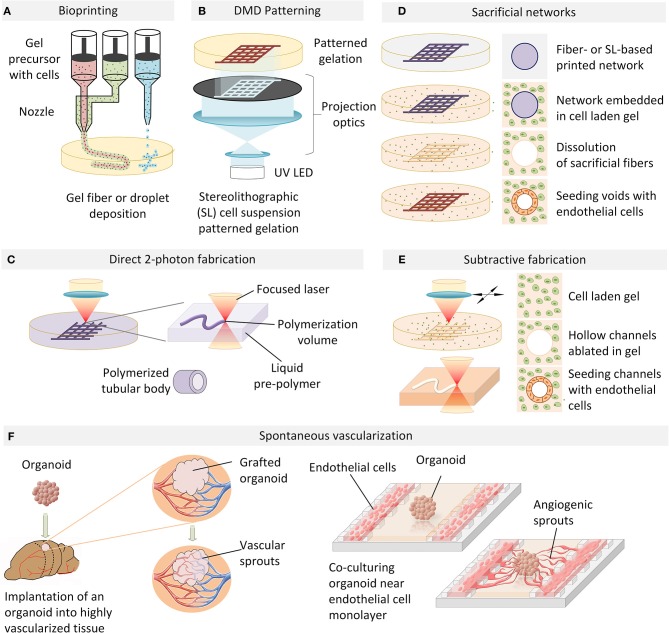
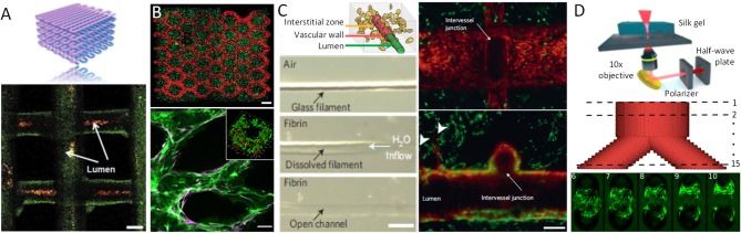
Verification
Claims the paper makes
The claims and statistics of this paper haven’t been checked yet.
Run the check — free, no account needed. It reads the open-access text and checks the sources it cites.
Claims other papers make about this one
Each is checked against this paper’s own text: does it say what the citing paper says it does?
The source backs part of the claim, not all of it — or backs it for a different population, condition or comparison.
In the citing paper
“Kidney organoids have been cultured in an OoC device which enables fluid flow to simulate shear stress107 and the generation of a vascular network108.”
From Organoids (2022) · cites this paper as [107]
Experimental — accuracy not yet measuredThe check wasn’t confident either way, so it gives no verdict — never disputed.
In the citing paper
“A more recent example shows how the presence of fluid flow enhances kidney organoids’ maturation and favors their vascularization in vitro107.”
From Organoids (2022) · cites this paper as [107]
Experimental — accuracy not yet measured
Self-check
Percentages, sums and reported differences re-computed from the paper’s own numbers.
The self-check needs the paper’s full text.
Citations
Cites
Designing materials for biology and medicine
Langer, R., & Tirrell, D. A. (2004) · Nature · doi:10.1038/nature02388 · Cited by 3,198
Modeling Development and Disease with Organoids
Clevers, H. (2016) · Cell · doi:10.1016/j.cell.2016.05.082 · Cited by 3,196
Capturing complex 3D tissue physiology in vitro
Griffith, L. G., & Swartz, M. A. (2006) · Nature Reviews Molecular Cell Biology · doi:10.1038/nrm1858 · Cited by 2,347
Vascularized and functional human liver from an iPSC-derived organ bud transplant
Takebe, T., Sekine, K., Enomura, M., Koike, H., Kimura, M., Ogaeri, T., Zhang, R.-R., Ueno, Y., Zheng, Y.-W., Koike, N., et al. (2013) · Nature · doi:10.1038/nature12271 · Cited by 2,170
Rapid casting of patterned vascular networks for perfusable engineered three-dimensional tissues
Miller, J. S., Stevens, K. R., Yang, M. T., Baker, B. M., Nguyen, D.-H. T., Cohen, D. M., Toro, E., Chen, A. A., Galie, P. A., Yu, X., et al. (2012) · Nature Materials · doi:10.1038/nmat3357 · Cited by 1,917
Matrigel: Basement membrane matrix with biological activity
Kleinman, H. K., & Martin, G. R. (2005) · Seminars in Cancer Biology · doi:10.1016/j.semcancer.2005.05.004 · Cited by 1,673
Three-dimensional bioprinting of thick vascularized tissues
Kolesky, D. B., Homan, K. A., Skylar-Scott, M. A., & Lewis, J. A. (2016) · Proceedings of the National Academy of Sciences · doi:10.1073/pnas.1521342113 · Cited by 1,492
Designer matrices for intestinal stem cell and organoid culture
Gjorevski, N., Sachs, N., Manfrin, A., Giger, S., Bragina, M. E., Ordóñez-Morán, P., Clevers, H., & Lutolf, M. P. (2016) · Nature · doi:10.1038/nature20168 · Cited by 1,426
Designing materials to direct stem-cell fate
Lütolf, M. P., Gilbert, P. M., & Blau, H. M. (2009) · Nature · doi:10.1038/nature08602 · Cited by 1,409
An in vivo model of functional and vascularized human brain organoids
Mansour, A. A., Gonçalves, J. T., Bloyd, C. W., Li, H., Fernandes, S., Quang, D., Johnston, S., Parylak, S. L., Jin, X., & Gage, F. H. (2018) · Nature Biotechnology · doi:10.1038/nbt.4127 · Cited by 1,211
Cited by
Sorted by citations
Zhao, Z., Chen, X., Dowbaj, A. M., Sljukic, A., Bratlie, K., Lin, L., Fong, E. L. S., Balachander, G. M., Chen, Z., Soragni, A., et al. (2022) · Nature Reviews Methods Primers · doi:10.1038/s43586-022-00174-y · Cited by 1,082
Basic and Therapeutic Aspects of Angiogenesis Updated
Eelen, G., Treps, L., Li, X., & Carmeliet, P. (2020) · Circulation Research · doi:10.1161/circresaha.120.316851 · Cited by 589
Engineering Stem Cell Self-organization to Build Better Organoids
Brassard, J. A., & Lütolf, M. P. (2019) · Cell stem cell · doi:10.1016/j.stem.2019.05.005 · Cited by 392
Vascularized organoids on a chip: strategies for engineering organoids with functional vasculature
Zhang, S., Wan, Z., & Kamm, R. D. (2021) · Lab on a Chip · doi:10.1039/d0lc01186j · Cited by 331
Tumor organoids: applications in cancer modeling and potentials in precision medicine
Xu, H., Jiao, D., Liu, A., & Wu, K. (2022) · Journal of Hematology & Oncology · doi:10.1186/s13045-022-01278-4 · Cited by 286
Bioengineering Approaches for the Advanced Organoid Research
Yi, S. A., Zhang, Y., Rathnam, C., Pongkulapa, T., & Lee, K. (2021) · Advanced Materials · doi:10.1002/adma.202007949 · Cited by 218
Advanced Bottom‐Up Engineering of Living Architectures
Gaspar, V. M., Lavrador, P., Borges, J., Oliveira, M. B., & Mano, J. F. (2019) · Advanced Materials · doi:10.1002/adma.201903975 · Cited by 207
The horizon of bone organoid: A perspective on construction and application
Chen, S., Chen, X., Geng, Z., & Su, J. (2022) · Bioactive Materials · doi:10.1016/j.bioactmat.2022.01.048 · Cited by 197
Engineering Vascularized Organoid-on-a-Chip Models
Shirure, V. S., Hughes, C. C. W., & George, S. C. (2021) · Annual Review of Biomedical Engineering · doi:10.1146/annurev-bioeng-090120-094330 · Cited by 200
Advances in Engineering Human Tissue Models
Moysidou, C., Barberio, C., & Owens, R. M. (2021) · Frontiers in Bioengineering and Biotechnology · doi:10.3389/fbioe.2020.620962 · Cited by 193
Mechanisms of radiation-induced endothelium damage: Emerging models and technologies
Wijerathne, H., Langston, J. C., Yang, Q., Sun, S., Miyamoto, C. T., Kilpatrick, L. E., & Kiani, M. F. (2021) · Radiotherapy and Oncology · doi:10.1016/j.radonc.2021.02.007 · Cited by 194
Review on the Vascularization of Organoids and Organoids-on-a-Chip
Zhao, X., Xu, Z., Xiao, L., Shi, T., Xiao, H., Wang, Y., Li, Y., Xue, F., & Zeng, W. (2021) · Frontiers in Bioengineering and Biotechnology · doi:10.3389/fbioe.2021.637048 · Cited by 167
Patient-Derived Organoids for Precision Cancer Immunotherapy
Grönholm, M., Feodoroff, M., Antignani, G., Martins, B., Hamdan, F., & Cerullo, V. (2021) · Cancer Research · doi:10.1158/0008-5472.can-20-4026 · Cited by 146
Large-scale perfused tissues via synthetic 3D soft microfluidics
Grebenyuk, S. E., Fattah, A. R. A., Kumar, M., Toprakhisar, B., Rustandi, G., Vananroye, A., Salmon, I., Verfaillie, C. M., Grillo, M. P., & Ranga, A. (2023) · Nature Communications · doi:10.1038/s41467-022-35619-1 · Cited by 160
Self-citationOrganoids as host models for infection biology – a review of methods
Aguilar, C., da Silva, M. A., Saraiva, M., Neyazi, M., Olsson, I. A. S., & Bartfeld, S. (2021) · Experimental & Molecular Medicine · doi:10.1038/s12276-021-00629-4 · Cited by 131
Harane, S. E., Zidi, B., Harane, N. E., Krause, K., Matthes, T., & Preynat‐Seauve, O. (2023) · Cells · doi:10.3390/cells12071001 · Cited by 135
Emulating Human Tissues and Organs: A Bioprinting Perspective Toward Personalized Medicine
Fonseca, A. C., Melchels, F. P. W., Ferreira, M. J. S., Moxon, S. R., Potjewyd, G., Dargaville, T. R., Kimber, S. J., & Domingos, M. A. N. (2020) · Chemical Reviews · doi:10.1021/acs.chemrev.0c00342 · Cited by 135
Advances in Spheroids and Organoids on a Chip
Fang, G., Chen, Y., Lu, H., & Jin, D. (2023) · Advanced Functional Materials · doi:10.1002/adfm.202215043 · Cited by 135
Human Blood Vessel Organoids Penetrate Human Cerebral Organoids and Form a Vessel-Like System
Ahn, Y., An, J.-H., Yang, H.-J., Lee, D. G., Kim, J., Koh, H., Park, Y., Song, B., Sim, B., Lee, H. J., et al. (2021) · Cells · doi:10.3390/cells10082036 · Cited by 121
In Vitro Strategies to Vascularize 3D Physiologically Relevant Models
Dellaquila, A., Le Bao, C., Letourneur, D., & Simon‐Yarza, T. (2021) · Advanced Science · doi:10.1002/advs.202100798 · Cited by 115
In vitro grafting of hepatic spheroids and organoids on a microfluidic vascular bed
Bonanini, F., Kurek, D., Previdi, S., Nicolas, A., Hendriks, D., de Ruiter, S., Meyer, M., Clapés Cabrer, M., Dinkelberg, R., García, S. B., et al. (2022) · Angiogenesis · doi:10.1007/s10456-022-09842-9 · Cited by 112
Microfluidic Tissue Engineering and Bio‐Actuation
Filippi, M., Buchner, T., Yaşa, Ö., Weirich, S., & Katzschmann, R. K. (2022) · Advanced Materials · doi:10.1002/adma.202108427 · Cited by 113
Velazquez, J. J., LeGraw, R., Moghadam, F., Tan, Y., Kilbourne, J. A., Maggiore, J. C., Hislop, J., Liu, S., Cats, D., Lopes, S. M. C. D. S., et al. (2020) · Cell Systems · doi:10.1016/j.cels.2020.11.002 · Cited by 103
El-Sadek, I. A., Miyazawa, A., Shen, L. T., Makita, S., Fukuda, S., Yamashita, T., Oka, Y., Mukherjee, P., Matsusaka, S., Oshika, T., et al. (2020) · Biomedical Optics Express · doi:10.1364/boe.404336 · Cited by 104
Barák, M., Fedorová, V., Pospíšilová, V., Raška, J., Vochyánová, S., Sedmík, J., Hříbková, H., Klímová, H., Váňová, T., & Bohačiaková, D. (2022) · Stem Cell Reviews and Reports · doi:10.1007/s12015-021-10254-3 · Cited by 102
Vascular organoids: unveiling advantages, applications, challenges, and disease modelling strategies
Naderi‐Meshkin, H., Cornelius, V. A., Eleftheriadou, M., Potel, K. N., Setyaningsih, W. A. W., & Margariti, A. (2023) · Stem Cell Research & Therapy · doi:10.1186/s13287-023-03521-2 · Cited by 101
Evolution of the Experimental Models of Cholangiocarcinoma
Massa, A., Varamo, C., Vita, F., Tavolari, S., Neia, C. P., Brandi, G., Rizzo, A., Cavalloni, G., & Aglietta, M. (2020) · Cancers · doi:10.3390/cancers12082308 · Cited by 96
Kim, S. K., Kim, Y. H., Park, S., & Cho, S. (2021) · Acta Biomaterialia · doi:10.1016/j.actbio.2021.03.002 · Cited by 96
Role of three-dimensional cell culture in therapeutics and diagnostics: an updated review
Biju, T. S., Priya, V. V., & Francis, A. P. (2023) · Drug Delivery and Translational Research · doi:10.1007/s13346-023-01327-6 · Cited by 98
Bertassoni, L. E. (2021) · Advanced Materials · doi:10.1002/adma.202101321 · Cited by 98
Patient-derived cell line, xenograft and organoid models in lung cancer therapy
Huo, K.-G., D’Arcangelo, E., & Tsao, M. (2020) · Translational Lung Cancer Research · doi:10.21037/tlcr-20-154 · Cited by 98
Synthetic scaffolds for 3D cell cultures and organoids: applications in regenerative medicine
Marchini, A., & Gelain, F. (2022) · Critical Reviews in Biotechnology · doi:10.1080/07388551.2021.1932716 · Cited by 91
Sex differences in the blood–brain barrier and neurodegenerative diseases
Weber, C. M., & Clyne, A. M. (2021) · APL Bioengineering · doi:10.1063/5.0035610 · Cited by 95
Past, Present, and Future of Brain Organoid Technology.
Koo, B., Choi, B., Park, H., & Yoon, K. (2019) · Europe PMC (PubMed Central) · doi:10.14348/molcells.2019.0162 · Cited by 93
Peking, P., Krisch, L. M., Wolf, M., Hochmann, S., Hoog, A., Vári, B., Muigg, K., Poupardin, R., Scharler, C., Schmidhuber, S. M., et al. (2021) · Theranostics · doi:10.7150/thno.59661 · Cited by 96
Zheng, F., Xiao, Y., Liu, H., Fan, Y., & Dao, M. (2021) · Advanced Biology · doi:10.1002/adbi.202000024 · Cited by 96
Strategies for re-vascularization and promotion of angiogenesis in trauma and disease
Gonçalves, R. C., Banfi, A., Oliveira, M. B., & Mano, J. F. (2020) · Biomaterials · doi:10.1016/j.biomaterials.2020.120628 · Cited by 90
iPSC-derived organ-on-a-chip models for personalized human genetics and pharmacogenomics studies
Palasantzas, V. E. J. M., Tamargo-Rubio, I., Le, K. T. T., Slager, J., Wijmenga, C., Jonkers, I. H., Kumar, V., Fu, J., & Withoff, S. (2023) · Trends in Genetics · doi:10.1016/j.tig.2023.01.002 · Cited by 94
3D tumor angiogenesis models: recent advances and challenges
Bhat, S. M., Badiger, V. A., Vasishta, S., Chakraborty, J., Seetharam, P., Ghosh, S., & Joshi, M. B. (2021) · Journal of Cancer Research and Clinical Oncology · doi:10.1007/s00432-021-03814-0 · Cited by 87
Organoid Models for Precision Cancer Immunotherapy
Sun, C., Lan, H., Fang, X., Yang, X., & Jin, K. (2022) · Frontiers in Immunology · doi:10.3389/fimmu.2022.770465 · Cited by 90
The endothelium, a key actor in organ development and hPSC-derived organoid vascularization
Vargas-Valderrama, A., Messina, A., Mitjavila‐Garcia, M. T., & Guénou, H. (2020) · Journal of Biomedical Science · doi:10.1186/s12929-020-00661-y · Cited by 86
Chaudhary, S., & Chakraborty, E. (2022) · Beni-Suef University Journal of Basic and Applied Sciences · doi:10.1186/s43088-021-00172-1 · Cited by 84
Bioengineered skin organoids: from development to applications
Hong, Z.-X., Zhu, S.-T., Li, H., Luo, J.-Z., Yang, Y., An, Y., Wang, X., & Wang, K. (2023) · Military Medical Research · doi:10.1186/s40779-023-00475-7 · Cited by 77
Hair follicle stem cells as a skin‐organizing signaling center during adult homeostasis
Li, K. N., & Tumbar, T. (2021) · The EMBO Journal · doi:10.15252/embj.2020107135 · Cited by 81
Advances in development and application of human organoids
Shankaran, A., Prasad, K., Chaudhari, S., Brand, A. M., & Satyamoorthy, K. (2021) · 3 Biotech · doi:10.1007/s13205-021-02815-7 · Cited by 77
Organoid Technology: Current Standing and Future Perspectives
Shariati, L., Esmaeili, Y., Javanmard, S. H., Bidram, E., & Amini, A. (2021) · Stem Cells · doi:10.1002/stem.3379 · Cited by 71
Preclinical Evaluation of CAR T Cell Function: In Vitro and In Vivo Models
Si, X., Xiao, L., Brown, C. E., & Wang, D. (2022) · International Journal of Molecular Sciences · doi:10.3390/ijms23063154 · Cited by 70
Geometric engineering of organoid culture for enhanced organogenesis in a dish
Park, S. E., Kang, S., Paek, J., Georgescu, A., Chang, J., Yi, A. Y., Wilkins, B. J., Karakasheva, T. A., Hamilton, K. E., & Huh, D. (2022) · Nature Methods · doi:10.1038/s41592-022-01643-8 · Cited by 67
Singh, R. K., & Nasonkin, I. O. (2020) · Frontiers in Cellular Neuroscience · doi:10.3389/fncel.2020.00179 · Cited by 67
Natural Hydrogels Support Kidney Organoid Generation and Promote In Vitro Angiogenesis
Garreta, E., Moya‐Rull, D., Marco, A., Amato, G., Ullate‐Agote, A., Tarantino, C., Gallo, M., Esporrín‐Ubieto, D., Centeno, A., Vilas‐Zornoza, A., et al. (2024) · Advanced Materials · doi:10.1002/adma.202400306 · Cited by 63
Teng, Y., Zhao, Z., Tasnim, F., Huang, X., & Yu, H. (2021) · Biomaterials · doi:10.1016/j.biomaterials.2021.120904 · Cited by 62
Advances in Microfluidic Technologies in Organoid Research
Liu, H., Gan, Z., Qin, X., Wang, Y., & Qin, J. (2023) · Advanced Healthcare Materials · doi:10.1002/adhm.202302686 · Cited by 68
Kim, D., Lim, H., Youn, J., Park, T., & Kim, D. S. (2024) · Nature Communications · doi:10.1038/s41467-024-53073-z · Cited by 66
Establishment of Colorectal Cancer Organoids in Microfluidic-Based System
Pinho, D., Santos, D. R. D., Vila, A., & Carvalho, S. (2021) · Micromachines · doi:10.3390/mi12050497 · Cited by 63
Biohybrid Actuators for Soft Robotics: Challenges in Scaling Up
Won, P., Ko, S. H., Majidi, C., Feinberg, A. W., & Webster‐Wood, V. A. (2020) · Actuators · doi:10.3390/act9040096 · Cited by 60
Pinton, L., Khedr, M., Lionello, V. M., Sarcar, S., Maffioletti, S. M., Dastidar, S., Négroni, E., Choi, S., Khokhar, N., Bigot, A., et al. (2023) · Nature Protocols · doi:10.1038/s41596-022-00790-8 · Cited by 59
Moor, L. D., Smet, J., Plovyt, M., Bekaert, B., Vercruysse, C., Asadian, M., Geyter, N. D., Vlierberghe, S. V., Dubruel, P., & Declercq, H. (2021) · Biofabrication · doi:10.1088/1758-5090/ac24de · Cited by 62
de Jongh, R., Spijkers, X. M., Pasteuning-Vuhman, S., Vulto, P., & Pasterkamp, R. J. (2020) · Journal of Neurochemistry · doi:10.1111/jnc.15289 · Cited by 64
Cassotta, M., Forbes-Hernández, T. Y., Iglesias, R. C., Ruíz, R., Elexpuru-Zabaleta, M., Giampieri, F., & Battino, M. A. (2020) · Nutrients · doi:10.3390/nu12061827 · Cited by 59
Advanced lung organoids for respiratory system and pulmonary disease modeling
Joo, H., Min, S., & Cho, S. (2024) · Journal of Tissue Engineering · doi:10.1177/20417314241232502 · Cited by 66
Yan, L., Moriarty, R. A., & Stroka, K. M. (2021) · Theranostics · doi:10.7150/thno.63195 · Cited by 58
Cancer organoids: A platform in basic and translational research
Ma, X., Wang, Q. L., Li, G., Li, H., Xu, S., & Pang, D. (2023) · Genes & Diseases · doi:10.1016/j.gendis.2023.02.052 · Cited by 62
Bioengineering in vitro models of embryonic development
Gupta, A., Lütolf, M. P., Hughes, A. J., & Sonnen, K. F. (2021) · Stem Cell Reports · doi:10.1016/j.stemcr.2021.04.005 · Cited by 53
An Air Bubble-Isolating Rotating Wall Vessel Bioreactor for Improved Spheroid/Organoid Formation
Phelan, M. A., Gianforcaro, A. L., Gerstenhaber, J. A., & Lelkes, P. I. (2019) · Tissue Engineering Part C Methods · doi:10.1089/ten.tec.2019.0088 · Cited by 55
Bodnar, B. H., Zhang, Y., Liu, J., Lin, Y., Wang, P., Wei, Z., Saribas, S., Zhu, Y., Li, F., Wang, X., et al. (2021) · Frontiers in Cellular Neuroscience · doi:10.3389/fncel.2021.682272 · Cited by 57
Engineered organoids for biomedical applications
de Barros, N. R., Wang, C., Maity, S., Peirsman, A., Nasiri, R., Herland, A., Ermis, M., Kawakita, S., de Carvalho, B. G., Kouchehbaghi, N. H., et al. (2023) · Advanced Drug Delivery Reviews · doi:10.1016/j.addr.2023.115142 · Cited by 60
Bhattacharya, A., Alam, K., Roy, N. S., Kaur, K., Kaity, S., Ravichandiran, V., & Roy, S. (2023) · Journal of Experimental & Clinical Cancer Research · doi:10.1186/s13046-023-02926-4 · Cited by 62
Generation of Skin Organoids: Potential Opportunities and Challenges
Sun, H., Zhang, Y., & Li, Y. (2021) · Frontiers in Cell and Developmental Biology · doi:10.3389/fcell.2021.709824 · Cited by 55
Vascularized human brain organoid on-chip
Tan, S. Y., Feng, X., Cheng, L. K., & Wu, A. R. (2023) · Lab on a Chip · doi:10.1039/d2lc01109c · Cited by 52
What can biofabrication do for space and what can space do for biofabrication?
Moroni, L., Tabury, K., Stenuit, H., Grimm, D. G., Baatout, S., & Mironov, V. A. (2021) · Trends in biotechnology · doi:10.1016/j.tibtech.2021.08.008 · Cited by 53
Acharya, P., Choi, N. Y., Shrestha, S., Jeong, S., & Lee, M. (2023) · Biotechnology and Bioengineering · doi:10.1002/bit.28606 · Cited by 50
Engineering the 3D structure of organoids
Moss, S. P., Bakırcı, E., & Feinberg, A. W. (2024) · Stem Cell Reports · doi:10.1016/j.stemcr.2024.11.009 · Cited by 52
Sound innovations for biofabrication and tissue engineering
Wu, M., Ma, Z., Tian, Z., Rich, J. T., He, X. B., Xia, J., He, Y., Yang, K., Yang, S., Leong, K. W., et al. (2024) · Microsystems & Nanoengineering · doi:10.1038/s41378-024-00759-5 · Cited by 53
Rajkumar, A. S., & Morrissey, J. P. (2020) · Microbial Cell Factories · doi:10.1186/s12934-020-01461-7 · Cited by 48
Human iPSC-Based Modeling of Central Nerve System Disorders for Drug Discovery
Lu, Q., & Julia, T. C. W. (2021) · International Journal of Molecular Sciences · doi:10.3390/ijms22031203 · Cited by 49
Organoid co‐culture models of the tumor microenvironment promote precision medicine
Gu, Z., Wu, Q., Shang, B., Zhang, K., & Zhang, W. (2024) · Cancer Innovation · doi:10.1002/cai2.101 · Cited by 47
A human induced pluripotent stem cell-based modular platform to challenge sensorineural hearing loss
Zine, A., Messat, Y., & Fritzsch, B. (2021) · Stem Cells · doi:10.1002/stem.3346 · Cited by 46
Toward the next generation of vascularized human neural organoids
Li, M., Gao, L., Zhao, L., Zou, T., & Xu, H. (2022) · Medicinal Research Reviews · doi:10.1002/med.21922 · Cited by 45
Wang, J., Chen, C., Wang, L., Xie, M., Ge, X., Wu, S., He, Y., Mou, X., Ye, C., & Sun, Y. (2022) · Frontiers in Oncology · doi:10.3389/fonc.2022.872531 · Cited by 46
Artificial Muscles and Soft Robotic Devices for Treatment of End‐Stage Heart Failure
Weymann, A., Foroughi, J., Vardanyan, R., Punjabi, P. P., Schmack, B., Aloko, S., Spinks, G. M., Wang, C., Rad, A. A., & Ruhparwar, A. (2022) · Advanced Materials · doi:10.1002/adma.202207390 · Cited by 44
Exploiting CRISPR Cas9 in Three-Dimensional Stem Cell Cultures to Model Disease
Gopal, S., Rodrigues, A. L., & Dordick, J. S. (2020) · Frontiers in Bioengineering and Biotechnology · doi:10.3389/fbioe.2020.00692 · Cited by 46
Mild formation of core–shell hydrogel microcapsules for cell encapsulation
Liu, Z., Zhang, H., Zhan, Z., Nan, H., Huang, N., Xu, T., Gong, X., & Hu, C. (2020) · Biofabrication · doi:10.1088/1758-5090/abd076 · Cited by 41
Fang, Y., Sun, W., Zhang, T., & Xiong, Z. (2022) · Biomaterials · doi:10.1016/j.biomaterials.2021.121298 · Cited by 40
hPSC-derived organoids: models of human development and disease
Frum, T., & Spence, J. R. (2020) · Journal of Molecular Medicine · doi:10.1007/s00109-020-01969-w · Cited by 43
Panoutsopoulos, A. A. (2020) · The Neuroscientist · doi:10.1177/1073858420960112 · Cited by 42
Vivarelli, S., Candido, S., Caruso, G., Falzone, L., & Libra, M. (2020) · Cancers · doi:10.3390/cancers12123636 · Cited by 41
Human neural organoids: Models for developmental neurobiology and disease
Guy, B., Zhang, J. S., Duncan, L. H., & Johnston, R. J. (2021) · Developmental Biology · doi:10.1016/j.ydbio.2021.06.012 · Cited by 43
Organoid transplant approaches for the liver
Reza, H. A., Okabe, R., & Takebe, T. (2021) · Transplant International · doi:10.1111/tri.14128 · Cited by 39
Patient‐Derived Microphysiological Systems for Precision Medicine
Ko, J., Song, J., Choi, N., & Kim, H. N. (2023) · Advanced Healthcare Materials · doi:10.1002/adhm.202303161 · Cited by 40
Shaping the Neurovascular Unit Exploiting Human Brain Organoids
Rizzuti, M., Melzi, V., Brambilla, L., Quetti, L., Sali, L., Ottoboni, L., Meneri, M., Ratti, A., Verde, F., Ticozzi, N., et al. (2024) · Molecular Neurobiology · doi:10.1007/s12035-024-03998-9 · Cited by 41
Designer cellular spheroids with DNA origami for drug screening
Wei, J., Sun, Y., Wang, H., Zhu, T., Li, L., Zhou, Y., Liu, Q., Dai, Z., Li, W., Yang, T., et al. (2024) · Science Advances · doi:10.1126/sciadv.ado9880 · Cited by 40
Mesenchymal Stem Cells as a Promising Cell Source for Integration in Novel In Vitro Models
Afflerbach, A., Kiri, M. D., Zur, T. D., & Maoz, B. M. (2020) · Biomolecules · doi:10.3390/biom10091306 · Cited by 40
Precision-based modeling approaches for necrotizing enterocolitis
Kovler, M. L., Sodhi, C. P., & Hackam, D. J. (2020) · Disease Models & Mechanisms · doi:10.1242/dmm.044388 · Cited by 37
Jacquet, A. D. R., Denis, H. L., Cicchetti, F., & Alpaugh, M. (2021) · Molecular Psychiatry · doi:10.1038/s41380-020-00999-7 · Cited by 36
Engineering a Vascularized 3D Hybrid System to Model Tumor-Stroma Interactions in Breast Cancer
Teixeira, F. C., Chaves, S., Torres, A. L., Barrias, C. C., & Bidarra, S. J. (2021) · Frontiers in Bioengineering and Biotechnology · doi:10.3389/fbioe.2021.647031 · Cited by 36
Torrens‐Mas, M., Perelló-Reus, C. M., Navas-Enamorado, C., Ibargüen-González, L., Sánchez-Polo, A., Segura‐Sampedro, J. J., Masmiquel, L., Barceló, C., & González‐Freire, M. (2021) · International Journal of Molecular Sciences · doi:10.3390/ijms221910547 · Cited by 38
Tricot, T., Verfaillie, C. M., & Kumar, M. (2022) · Cells · doi:10.3390/cells11030442 · Cited by 36
Advances in the Model Structure of In Vitro Vascularized Organ-on-a-Chip
Yin, H., Wang, Y., Liu, N., Zhong, S., Li, L., Zhang, Q., Liu, Z., & Yue, T. (2024) · Cyborg and Bionic Systems · doi:10.34133/cbsystems.0107 · Cited by 37
Reverse engineering human brain evolution using organoid models
Mostajo-Radji, M. A., Schmitz, M. T., Torres, S., & Pollen, A. A. (2019) · Brain Research · doi:10.1016/j.brainres.2019.146582 · Cited by 36
Sharma, S., Venzac, B., Burgers, T. C. Q., Gac, S. L., & Schlatt, S. (2020) · Molecular Human Reproduction · doi:10.1093/molehr/gaaa006 · Cited by 34
Metadata
- Authors
- Grebenyuk, S., & Ranga, A.
- Affiliations
- KU Leuven (BE)
- Corresponding author
- Ranga, Adrian
- Venue
- Frontiers in Bioengineering and Biotechnology
- Publisher
- Frontiers Media SA
- Published
- 2019-03-19
- Volume
- 7
- Pages
- 39-39
- ISSN
- 2296-4185 (ISSN-L)
- PMC
- PMC6433749
- OpenAlex
- W2923099616
- Cited by
- 295
- References
- 80
- Citations by year
- 2026: 27 · 2025: 25 · 2024: 35 · 2023: 47 · 2022: 42 · 2021: 73 · 2020: 38 · 2019: 8
- FWCI
- 3.24
- Type
- journal article
- Language
- en
- Open access
- gold
- doi.org
- Licence
- Locations
- Frontiers in Bioengineering and Biotechnology (publishedVersion, cc-by)
- Europe PMC (PubMed Central) (publishedVersion, cc-by)
- PubMed Central (publishedVersion, cc-by)
- PubMed (publishedVersion)
- Lirias (KU Leuven) (submittedVersion, other-oa)
- DOAJ (DOAJ: Directory of Open Access Journals) (submittedVersion, cc-by-sa)
- Funders
- Fonds Wetenschappelijk Onderzoek (G087018N, C14/17/111)
- KU Leuven (C32/17/027, C14/17/111)
- Vlaamse regering
- Interreg
- Topics
- Physical Sciences › Engineering › Biomedical Engineering › 3D Printing in Biomedical Research
- Health Sciences › Medicine › Surgery › Tissue Engineering and Regenerative Medicine
- Life Sciences › Biochemistry, Genetics and Molecular Biology › Molecular Biology › Angiogenesis and VEGF in Cancer
- Keywords
- organoids, organoid vascularization, vascular system, tissue engineering, organoid development, tissue organization, drug discovery, regenerative medicine
About these checks
Registry, retraction, Cites and statistics results are computed, not judged, with one exception: a printed reference that no index lists is read once by a model before it counts as a citation error. Claims are extracted by a model and kept only when their quote appears verbatim in the text; each cited source is then checked, with a model’s help, for whether it says what the claim relies on. Those verdicts can be wrong — read the claim’s quote and the cited source before relying on one.
Statistics and arithmetic are recomputed, not judged.
Confidence bands are shown only for fields calibrated on our annotated test set. None is calibrated yet, so no band is shown.
What counts as a claim ›How we measure confidence ›How we check this ›
Are you an author, or do you see a problem with this page? Request a review
