

The DOI matches its Crossref record; 7 of the 7 references it cites were found in a registry or an index.
Review on Recent Progress in Magnetic Nanoparticles: Synthesis, Characterization, and Diverse Applications
Abstract
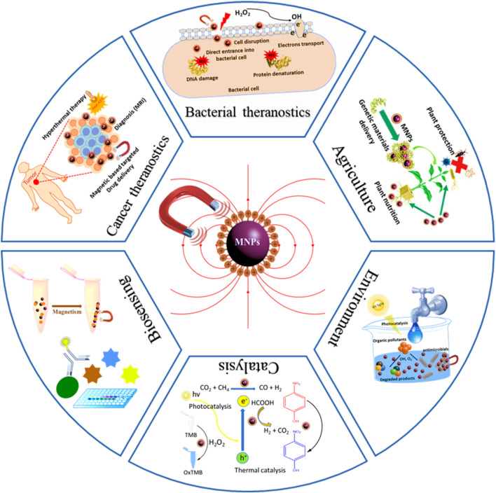
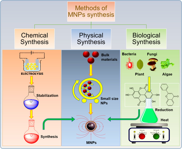
Diverse applications of nanoparticles (NPs) have revolutionized various sectors in society. In the recent decade, particularly magnetic nanoparticles (MNPs) have gained enormous interest owing to their applications in specialized areas such as medicine, cancer theranostics, biosensing, catalysis, agriculture, and the environment. Controlled surface engineering for the design of multi-functional MNPs is vital for achieving desired application. The MNPs have demonstrated great efficacy as thermoelectric materials, imaging agents, drug delivery vehicles, and biosensors. In the present review, first we have briefly discussed main synthetic methods of MNPs, followed by their characterizations and composition. Then we have discussed the potential applications of MNPs in different with representative examples. At the end, we gave an overview on the current challenges and future prospects of MNPs. This comprehensive review not only provides the mechanistic insight into the synthesis, functionalization, and application of MNPs but also outlines the limits and potential prospects.
Overview
The claims and statistics of this paper haven’t been checked yet.
Run the check — free, no account needed. It reads the open-access text and checks the sources it cites.
Integrity signals
Registry
Cites
Statistics
Figures
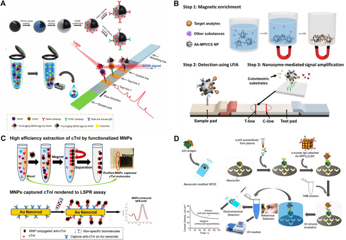
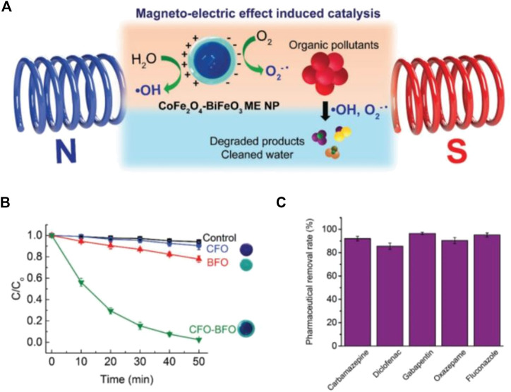
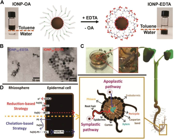
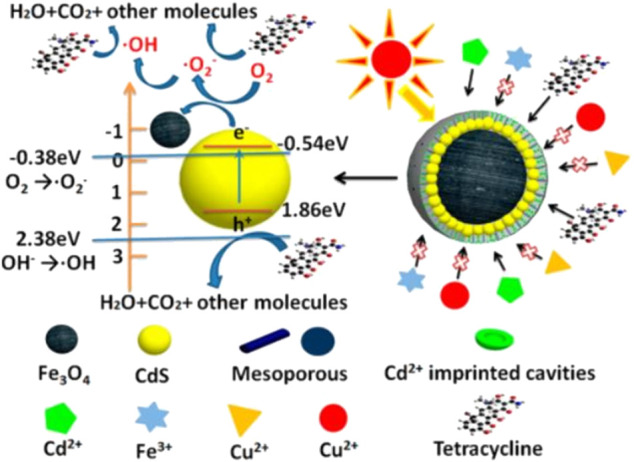
Verification
Claims the paper makes
The claims and statistics of this paper haven’t been checked yet.
Run the check — free, no account needed. It reads the open-access text and checks the sources it cites.
Claims other papers make about this one
Each is checked against this paper’s own text: does it say what the citing paper says it does?
In the citing paper
“Ensuring batch-to-batch uniformity is critical for reproducibility and regulatory approval [94].”
From Advances in green-synthesized magnetic nanoparticles for targeted cancer therapy: mechanisms, applications, and future perspectives (2026) · cites this paper as [94]
In the citing paper
“Minor variations in temperature, pH, or precursor concentration can lead to significant inconsistencies.”
From Advances in green-synthesized magnetic nanoparticles for targeted cancer therapy: mechanisms, applications, and future perspectives (2026) · cites this paper as [94]
In the citing paper
“The physicochemical properties of MNPs, including size, shape, surface charge, and magnetic behavior, are highly sensitive to synthesis conditions.”
From Advances in green-synthesized magnetic nanoparticles for targeted cancer therapy: mechanisms, applications, and future perspectives (2026) · cites this paper as [94]
In the citing paper
“One major challenge is the reproducibility of synthesis.”
From Advances in green-synthesized magnetic nanoparticles for targeted cancer therapy: mechanisms, applications, and future perspectives (2026) · cites this paper as [94]
Self-check
Percentages, sums and reported differences re-computed from the paper’s own numbers.
The self-check needs the paper’s full text.
Citations
Cites
Antimicrobial peptides of multicellular organisms
Zasloff, M. (2002) · Nature · doi:10.1038/415389a · Cited by 8,457
Targeting cancer cells by ROS-mediated mechanisms: a radical therapeutic approach?
Trachootham, D., Alexandre, J., & Huang, P. (2009) · Nature Reviews Drug Discovery · doi:10.1038/nrd2803 · Cited by 5,452
Magnetic iron oxide nanoparticles: synthesis and surface functionalization strategies
Wu, W., He, Q., & Jiang, C. (2008) · Nanoscale Research Letters · doi:10.1007/s11671-008-9174-9 · Cited by 2,300
Water Analysis: Emerging Contaminants and Current Issues
Richardson, S. D., & Ternes, T. A. (2014) · Analytical Chemistry · doi:10.1021/ac500508t · Cited by 2,144
Treatment strategies for cryptococcal infection: challenges, advances and future outlook
Iyer, K. R., Revie, N. M., Fu, C., Robbins, N., & Cowen, L. E. (2021) · Nature Reviews Microbiology · doi:10.1038/s41579-021-00511-0 · Cited by 363
Environmental transformations and ecological effects of iron-based nanoparticles
Lei, C., Sun, Y., Tsang, D. C. W., & Lin, D. (2018) · Environmental Pollution · doi:10.1016/j.envpol.2017.09.052 · Cited by 347
Systematic review of the preparation techniques of iron oxide magnetic nanoparticles
F. Hasany, S., Ahmed, I., J, R., & Rehman, A. (2013) · Nanoscience and Nanotechnology · doi:10.5923/j.nn.20120206.01 · Cited by 318
Research works printed but not linked: 240
- Adeleye, 2016, Engineered Nanomaterials for Water Treatment and Remediation: Costs, Benefits, and Applicability, Chem. Eng. J., 286, 640, 10.1016/j.cej.2015.10.105 · DOI
- Ali, 2020, Safety Assessment of Nanomaterials for Antimicrobial Applications, Chem. Res. Toxicol., 33, 1082, 10.1021/acs.chemrestox.9b00519 · DOI
- Alvarez, 2018, Emerging Opportunities for Nanotechnology to Enhance Water Security, Nat. Nanotech., 13, 634, 10.1038/s41565-018-0203-2 · DOI
- Amendola, 2009, Laser Ablation Synthesis in Solution and Size Manipulation of noble Metal Nanoparticles, Phys. Chem. Chem. Phys., 11, 3805, 10.1039/b900654k · DOI
- Andersson, 2013, Modeling and Characterization of Magnetic Nanoparticles Intended for Cancer Treatment
- Ansari, 2019, Magnetic Iron Oxide Nanoparticles: Synthesis, Characterization and Functionalization for Biomedical Applications in the central Nervous System, Materials, 12, 465, 10.3390/ma12030465 · DOI
- Arakha, 2015, Antimicrobial Activity of Iron Oxide Nanoparticle upon Modulation of Nanoparticle-Bacteria Interface, Scientific Rep., 5, 14813, 10.1038/srep14813 · DOI
- Arciola, 2018, Implant Infections: Adhesion, Biofilm Formation and Immune Evasion, Nat. Rev. Microbiol., 16, 397, 10.1038/s41579-018-0019-y · DOI
- Arias, 2009, Antibiotic-Resistant Bugs in the 21st Century - A Clinical Super-Challenge, N. Engl. J. Med., 360, 439, 10.1056/nejmp0804651 · DOI
- Assa, 2016, A Biotechnological Perspective on the Application of Iron Oxide Nanoparticles, Nano. Res., 9, 2203, 10.1007/s12274-016-1131-9 · DOI
- Babes, 1999, Synthesis of Iron Oxide Nanoparticles Used as MRI Contrast Agents: a Parametric Study, J. Colloid Interf. Sci., 212, 474, 10.1006/jcis.1998.6053 · DOI
- Bai, 2005, High-magnetic-moment Core-shell-type FeCo-Au∕Ag Nanoparticles, Appl. Phys. Lett., 87, 152502, 10.1063/1.2089171 · DOI
- Bansal, 2017, Relaxin-coated Superparamagnetic Iron-Oxide Nanoparticles as a Novel Theranostic Approach for the Diagnosis and Treatment of Liver Fibrosis, J. Hepatol., 66, S43, 10.1016/s0168-8278(17)30348-3 · DOI
- Benjamin, 1970, Dispersion Strengthened Superalloys by Mechanical Alloying, Metallurgical Trans., 1, 2943, 10.1007/BF03037835 · DOI
- Bhalerao, 2019, Magnetic Nanostructures: Environmental and Agricultural Applications, 213, 10.1007/978-3-030-16439-3_11 · DOI
- Biehl, 2018, Synthesis, Characterization, and Applications of Magnetic Nanoparticles Featuring Polyzwitterionic Coatings, Polymers, 10, 91, 10.3390/polym10010091 · DOI
- Bohara, 2015, Innovative Developments in Bacterial Detection with Magnetic Nanoparticles, Appl. Biochem. Biotechnol., 176, 1044, 10.1007/s12010-015-1628-9 · DOI
- Borysiuk, 2008, Structure and Magnetic Properties of Carbon Encapsulated Fe Nanoparticles Obtained by Arc Plasma and Combustion Synthesis, Carbon, 46, 1693, 10.1016/j.carbon.2008.07.011 · DOI
- Boudarel, 2018, Towards Standardized Mechanical Characterization of Microbial Biofilms: Analysis and Critical Review, NPJ biofilms and microbiomes, 4, 1, 10.1038/s41522-018-0062-5 · DOI
- Cardoso, 2018, Advances in Magnetic Nanoparticles for Biomedical Applications, Adv. Healthc. Mater., 7, 1700845, 10.1002/adhm.201700845 · DOI
- Chekli, 2016, Analytical Characterisation of Nanoscale Zero-Valent Iron: A Methodological Review, Analytica Chim. Acta, 903, 13, 10.1016/j.aca.2015.10.040 · DOI
- Chen, 2016, Nanochemistry and Nanomedicine for Nanoparticle-Based Diagnostics and Therapy, Chem. Rev., 116, 2826, 10.1021/acs.chemrev.5b00148 · DOI
- Chen, 2017, Rethinking Cancer Nanotheranostics, Nat. Rev. Mater., 2, 1, 10.1038/natrevmats.2017.24 · DOI
- Chen, 1996, Size-dependent Magnetic Properties ofMnFe2O4fine Particles Synthesized by Coprecipitation, Phys. Rev. B, 54, 9288, 10.1103/physrevb.54.9288 · DOI
- Chen, 1999, Synthesis of Superparamagnetic MgFe2O4 Nanoparticles by Coprecipitation, J. Magnetism Magn. Mater., 194, 1, 10.1016/s0304-8853(98)00585-x · DOI
- Cheng, 2015, Curcumin-conjugated Magnetic Nanoparticles for Detecting Amyloid Plaques in Alzheimer's Disease Mice Using Magnetic Resonance Imaging (MRI), Biomaterials, 44, 155, 10.1016/j.biomaterials.2014.12.005 · DOI
- Cuenya, 2010, Synthesis and Catalytic Properties of Metal Nanoparticles: Size, Shape, Support, Composition, and Oxidation State Effects, Thin Solid Films, 518, 3127, 10.1016/j.tsf.2010.01.018 · DOI
- De La Presa, 2007, Synthesis and Characterization of FePt/Au Core-Shell Nanoparticles, J. Magnetism Magn. Mater., 316, e753, 10.1016/j.jmmm.2007.03.084 · DOI
- DeCastro, 2002, Nanoparticles from Mechanical Attrition, Synthesis, Functionalization, and Surface Treatment of Nanoparticles
- Del Sol-Fernández, 2019, Flower-like Mn-Doped Magnetic Nanoparticles Functionalized with αvβ3-Integrin-Ligand to Efficiently Induce Intracellular Heat after Alternating Magnetic Field Exposition, Triggering Glioma Cell Death, ACS Appl. Mater. Inter., 11, 26648, 10.1021/acsami.9b08318 · DOI
- Deng, 2017, A Magnetic Nanoparticle-Supported N-Heterocyclic Carbene-Palladacycle: an Efficient and Recyclable Solid Molecular Catalyst for Suzuki-Miyaura Cross-Coupling of 9-chloroacridine, Chem. Commun., 53, 13063, 10.1039/c7cc06958h · DOI
- Dong, 2021, Magnetic Microswarm Composed of Porous Nanocatalysts for Targeted Elimination of Biofilm Occlusion, ACS nano, 15, 5056, 10.1021/acsnano.0c10010 · DOI
- Duan, 2018, Recent Progress in Magnetic Nanoparticles: Synthesis, Properties, and Applications, Nanotechnology, 29, 452001, 10.1088/1361-6528/aadcec · DOI
- Duguet, 2006, Towards a Versatile Platform Based on Magnetic Nanoparticles for In Vivo Applications, Bull. Mater. Sci., 29, 581, 10.1007/s12034-006-0007-0 · DOI
- Effenberger, 2017, Economically Attractive Route for the Preparation of High Quality Magnetic Nanoparticles by the thermal Decomposition of Iron(III) Acetylacetonate, Nanotechnology, 28, 115603, 10.1088/1361-6528/aa5ab0 · DOI
- El-Eskandarany, 2001, Mechanical Alloying: For Fabrication of Advanced Engineering Materials
- El-Temsah, 2016, DDT Degradation Efficiency and Ecotoxicological Effects of Two Types of Nano-Sized Zero-Valent Iron (nZVI) in Water and Soil, Chemosphere, 144, 2221, 10.1016/j.chemosphere.2015.10.122 · DOI
- Espinosa, 2018, Magnetic (Hyper)Thermia or Photothermia? Progressive Comparison of Iron Oxide and Gold Nanoparticles Heating in Water, in Cells, and In Vivo, Adv. Funct. Mater., 28, 1803660, 10.1002/adfm.201803660 · DOI
- Etayash, 2014, Impedimetric Detection of Pathogenic Gram-Positive Bacteria Using an Antimicrobial Peptide from Class IIa Bacteriocins, Anal. Chem., 86, 1693, 10.1021/ac4034938 · DOI
- Faraji, 2010, Magnetic Nanoparticles: Synthesis, Stabilization, Functionalization, Characterization, and Applications, Jics, 7, 1, 10.1007/bf03245856 · DOI
- Farjadian, 2017, Thin Chitosan Films Containing Super-paramagnetic Nanoparticles with Contrasting Capability in Magnetic Resonance Imaging, J. Mater. Sci. Mater. Med., 28, 47, 10.1007/s10856-017-5854-2 · DOI
- Farzin, 2020, Magnetic Nanoparticles in Cancer Therapy and Diagnosis, Adv. Healthc. Mater., 9, 1901058, 10.1002/adhm.201901058 · DOI
- Fatima, 2017, Magnetic Nanoparticles for Bioseparation, Korean J. Chem. Eng., 34, 589, 10.1007/s11814-016-0349-2 · DOI
- Fecht, 1990, Nanocrystalline Metals Prepared by High-Energy ball Milling, Mta, 21, 2333, 10.1007/bf02646980 · DOI
- Fenton, 1894, LXXIII.-Oxidation of Tartaric Acid in Presence of Iron, J. Chem. Soc. Trans., 65, 899, 10.1039/ct8946500899 · DOI · Paper page
- Frey, 2009, Magnetic Nanoparticles: Synthesis, Functionalization, and Applications in Bioimaging and Magnetic Energy Storage, Chem. Soc. Rev., 38, 2532, 10.1039/b815548h · DOI
- Fu, 2009, Protein-functionalized Synthetic Antiferromagnetic Nanoparticles for Biomolecule Detection and Magnetic Manipulation, Angew. Chem. Int. Edition, 48, 1620, 10.1002/anie.200803994 · DOI
- Gabbasov, 2015, Mössbauer, Magnetization and X-ray Diffraction Characterization Methods for Iron Oxide Nanoparticles, J. Magnetism Magn. Mater., 380, 111, 10.1016/j.jmmm.2014.11.032 · DOI
- Gal-Mor, 2014, Same Species, Different Diseases: How and Why Typhoidal and Non-typhoidal Salmonella enterica Serovars Differ, Front. Microbiol., 5, 391, 10.3389/fmicb.2014.00391 · DOI
- Galloway, 2015, Developing Biotemplated Data Storage: Room Temperature Biomineralization of L10CoPt Magnetic Nanoparticles, Adv. Funct. Mater., 25, 4590, 10.1002/adfm.201501090 · DOI
- Gao, 2018, Functionalized MoS2Nanovehicle with Near-Infrared Laser-Mediated Nitric Oxide Release and Photothermal Activities for Advanced Bacteria-Infected Wound Therapy, Small, 14, 1802290, 10.1002/smll.201802290 · DOI
- Goel, 2017, Positron Emission Tomography and Nanotechnology: A Dynamic Duo for Cancer Theranostics, Adv. Drug Deliv. Rev., 113, 157, 10.1016/j.addr.2016.08.001 · DOI
- Gong, 2020, Tumor Microenvironment-Responsive Intelligent Nanoplatforms for Cancer Theranostics, Nano Today, 32, 100851, 10.1016/j.nantod.2020.100851 · DOI
- González-Melendi, 2008, Nanoparticles as Smart Treatment-Delivery Systems in Plants: Assessment of Different Techniques of Microscopy for Their Visualization in Plant Tissues, Ann. Bot., 101, 187, 10.1093/aob/mcm283 · DOI
- Govan, 2020, Recent Advances in Magnetic Nanoparticles and Nanocomposites for the Remediation of Water Resources, Magnetochemistry, 6, 49, 10.3390/magnetochemistry6040049 · DOI
- Gu, 2006, Biofunctional Magnetic Nanoparticles for Protein Separation and Pathogen Detection, Chem. Commun., 37, 941, 10.1039/b514130c · DOI
- Gul, 2019, A Comprehensive Review of Magnetic Nanomaterials Modern Day Theranostics, Front. Mater., 6, 179, 10.3389/fmats.2019.00179 · DOI
- Guo, 2019, Magnetic-responsive and Targeted Cancer Nanotheranostics by PA/MR Bimodal Imaging-Guided Photothermally Triggered Immunotherapy, Biomaterials, 219, 119370, 10.1016/j.biomaterials.2019.119370 · DOI
- Häffner, 2017, Membrane Interactions and Antimicrobial Effects of Inorganic Nanoparticles, Adv. Colloid. Interf. Sci., 248, 105, 10.1016/j.cis.2017.07.029 · DOI
- Han, 2020, Metformin-induced Stromal Depletion to Enhance the Penetration of Gemcitabine-Loaded Magnetic Nanoparticles for Pancreatic Cancer Targeted Therapy, J. Am. Chem. Soc., 142, 4944, 10.1021/jacs.0c00650 · DOI
- Hao, 2010, Synthesis, Functionalization, and Biomedical Applications of Multifunctional Magnetic Nanoparticles, Adv. Mater., 22, 2729, 10.1002/adma.201000260 · DOI
- Harms, 2016, Mechanisms of Bacterial Persistence during Stress and Antibiotic Exposure, Science, 354, aaf4268, 10.1126/science.aaf4268 · DOI
- Haun, 2010, Magnetic Nanoparticle Biosensors, WIREs Nanomed. Nanobiotechnol., 2, 291, 10.1002/wnan.84 · DOI
- Hodges, 2018, Challenges and Prospects of Advanced Oxidation Water Treatment Processes Using Catalytic Nanomaterials, Nat. Nanotech., 13, 642, 10.1038/s41565-018-0216-x · DOI
- Hou, 2004, Solvothermal Reduction Synthesis and Magnetic Properties of Polymer Protected Iron and Nickel Nanocrystals, J. alloys Compd., 365, 112, 10.1016/s0925-8388(03)00651-0 · DOI
- Hou, 2003, Monodisperse Nickel Nanoparticles Prepared from a Monosurfactant System and Their Magnetic propertiesElectronic Supplementary Information (ESI) Available: XPS of Ni Nanoparticles; Plot of Magnetization vs. Applied Field. See http://www.rsc.Org/suppdata/jm/b3/b303226d/, J. Mater. Chem., 13, 1510, 10.1039/b303226d · DOI
- Hou, 2004, Preparation and Characterization of Monodisperse FePd Nanoparticles, Chem. Mater., 16, 5149, 10.1021/cm048902c · DOI
- Hou, 2005, High-yield Preparation of Uniform Cobalt Hydroxide and Oxide Nanoplatelets and Their Characterization, J. Phys. Chem. B, 109, 19094, 10.1021/jp0521149 · DOI
- Hou, SmCo5∕Fe Nanocomposites Synthesized from Reductive Annealing of Oxide Nanoparticles, Appl. Phys. Lett., 91, 153117, 10.1063/1.2799170 · DOI
- Hou, Controlled Synthesis and Chemical Conversions of FeO Nanoparticles, Angew. Chem., 119, 6445, 10.1002/ange.200701694 · DOI
- Hou, 2003, Solvothermal Reduction Synthesis and Characterization of Superparamagnetic Magnetite nanoparticlesElectronic Supplementary Information (ESI) Available: Size Distributions of Samples Modified with TOPO + PVP, HDA + PVP, and PVP Only. See http://www.rsc.Org/suppdata/jm/b3/b305526d/, J. Mater. Chem., 13, 1983, 10.1039/b305526d · DOI
- Houry, 2012, Bacterial Swimmers that Infiltrate and Take over the Biofilm Matrix, Proc. Natl. Acad. Sci., 109, 13088, 10.1073/pnas.1200791109 · DOI
- Huang, 2019, A Facile Fabrication of Novel Stuff with Antibacterial Property and Osteogenic Promotion Utilizing Red Phosphorus and Near-Infrared Light, Bioactive Mater., 4, 17, 10.1016/j.bioactmat.2018.11.002 · DOI
- Huang, 2014, Antimicrobial Applications of Water-Dispersible Magnetic Nanoparticles in Biomedicine, Cmc, 21, 3312, 10.2174/0929867321666140304101752 · DOI
- Huang, 2016, Membrane-based Lateral Flow Immunochromatographic Strip with Nanoparticles as Reporters for Detection: A Review, Biosens. Bioelectron., 75, 166, 10.1016/j.bios.2015.08.032 · DOI
- Huber, 2005, Synthesis, Properties, and Applications of Iron Nanoparticles, Small, 1, 482, 10.1002/smll.200500006 · DOI
- Ibrahim, 2016, Environmental Application of Nanotechnology: Air, Soil, and Water, Environ. Sci. Pollut. Res., 23, 13754, 10.1007/s11356-016-6457-z · DOI
- Ida, 2005, Analysis of Painted Steel by a Hand-Held X-ray Fluorescence Spectrometer, Spectrochimica Acta B: At. Spectrosc., 60, 249, 10.1016/j.sab.2004.12.010 · DOI
- Indira, 2010, Magnetic Nanoparticles–A Review, Int. J. Pharm. Sci. Nanotechnology, 3, 1035, 10.37285/ijpsn.2010.3.3.1 · DOI
- Issa, 2013, Magnetic Nanoparticles: Surface Effects and Properties Related to Biomedicine Applications, Int. J. Mol. Sci., 14, 21266, 10.3390/ijms141121266 · DOI
- Jana, 2004, Size- and Shape-Controlled Magnetic (Cr, Mn, Fe, Co, Ni) Oxide Nanocrystals via a Simple and General Approach, Chem. Mater., 16, 3931, 10.1021/cm049221k · DOI
- Jendrzej, 2017, How Size Determines the Value of Gold: Economic Aspects of Wet Chemical and Laser-Based Metal Colloid Synthesis, ChemPhysChem, 18, 1012, 10.1002/cphc.201601139 · DOI · Paper page
- Jiang, 2004, Preparation and Properties of Superparamagnetic Nanoparticles with Narrow Size Distribution and Biocompatible, J. Magnetism Magn. Mater., 283, 210, 10.1016/j.jmmm.2004.05.022 · DOI
- Jin, 2014, Efficient Bacterial Capture with Amino Acid Modified Magnetic Nanoparticles, Water Res., 50, 124, 10.1016/j.watres.2013.11.045 · DOI
- Jones, 1999, Persistent Organic Pollutants (POPs): State of the Science, Environ. Pollut., 100, 209, 10.1016/s0269-7491(99)00098-6 · DOI
- Ju, 2019, Exploring the Chelation-Based Plant Strategy for Iron Oxide Nanoparticle Uptake in Garden Cress (Lepidium Sativum) Using Magnetic Particle Spectrometry, Nanoscale, 11, 18582, 10.1039/c9nr05477d · DOI
- Jurgons, 2006, Drug Loaded Magnetic Nanoparticles for Cancer Therapy, J. Phys. Condens. Matter, 18, S2893, 10.1088/0953-8984/18/38/s24 · DOI
- Kang, 2017, Surface Design of Magnetic Nanoparticles for Stimuli-Responsive Cancer Imaging and Therapy, Biomaterials, 136, 98, 10.1016/j.biomaterials.2017.05.013 · DOI
- Karabelli, 2008, Batch Removal of Aqueous Cu2+Ions Using Nanoparticles of Zero-Valent Iron: A Study of the Capacity and Mechanism of Uptake, Ind. Eng. Chem. Res., 47, 4758, 10.1021/ie800081s · DOI
- Kawamura, 2015, Production of Oxidation-Resistant Cu-Based Nanoparticles by Wire Explosion, Scientific Rep., 5, 1, 10.1038/srep18333 · DOI
- Kefeni, 2017, Ferrite Nanoparticles: Synthesis, Characterisation and Applications in Electronic Device, Mater. Sci. Eng. B, 215, 37, 10.1016/j.mseb.2016.11.002 · DOI
- Kilianová, 2013, Remarkable Efficiency of Ultrafine Superparamagnetic Iron(III) Oxide Nanoparticles toward Arsenate Removal from Aqueous Environment, Chemosphere, 93, 2690, 10.1016/j.chemosphere.2013.08.071 · DOI
- Kim, Pt-decorated Magnetic Nanozymes for Facile and Sensitive point-of-care Bioassay, ACS Appl. Mater. Inter., 9, 35133, 10.1021/acsami.7b12326 · DOI
- Kim, Pt-Decorated Magnetic Nanozymes for Facile and Sensitive Point-of-Care Bioassay, ACS Appl. Mater. Inter., 9, 35133, 10.1021/acsami.7b12326 · DOI
- Kim, 2018, Ultrasensitive Detection of Escherichia coli O157:H7 by Immunomagnetic Separation and Selective Filtration with Nitroblue Tetrazolium/5-Bromo-4-Chloro-3-Indolyl Phosphate Signal Amplification, J. Agric. Food Chem., 66, 4941, 10.1021/acs.jafc.8b00973 · DOI
- Kircher, 2012, Molecular Body Imaging: MR Imaging, CT, and US. Part I. Principles, Radiology, 263, 633, 10.1148/radiol.12102394 · DOI
- Komeili, 2012, Molecular Mechanisms of Compartmentalization and Biomineralization in Magnetotactic Bacteria, FEMS Microbiol. Rev., 36, 232, 10.1111/j.1574-6976.2011.00315.x · DOI
- Kong, 2011, Experimental Considerations on the Cytotoxicity of Nanoparticles, Nanomedicine, 6, 929, 10.2217/nnm.11.77 · DOI
- Koo, 2017, Targeting Microbial Biofilms: Current and Prospective Therapeutic Strategies, Nat. Rev. Microbiol., 15, 740, 10.1038/nrmicro.2017.99 · DOI
- Kotov, 2003, Electric Explosion of Wires as a Method for Preparation of Nanopowders, J. Nanoparticle Res., 5, 539, 10.1023/b:nano.0000006069.45073.0b · DOI
- Kruijshaar, 2008, Increasing Antituberculosis Drug Resistance in the United Kingdom: Analysis of National Surveillance Data, Bmj, 336, 1231, 10.1136/bmj.39546.573067.25 · DOI
- Krzyminiewski, 2018, Focusing of Fe3O4 Nanoparticles Using a Rotating Magnetic Field in Various Environments, Phys. Lett. A, 382, 3192, 10.1016/j.physleta.2018.07.051 · DOI
- Kudr, 2017, Magnetic Nanoparticles: From Design and Synthesis to Real World Applications, Nanomaterials, 7, 243, 10.3390/nano7090243 · DOI
- Kumar, 2011, Magnetic Nanomaterials for Hyperthermia-Based Therapy and Controlled Drug Delivery, Adv. Drug Delivery Rev., 63, 789, 10.1016/j.addr.2011.03.008 · DOI
- Kurland, 2007, Magnetic Iron Oxide Nanopowders Produced by CO2 Laser Evaporation, J. Magnetism Magn. Mater., 311, 73, 10.1016/j.jmmm.2006.10.1166 · DOI
- Laconte, 2005, Magnetic Nanoparticle Probes, Mater. Today, 8, 32, 10.1016/s1369-7021(05)00893-x · DOI
- Lee, 2008, Simple Synthesis of Functionalized Superparamagnetic Magnetite/silica Core/shell Nanoparticles and Their Application as Magnetically Separable High-Performance Biocatalysts, Small, 4, 143, 10.1002/smll.200700456 · DOI
- Lenders, 2014, A Bioinspired Coprecipitation Method for the Controlled Synthesis of Magnetite Nanoparticles, Cryst. Growth Des., 14, 5561, 10.1021/cg500816z · DOI
- Lewinski, 2008, Cytotoxicity of Nanoparticles, Small, 4, 26, 10.1002/smll.200700595 · DOI
- Li, 2008, Preparation and Properties of Magnetic Fe3O4-Chitosan Nanoparticles, J. alloys Compd., 466, 451, 10.1016/j.jallcom.2007.11.100 · DOI
- Li, 2013, Polyethyleneimine-mediated Synthesis of Folic Acid-Targeted Iron Oxide Nanoparticles for In Vivo Tumor MR Imaging, Biomaterials, 34, 8382, 10.1016/j.biomaterials.2013.07.070 · DOI
- Li, 2020, Clinical Development and Potential of Photothermal and Photodynamic Therapies for Cancer, Nat. Rev. Clin. Oncol., 17, 657, 10.1038/s41571-020-0410-2 · DOI · Paper page
- Lin, 2017, Magnetic Sensing Platform Technologies for Biomedical Applications, Lab. Chip, 17, 1884, 10.1039/c7lc00026j · DOI
- Liu, 2016, Catalytic Conversion of Biomass into Chemicals and Fuels over Magnetic Catalysts, ACS Catal., 6, 326, 10.1021/acscatal.5b02094 · DOI
- Liu, Exchange-coupled Nanocomposites: Chemical Synthesis, Characterization and Applications, Chem. Soc. Rev., 43, 8098, 10.1039/c4cs00162a · DOI
- Liu, Building Nanocomposite Magnets by Coating a Hard Magnetic Core with a Soft Magnetic Shell, Angew. Chem. Int. Ed., 53, 2176, 10.1002/anie.201309723 · DOI
- Liu, 2020, Comprehensive Understanding of Magnetic Hyperthermia for Improving Antitumor Therapeutic Efficacy, Theranostics, 10, 3793, 10.7150/thno.40805 · DOI
- Liu, 2019, Multifunctional Magnetic Copper Ferrite Nanoparticles as fenton-like Reaction and Near-Infrared Photothermal Agents for Synergetic Antibacterial Therapy, ACS Appl. Mater. Inter., 11, 31649, 10.1021/acsami.9b10096 · DOI
- López Pérez, 1997, Advances in the Preparation of Magnetic Nanoparticles by the Microemulsion Method, J. Phys. Chem. B, 101, 8045, 10.1021/jp972046t · DOI
- Lu, 2007, Magnetic Nanoparticles: Synthesis, Protection, Functionalization, and Application, Angew. Chem. Int. Ed., 46, 1222, 10.1002/anie.200602866 · DOI
- Lu, 2013, Surfactant Effects on the Microstructures of Fe3O4 Nanoparticles Synthesized by Microemulsion Method, Colloids Surf. A: Physicochemical Eng. Aspects, 436, 675, 10.1016/j.colsurfa.2013.08.004 · DOI
- Lu, 2017, Iron Oxide Nanoclusters for T 1 Magnetic Resonance Imaging of Non-Human Primates, Nat. Biomed. Eng., 1, 637, 10.1038/s41551-017-0116-7 · DOI
- Lu, 2019, Magnetic Hierarchical Photocatalytic Nanoreactors: toward Highly Selective Cd2+ Removal with Secondary Pollution Free Tetracycline Degradation, ACS Appl. Nano Mater., 2, 1664, 10.1021/acsanm.9b00113 · DOI
- Luo, 2010, Self-catalyzed, Self-Limiting Growth of Glucose Oxidase-Mimicking Gold Nanoparticles, Acs Nano, 4, 7451, 10.1021/nn102592h · DOI
- Ma, 2020, Spatio‐Temporally Reporting Dose‐Dependent Chemotherapy via Uniting Dual‐Modal MRI/NIR Imaging, Angew. Chem. Int. Ed., 59, 21143, 10.1002/anie.202009380 · DOI
- Mackowiak, 1981, Direct Effects of Hyperthermia on Pathogenic Microorganisms: Teleologic Implications with Regard to Fever, Clin. Infect. Dis., 3, 508, 10.1093/clinids/3.3.508 · DOI
- Majetich, 1999, Magnetization Directions of Individual Nanoparticles, Science, 284, 470, 10.1126/science.284.5413.470 · DOI
- Majid, 2018, Synthesis and Characterisation of Magnetic Nanoparticles in Medicine, 413, 10.1007/978-3-319-67132-1_14 · DOI
- Mao, 2019, Local Photothermal/photodynamic Synergistic Therapy by Disrupting Bacterial Membrane to Accelerate Reactive Oxygen Species Permeation and Protein Leakage, ACS Appl. Mater. Inter., 11, 17902, 10.1021/acsami.9b05787 · DOI
- Martínez-Edo, 2018, Functionalized Ordered Mesoporous Silicas (MCM-41): Synthesis and Applications in Catalysis, Catalysts, 8, 617, 10.3390/catal8120617 · DOI
- Masud, Gold-loaded Nanoporous Ferric Oxide Nanocubes with Peroxidase-Mimicking Activity for Electrocatalytic and Colorimetric Detection of Autoantibody, Anal. Chem., 89, 11005, 10.1021/acs.analchem.7b02880 · DOI
- Masud, Gold-Loaded Nanoporous Ferric Oxide Nanocubes with Peroxidase-Mimicking Activity for Electrocatalytic and Colorimetric Detection of Autoantibody, Anal. Chem., 89, 11005, 10.1021/acs.analchem.7b02880 · DOI
- Mireles, 2016, A Comparative Physicochemical, Morphological and Magnetic Study of Silane-Functionalized Superparamagnetic Iron Oxide Nanoparticles Prepared by Alkaline Coprecipitation, Int. J. Biochem. Cel. Biol., 75, 203, 10.1016/j.biocel.2015.12.002 · DOI
- Mishra, 2017, Integrated Approach of Agri-Nanotechnology: Challenges and Future Trends, Front. Plant Sci., 8, 471, 10.3389/fpls.2017.00471 · DOI
- Mohamed, 2019, Nanoparticles: Magnetism and Applications, 1, 10.1007/978-3-030-16439-3_1 · DOI
- Mohammed, 2017, Magnetic Nanoparticles for Environmental and Biomedical Applications: A review, Particuology, 30, 1, 10.1016/j.partic.2016.06.001 · DOI · Paper page
- Mondal, 2020, Green Synthesis and Environmental Application of Iron-Based Nanomaterials and Nanocomposite: A Review, Chemosphere, 259, 127509, 10.1016/j.chemosphere.2020.127509 · DOI
- Mornet, 2006, Magnetic Nanoparticle Design for Medical Applications, Prog. Solid State. Chem., 34, 237, 10.1016/j.progsolidstchem.2005.11.010 · DOI
- Moros, 2019, Triggering Antitumoural Drug Release and Gene Expression by Magnetic Hyperthermia, Adv. Drug Deliv. Rev., 138, 326, 10.1016/j.addr.2018.10.004 · DOI
- Mosayebi, 2017, Synthesis, Functionalization, and Design of Magnetic Nanoparticles for Theranostic Applications, Adv. Healthc. Mater., 6, 1700306, 10.1002/adhm.201700306 · DOI
- Mushtaq, 2019, Magnetoelectrically Driven Catalytic Degradation of Organics, Adv. Mater., 31, 1901378, 10.1002/adma.201901378 · DOI
- Nam, 2019, Cancer Nanomedicine for Combination Cancer Immunotherapy, Nat. Rev. Mater., 4, 398, 10.1038/s41578-019-0108-1 · DOI
- Namboodiri, 2001, Microwave-accelerated Suzuki Cross-Coupling Reaction in Polyethylene Glycol (PEG), Green. Chem., 3, 146, 10.1039/b102337n · DOI
- Narayanan, 2010, Biological Synthesis of Metal Nanoparticles by Microbes, Adv. Colloid Interf. Sci., 156, 1, 10.1016/j.cis.2010.02.001 · DOI
- Noh, 2017, Recent Advances of Magneto-thermal Capabilities of Nanoparticles: From Design Principles to Biomedical Applications, Nano Today, 13, 61, 10.1016/j.nantod.2017.02.006 · DOI
- Nosrati, 2017, New Advances Strategies for Surface Functionalization of Iron Oxide Magnetic Nano Particles (IONPs), Res. Chem. Intermed., 43, 7423, 10.1007/s11164-017-3084-3 · DOI
- Okoli, 2011, Application of Magnetic Iron Oxide Nanoparticles Prepared from Microemulsions for Protein Purification, J. Chem. Technol. Biotechnol., 86, 1386, 10.1002/jctb.2704 · DOI
- Overchuk, 2018, Overcoming Obstacles in the Tumor Microenvironment: Recent Advancements in Nanoparticle Delivery for Cancer Theranostics, Biomaterials, 156, 217, 10.1016/j.biomaterials.2017.10.024 · DOI
- Pathak, 2019, Improved thermal Performance of Annular Fin-Shell Tube Storage System Using Magnetic Fluid, Appl. Energ., 239, 1524, 10.1016/j.apenergy.2019.01.098 · DOI
- Pathak, 2017, Magnetic Fluid Based High Precision Temperature Sensor, IEEE Sensors J., 17, 2670, 10.1109/jsen.2017.2675440 · DOI
- Pathak, 2021, Spin Dynamics Investigations of Multifunctional Ambient Scalable Fe 3 O 4 Surface Decorated ZnO Magnetic Nanocomposite Using FMR, Scientific Rep., 11, 1, 10.1038/s41598-021-83394-8 · DOI
- Patsula, 2016, Superparamagnetic Fe3O4 Nanoparticles: Synthesis by Thermal Decomposition of Iron(III) Glucuronate and Application in Magnetic Resonance Imaging, ACS Appl. Mater. Inter., 8, 7238, 10.1021/acsami.5b12720 · DOI
- Pazos-Perez, 2016, Ultrasensitive Multiplex Optical Quantification of Bacteria in Large Samples of Biofluids, Scientific Rep., 6, 1, 10.1038/srep29014 · DOI
- Prucek, 2011, The Targeted Antibacterial and Antifungal Properties of Magnetic Nanocomposite of Iron Oxide and Silver Nanoparticles, Biomaterials, 32, 4704, 10.1016/j.biomaterials.2011.03.039 · DOI
- Purbia, 2015, Yolk/shell Nanoparticles: Classifications, Synthesis, Properties, and Applications, Nanoscale, 7, 19789, 10.1039/c5nr04729c · DOI
- Quan, Artificial Channels in an Infectious Biofilm Created by Magnetic Nanoparticles Enhanced Bacterial Killing by Antibiotics, Small, 15, e1902313, 10.1002/smll.201902313 · DOI
- Quan, Artificial Channels in an Infectious Biofilm Created by Magnetic Nanoparticles Enhanced Bacterial Killing by Antibiotics, Small, 15, 1902313, 10.1002/smll.201902313 · DOI
- Ramsey, 2006, ROS as a Tumour Suppressor?, Nat. Cel. Biol., 8, 1213, 10.1038/ncb1106-1213 · DOI · Paper page
- Reddy, 2012, Magnetic Nanoparticles: Design and Characterization, Toxicity and Biocompatibility, Pharmaceutical and Biomedical Applications, Chem. Rev., 112, 5818, 10.1021/cr300068p · DOI · Paper page
- Ren, 2019, Preparation of Au Nanoparticles on a Magnetically Responsive Support via Pyrolysis of a Prussian Blue Composite, J. Colloid Interf. Sci., 540, 563, 10.1016/j.jcis.2019.01.027 · DOI
- Rocha-Santos, 2014, Sensors and Biosensors Based on Magnetic Nanoparticles, Trac Trends Anal. Chem., 62, 28, 10.1016/j.trac.2014.06.016 · DOI
- Rodriguez-Narvaez, 2017, Treatment Technologies for Emerging Contaminants in Water: a Review, Chem. Eng. J., 323, 361, 10.1016/j.cej.2017.04.106 · DOI
- Rout, 2015, Role of Iron in Plant Growth and Metabolism, Ras, 3, 1, 10.7831/ras.3.1 · DOI
- Rui, 2016, Iron Oxide Nanoparticles as a Potential Iron Fertilizer for Peanut (Arachis hypogaea), Front. Plant Sci., 7, 815, 10.3389/fpls.2016.00815 · DOI
- Sánchez-Alcalá, 2014, Evaluation of Preflooding Effects on Iron Extractability and Phytoavailability in Highly Calcareous Soil in Containers, Z. Pflanzenernähr. Bodenk., 177, 150, 10.1002/jpln.201200302 · DOI
- Sandeep Kumar, 2013, Magnetic Nanoparticles-Based Biomedical and Bioanalytical Applications, J. Nanomed. Nanotechol., 4, e130, 10.4172/2157-7439.1000e130 · DOI
- Sarwat, 2017, Contamination in Wet-ball Milling, Powder Metall., 60, 267, 10.1080/00325899.2017.1280647 · DOI
- Schenkman, 2011, Second Thoughts about CT Imaging, Am. Assoc. Adv. Sci., 331, 1002, 10.1126/science.331.6020.1002 · DOI
- Schmidt, 2020, Engineering Microrobots for Targeted Cancer Therapies from a Medical Perspective, Nat. Commun., 11, 1, 10.1038/s41467-020-19322-7 · DOI
- Seo, 2006, FeCo/graphitic-shell Nanocrystals as Advanced Magnetic-Resonance-Imaging and Near-Infrared Agents, Nat. Mater, 5, 971, 10.1038/nmat1775 · DOI
- Sharma, 2019, Nanoparticles-based Magnetic and Photo Induced Hyperthermia for Cancer Treatment, Nano Today, 29, 100795, 10.1016/j.nantod.2019.100795 · DOI
- Shen, 2014, Facile Co-precipitation Synthesis of Shape-Controlled Magnetite Nanoparticles, Ceramics Int., 40, 1519, 10.1016/j.ceramint.2013.07.037 · DOI
- Shi, 2019, In Situ structure and Assembly of the Multidrug Efflux Pump AcrAB-TolC, Nat. Commun., 10, 1, 10.1038/s41467-019-10512-6 · DOI
- Shin, 2004, On the Iron Oxide Neutral Cluster Distribution in the Gas Phase. II. Detection through 118 Nm Single Photon Ionization, J. Chem. Phys., 120, 4157, 10.1063/1.1643733 · DOI
- Simoes, 2017, Novel Antibacterial Agents: an Emergent Need to Win the Battle against Infections, Mini Rev. Med. Chem., 17, 1364, 10.2174/1389557516666160907151454 · DOI
- Singamaneni, 2011, Magnetic Nanoparticles: Recent Advances in Synthesis, Self-Assembly and Applications, J. Mater. Chem., 21, 16819, 10.1039/c1jm11845e · DOI
- Singh, 2017, Shape and Size-dependent Magnetic Properties of Fe 3 O 4 Nanoparticles Synthesized Using Piperidine, Nanoscale Res. Lett., 12, 1, 10.1186/s11671-017-2039-3 · DOI
- Skomski, 1994, Exchange Coupling and Energy Product in Random Two-phase Aligned Magnets, IEEE Trans. Magn., 30, 607, 10.1109/20.312350 · DOI
- Smith, 1980, Colloidal Iron Dispersions Prepared via the Polymer-Catalyzed Decomposition of Iron Pentacarbonyl, J. Phys. Chem., 84, 1621, 10.1021/j100449a037 · DOI
- Song, 2013, Magnetic Iron Oxide Nanoparticles Prepared by Electrical Wire Explosion for Arsenic Removal, Powder Technol., 246, 572, 10.1016/j.powtec.2013.06.023 · DOI
- Suaifan, 2013, Ultra-rapid Colorimetric Assay for Protease Detection Using Magnetic Nanoparticle-Based Biosensors, Analyst, 138, 3735, 10.1039/c3an36881e · DOI
- Sudarsanam, 2018, Functionalised Heterogeneous Catalysts for Sustainable Biomass Valorisation, Chem. Soc. Rev., 47, 8349, 10.1039/c8cs00410b · DOI
- Sun, 1999, Synthesis of Monodisperse Cobalt Nanocrystals and Their Assembly into Magnetic Superlattices (Invited), J. Appl. Phys., 85, 4325, 10.1063/1.370357 · DOI
- Sun, 2000, Monodisperse FePt Nanoparticles and Ferromagnetic FePt Nanocrystal Superlattices, Science, 287, 1989, 10.1126/science.287.5460.1989 · DOI · Paper page
- Sun, 2002, Size-controlled Synthesis of Magnetite Nanoparticles, J. Am. Chem. Soc., 124, 8204, 10.1021/ja026501x · DOI
- Sun, 2007, A Method for the Preparation of Stable Dispersion of Zero-Valent Iron Nanoparticles, Colloids Surf. A: Physicochemical Eng. Aspects, 308, 60, 10.1016/j.colsurfa.2007.05.029 · DOI
- Suslick, 1996, Sonochemical Synthesis of Iron Colloids, J. Am. Chem. Soc., 118, 11960, 10.1021/ja961807n · DOI
- Tang, 2013, Magnetic Nanoparticle Mediated Enhancement of Localized Surface Plasmon Resonance for Ultrasensitive Bioanalytical Assay in Human Blood Plasma, Anal. Chem., 85, 1431, 10.1021/ac302422k · DOI
- Thorat, 2017, Effective Cancer Theranostics with Polymer Encapsulated Superparamagnetic Nanoparticles: Combined Effects of Magnetic Hyperthermia and Controlled Drug Release, ACS Biomater. Sci. Eng., 3, 1332, 10.1021/acsbiomaterials.6b00420 · DOI
- Tran, 2010, Gadonanotubes as Magnetic Nanolabels for Stem Cell Detection, Biomaterials, 31, 9482, 10.1016/j.biomaterials.2010.08.034 · DOI
- Tsuchido, 1985, Destruction of the Outer Membrane Permeability Barrier of Escherichia coli by Heat Treatment, Appl. Environ. Microbiol., 50, 298, 10.1128/aem.50.2.298-303.1985 · DOI
- Unsoy, 2012, Synthesis Optimization and Characterization of Chitosan-Coated Iron Oxide Nanoparticles Produced for Biomedical Applications, J. Nanoparticle Res., 14, 1, 10.1007/s11051-012-0964-8 · DOI
- Vandenberg, 2020, Considerations for Diagnostic COVID-19 Tests, Nat. Rev. Microbiol., 19, 1, 10.1038/s41579-020-00461-z · DOI
- Váradi, 2017, Methods for the Detection and Identification of Pathogenic Bacteria: Past, Present, and Future, Chem. Soc. Rev., 46, 4818, 10.1039/c6cs00693k · DOI
- Verma, 2021, ZnO Nanomaterials: Green Synthesis, Toxicity Evaluation and New Insights in Biomedical Applications, J. Alloys Comp., 876, 160175, 10.1016/j.jallcom.2021.160175 · DOI
- Vidal-Vidal, 2006, Synthesis of Monodisperse Maghemite Nanoparticles by the Microemulsion Method, Colloids Surf. A: Physicochemical Eng. Aspects, 288, 44, 10.1016/j.colsurfa.2006.04.027 · DOI
- Wang, 2007, Solid-phase Synthesis of Carbon-Encapsulated Magnetic Nanoparticles, J. Phys. Chem. C, 111, 6303, 10.1021/jp0707283 · DOI
- Wang, 2019, Magnetic SERS Strip for Sensitive and Simultaneous Detection of Respiratory Viruses, ACS Appl. Mater. Inter., 11, 19495, 10.1021/acsami.9b03920 · DOI
- Wang, Exerting Enhanced Permeability and Retention Effect Driven Delivery by Ultrafine Iron Oxide Nanoparticles with T1-T2 Switchable Magnetic Resonance Imaging Contrast, Acs Nano, 11, 4582, 10.1021/acsnano.7b00038 · DOI
- Wang, 2021, Ultrasound Doppler-Guided Real-Time Navigation of a Magnetic Microswarm for Active Endovascular Delivery, Sci. Adv., 7, eabe5914, 10.1126/sciadv.abe5914 · DOI
200 of 240 shown
Cited by
Sorted by citations
Hussein, E. B., Rasheed, F. A., Mohammed, A. S., & Kayani, K. F. (2025) · RSC Advances · doi:10.1039/d5ra06914a · Cited by 86
Yanar, F., Carugo, D., & Zhang, X. (2023) · Molecules · doi:10.3390/molecules28155694 · Cited by 80
El-khawaga, A. M., Youssef, O. I., El-dydamoni, O. A., Ragab, W. M., & Mohammed, R. A. E. (2026) · European Journal of Medical Research · doi:10.1186/s40001-025-03804-9 · Cited by 14
Pasupuleti, R. R., & Huang, Y. (2023) · Journal of the Chinese Chemical Society · doi:10.1002/jccs.202300029 · Cited by 11
Hashim, N. M., Yahaya, N., & Zain, N. N. M. (2024) · Green Analytical Chemistry · doi:10.1016/j.greeac.2024.100132 · Cited by 10
Metadata
- Authors
- Ali, A., Shah, T., Ullah, R., Zhou, P., Guo, M., Ovais, M., Tan, Z., & Rui, Y.
- Venue
- Frontiers in Chemistry
- Publisher
- Frontiers Media SA
- Published
- 2021-07-13
- Volume
- 9
- ISSN
- 2296-2646
- PMC
- PMC8314212
- Cited by
- 650
- References
- 7
About these checks
Registry, retraction, Cites and statistics results are computed, not judged, with one exception: a printed reference that no index lists is read once by a model before it counts as a citation error. Claims are extracted by a model and kept only when their quote appears verbatim in the text; each cited source is then checked, with a model’s help, for whether it says what the claim relies on. Those verdicts can be wrong — read the claim’s quote and the cited source before relying on one.
Statistics and arithmetic are recomputed, not judged.
Confidence bands are shown only for fields calibrated on our annotated test set. None is calibrated yet, so no band is shown.
What counts as a claim ›How we measure confidence ›How we check this ›
Are you an author, or do you see a problem with this page? Request a review
