

The DOI matches its Crossref record; 7 of the 7 references it cites were found in a registry or an index.
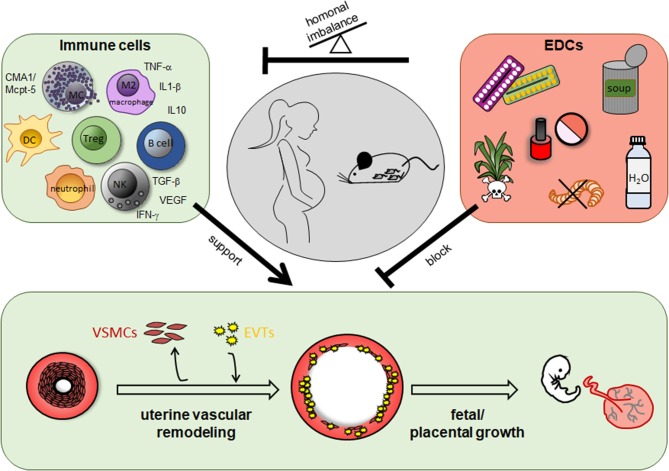
Immune Cells in the Uterine Remodeling: Are They the Target of Endocrine Disrupting Chemicals?
Overview
The claims and statistics of this paper haven’t been checked yet.
Run the check — free, no account needed. It reads the open-access text and checks the sources it cites.
Integrity signals
Registry
Cites
Statistics
Figures
Verification
Claims the paper makes
The claims and statistics of this paper haven’t been checked yet.
Run the check — free, no account needed. It reads the open-access text and checks the sources it cites.
Claims other papers make about this one
Each is checked against this paper’s own text: does it say what the citing paper says it does?
In the citing paper
“In the resting, non‐pregnant uterus, the blood vessels are thick‐walled. Early during pregnancy, these are converted into thin‐walled vessels with large lumens. 121 This allows for effective exchange of oxygen and nutrients to meet the increased demands to the developing fetus.”
From Mast cell ontogeny: From fetal development to life‐long health and disease (2023) · cites this paper as [121]
Self-check
Percentages, sums and reported differences re-computed from the paper’s own numbers.
The self-check needs the paper’s full text.
Citations
Cites
Effect of In Utero and Early-Life Conditions on Adult Health and Disease
Gluckman, P. D., Hanson, M. A., Cooper, C., & Thornburg, K. L. (2008) · New England Journal of Medicine · doi:10.1056/nejmra0708473 · Cited by 3,898
Hormones and endocrine-disrupting chemicals: low-dose effects and nonmonotonic dose responses
Vandenberg, L. N., Colborn, T., Hayes, T. B., Heindel, J. J., Jacobs, D. R., Lee, D.-H., Shioda, T., Soto, A. M., vom Saal, F. S., Welshons, W. V., et al. (2012) · Endocrine Reviews · doi:10.1210/er.2011-1050 · Cited by 3,209
Mast cells are essential intermediaries in regulatory T-cell tolerance
Lu, L.-F., Lind, E. F., Gondek, D. C., Bennett, K. A., Gleeson, M. W., Pino-Lagos, K., Scott, Z. A., Coyle, A. J., Reed, J. L., Van Snick, J., et al. (2006) · Nature · doi:10.1038/nature05010 · Cited by 751
The Consortium on Thyroid and Pregnancy—Study Group on Preterm Birth, Korevaar, T. I. M., Derakhshan, A., Taylor, P. N., Meima, M., Chen, L., Bliddal, S., Carty, D. M., Meems, M., Vaidya, B., et al. (2019) · JAMA · doi:10.1001/jama.2019.10931 · Cited by 378
Jensen, F., Woudwyk, M., Teles, A., Woidacki, K., Taran, F., Costa, S., Malfertheiner, S. F., & Zenclussen, A. C. (2010) · PLoS ONE · doi:10.1371/journal.pone.0014409 · Cited by 145 · cited 1×
“They are subjected to the hormonal influence of progesterone and estrogen as they express hormone receptors (102).”
BackgroundSelf-citationZhang, Y., Wang, H., Pan, X., Teng, W., & Shan, Z. (2017) · PLOS ONE · doi:10.1371/journal.pone.0175708 · Cited by 143
Keogh, R. J., Harris, L. K., Freeman, A., Baker, P. N., Aplin, J. D., Whitley, G. S., & Cartwright, J. E. (2007) · Circulation Research · doi:10.1161/01.res.0000261352.81736.37 · Cited by 114
Research works printed but not linked: 189
- Calkins, 2011, Fetal origins of adult disease, Curr Probl Pediatr Adolesc Health Care, 41, 158, 10.1016/j.cppeds.2011.01.001 · DOI · Paper page
- Cartwright, 2010, Remodelling at the maternal-fetal interface: relevance to human pregnancy disorders, Reproduction, 140, 803, 10.1530/REP-10-0294 · DOI
- Maybin, 2015, Menstrual physiology: implications for endometrial pathology and beyond, Hum Reprod Update, 21, 748, 10.1093/humupd/dmv038 · DOI
- Zenclussen, 2015, Cellular regulation of the uterine microenvironment that enables embryo implantation, Front Immunol., 6, 321, 10.3389/fimmu.2015.00321 · DOI
- Grzechocinska, 2017, The role of metalloproteinases in endometrial remodelling during menstrual cycle, Ginekol Pol., 88, 337, 10.5603/GP.a2017.0063 · DOI
- Nair, 2017, The Mechanism of Menstruation.
- Berbic, 2013, Immunology of normal and abnormal menstruation, 387
- Jones, 2004, Identification of chemokines important for leukocyte recruitment to the human endometrium at the times of embryo implantation and menstruation, J Clin Endocrinol Metab., 89, 6155, 10.1210/jc.2004-0507 · DOI
- Berbic, 2014, Inflammation and endometrial bleeding, Climacteric, 17, 47, 10.3109/13697137.2014.963964 · DOI
- Jabbour, 2006, Endocrine regulation of menstruation, Endocr Rev., 27, 17, 10.1210/er.2004-0021 · DOI · Paper page
- Osol, 2009, Maternal uterine vascular remodeling during pregnancy, Physiology., 24, 58, 10.1152/physiol.00033.2008 · DOI
- Boss, 2018, Placental formation in early pregnancy: how is the centre of the placenta made?, Hum Reprod Update, 24, 750, 10.1093/humupd/dmy030 · DOI
- Burke, 2010, Uterine NK cells, spiral artery modification and the regulation of blood pressure during mouse pregnancy, Am J Reprod Immunol, 63, 472, 10.1111/j.1600-0897.2010.00818.x · DOI
- Whitley, 2010, Cellular and molecular regulation of spiral artery remodelling: lessons from the cardiovascular field, Placenta, 31, 465, 10.1016/j.placenta.2010.03.002 · DOI
- Cecchettini, 2011, Vascular smooth-muscle-cell activation: proteomics point of view, Int Rev Cell Mol Biol., 288, 43, 10.1016/B978-0-12-386041-5.00002-9 · DOI
- Wanjare, 2013, Derivation and maturation of synthetic and contractile vascular smooth muscle cells from human pluripotent stem cells, Cardiovasc Res., 97, 321, 10.1093/cvr/cvs315 · DOI
- Lacolley, 2012, The vascular smooth muscle cell in arterial pathology: a cell that can take on multiple roles, Cardiovasc Res., 95, 194, 10.1093/cvr/cvs135 · DOI
- Rensen, 2007, Regulation and characteristics of vascular smooth muscle cell phenotypic diversity, Neth Heart J., 15, 100, 10.1007/BF03085963 · DOI
- Louis, 2010, Vascular smooth muscle cell motility: from migration to invasion, Exp Clin Cardiol., 15, e75
- Bulmer, 2012, The role of vascular smooth muscle cell apoptosis and migration during uterine spiral artery remodeling in normal human pregnancy, FASEB J., 26, 2975, 10.1096/fj.12-203679 · DOI
- Liu, 2017, dNK derived IFN-gamma mediates VSMC migration and apoptosis via the induction of LncRNA MEG3: a role in utero-vascular transformation, Placenta, 50, 32, 10.1016/j.placenta.2016.12.023 · DOI
- Kaufmann, 2003, Endovascular trophoblast invasion: implications for the pathogenesis of intrauterine growth retardation and preeclampsia, Biol Reprod., 69, 1, 10.1095/biolreprod.102.014977 · DOI
- Whitley, 2009, Trophoblast-mediated spiral artery remodelling: a role for apoptosis, J Anat., 215, 21, 10.1111/j.1469-7580.2008.01039.x · DOI
- Gellersen, 2014, Cyclic decidualization of the human endometrium in reproductive health and failure, Endocr Rev., 35, 851, 10.1210/er.2014-1045 · DOI
- Vinketova, 2016, Human decidual stromal cells as a component of the implantation niche and a modulator of maternal immunity, J Pregnancy., 2016, 8689436, 10.1155/2016/8689436 · DOI
- Pollard, 1998, Complexity in uterine macrophage responses to cytokines in mice, Biol Reprod., 58, 1469, 10.1095/biolreprod58.6.1469 · DOI
- Huang, 2006, Regulation of chemokine production in response to pro-inflammatory cytokines in first trimester decidual cells, J Reprod Immunol., 72, 60, 10.1016/j.jri.2006.03.002 · DOI
- Sentman, 2004, Recruitment of uterine NK cells: induction of CXC chemokine ligands 10 and 11 in human endometrium by estradiol and progesterone, J Immunol., 173, 6760, 10.4049/jimmunol.173.11.6760 · DOI
- Kitaya, 2005, Central role of interleukin-15 in postovulatory recruitment of peripheral blood CD16(-) natural killer cells into human endometrium, J Clin Endocrinol Metab., 90, 2932, 10.1210/jc.2004-2447 · DOI
- Carlino, 2008, Recruitment of circulating NK cells through decidual tissues: a possible mechanism controlling NK cell accumulation in the uterus during early pregnancy, Blood, 111, 3108, 10.1182/blood-2007-08-105965 · DOI
- Magatti, 2019, Perinatal mesenchymal stromal cells and their possible contribution to fetal-maternal tolerance, Cells, 8, 1401, 10.3390/cells8111401 · DOI
- Klemmt, 2006, Stromal cells from endometriotic lesions and endometrium from women with endometriosis have reduced decidualization capacity, Fertil Steril., 85, 564, 10.1016/j.fertnstert.2005.08.046 · DOI
- Giuliani, 2014, Characterization of uterine NK cells in women with infertility or recurrent pregnancy loss and associated endometriosis, Am J Reprod Immunol, 72, 262, 10.1111/aji.12259 · DOI
- Salker, 2010, Natural selection of human embryos: impaired decidualization of endometrium disables embryo-maternal interactions and causes recurrent pregnancy loss, PLoS ONE., 5, e10287, 10.1371/journal.pone.0010287 · DOI
- Kuo, 2019, Trophoblast-endothelium signaling involves angiogenesis and apoptosis in a dynamic bioprinted placenta model, Biotechnol Bioeng., 116, 181, 10.1002/bit.26850 · DOI
- Harris, 2010, Review: trophoblast-vascular cell interactions in early pregnancy: how to remodel a vessel, Placenta, 31, S93, 10.1016/j.placenta.2009.12.012 · DOI
- Harris, 2006, Invasive trophoblasts stimulate vascular smooth muscle cell apoptosis by a fas ligand-dependent mechanism, Am J Pathol., 169, 1863, 10.2353/ajpath.2006.060265 · DOI · Paper page
- Ashton, 2005, Uterine spiral artery remodeling involves endothelial apoptosis induced by extravillous trophoblasts through Fas/FasL interactions, Arterioscler Thromb Vasc Biol., 25, 102, 10.1161/01.ATV.0000148547.70187.89 · DOI
- James, 2010, Pre-eclampsia: fitting together the placental, immune and cardiovascular pieces, J Pathol., 221, 363, 10.1002/path.2719 · DOI
- Khong, 1986, Inadequate maternal vascular response to placentation in pregnancies complicated by pre-eclampsia and by small-for-gestational age infants, Br J Obstet Gynaecol., 93, 1049, 10.1111/j.1471-0528.1986.tb07830.x · DOI · Paper page
- van der Heijden, 2005, Uterine artery remodeling in pseudopregnancy is comparable to that in early pregnancy, Biol Reprod., 73, 1289, 10.1095/biolreprod.105.044438 · DOI
- Stieglitz, 2019, The microstructure in the placenta is influenced by the functional diversity of HLA-G allelic variants, Immunogenetics, 71, 455, 10.1007/s00251-019-01121-0 · DOI
- Fu, 2017, Natural Killer cells promote fetal development through the secretion of growth-promoting factors, Immunity, 47, 1100, 10.1016/j.immuni.2017.11.018 · DOI
- Hauk, 2019, Trophoblast VIP deficiency entails immune homeostasis loss and adverse pregnancy outcome in mice, FASEB J., 33, 1801, 10.1096/fj.201800592RR · DOI
- Gohner, 2017, Immune-modulatory effects of syncytiotrophoblast extracellular vesicles in pregnancy and preeclampsia, Placenta., 60, S41, 10.1016/j.placenta.2017.06.004 · DOI
- Croy, 2006, Uterine natural killer cells: a specialized differentiation regulated by ovarian hormones, Immunol Rev., 214, 161, 10.1111/j.1600-065X.2006.00447.x · DOI
- Vacca, 2011, Origin, phenotype and function of human natural killer cells in pregnancy, Trends Immunol., 32, 517, 10.1016/j.it.2011.06.013 · DOI
- Beagley, 2003, Regulation of innate and adaptive immunity by the female sex hormones oestradiol and progesterone, FEMS Immunol Med Microbiol., 38, 13, 10.1016/S0928-8244(03)00202-5 · DOI
- Gibson, 2015, Estrogen-dependent regulation of human uterine natural killer cells promotes vascular remodelling via secretion of CCL2, Hum Reprod., 30, 1290, 10.1093/humrep/dev067 · DOI · Paper page
- Ledee-Bataille, 2005, Role of the endometrial tripod interleukin-18,−15, and−12 in inadequate uterine receptivity in patients with a history of repeated in vitro fertilization-embryo transfer failure, Fertil Steril., 83, 598, 10.1016/j.fertnstert.2004.11.021 · DOI
- Cooper, 2002, In vivo evidence for a dependence on interleukin 15 for survival of natural killer cells, Blood, 100, 3633, 10.1182/blood-2001-12-0293 · DOI
- Beuneu, 2009, Dynamic behavior of NK cells during activation in lymph nodes, Blood, 114, 3227, 10.1182/blood-2009-06-228759 · DOI
- Croy, 2003, Uterine natural killer cells: insights into their cellular and molecular biology from mouse modelling, Reproduction, 126, 149, 10.1530/rep.0.1260149 · DOI
- Croy, 2012, Imaging of vascular development in early mouse decidua and its association with leukocytes and trophoblasts, Biol Reprod., 87, 125, 10.1095/biolreprod.112.102830 · DOI
- Ratsep, 2015, BA. Uterine natural killer cells: supervisors of vasculature construction in early decidua basalis, Reproduction., 149, R91, 10.1530/REP-14-0271 · DOI
- King, 2000, Recognition of trophoblast HLA class I molecules by decidual NK cell receptors–a review, Placenta, 21, S81, 10.1053/plac.1999.0520 · DOI
- Sojka, 2019, Uterine natural killer cells, Front Immunol., 10, 960, 10.3389/fimmu.2019.00960 · DOI
- Vacca, 2019, Heterogeneity of NK cells and other innate lymphoid cells in human and murine decidua, Front Immunol., 10, 170, 10.3389/fimmu.2019.00170 · DOI
- Ashkar, 2003, Assessment of requirements for IL-15 and IFN regulatory factors in uterine NK cell differentiation and function during pregnancy, J Immunol., 171, 2937, 10.4049/jimmunol.171.6.2937 · DOI
- Meyer, 2017, Safeguarding of fetal growth by mast cells and natural killer cells: deficiency of one is counterbalanced by the other, Front Immunol., 8, 711, 10.3389/fimmu.2017.00711 · DOI · Paper page
- Greenwood, 2000, Ultrastructural studies of implantation sites from mice deficient in uterine natural killer cells, Placenta, 21, 693, 10.1053/plac.2000.0556 · DOI
- Guimond, 1998, Engraftment of bone marrow from severe combined immunodeficient (SCID) mice reverses the reproductive deficits in natural killer cell-deficient tg epsilon 26 mice, J Exp Med., 187, 217, 10.1084/jem.187.2.217 · DOI
- Meyer, 2017, Chymase-producing cells of the innate immune system are required for decidual vascular remodeling and fetal growth, Sci Rep., 7, 45106, 10.1038/srep45106 · DOI · Paper page
- Renaud, 2017, Natural Killer-cell deficiency alters placental development in rats, Biol Reprod., 96, 145, 10.1095/biolreprod.116.142752 · DOI
- Ashkar, 2000, Interferon gamma contributes to initiation of uterine vascular modification, decidual integrity, and uterine natural killer cell maturation during normal murine pregnancy, J Exp Med., 192, 259, 10.1084/jem.192.2.259 · DOI
- Chen, 2015, Expression of interferon gamma by decidual cells and natural killer cells at the human implantation site: implications for preeclampsia, spontaneous abortion, and intrauterine growth restriction, Reprod Sci., 22, 1461, 10.1177/1933719115585148 · DOI
- Lockwood, 2013, Decidual cell regulation of natural killer cell-recruiting chemokines: implications for the pathogenesis and prediction of preeclampsia, Am J Pathol., 183, 841, 10.1016/j.ajpath.2013.05.029 · DOI
- Lockwood, 2014, Interferon-gamma protects first-trimester decidual cells against aberrant matrix metalloproteinases 1. 3, and 9 expression in preeclampsia, Am J Pathol, 184, 2549, 10.1016/j.ajpath.2014.05.025 · DOI
- Choudhury, 2017, Extravillous trophoblast and endothelial cell crosstalk mediates leukocyte infiltration to the early remodeling decidual spiral arteriole wall, J Immunol., 198, 4115, 10.4049/jimmunol.1601175 · DOI
- Xu, 2016, An M1-like macrophage polarization in decidual tissue during spontaneous preterm labor that is attenuated by rosiglitazone treatment, J Immunol., 196, 2476, 10.4049/jimmunol.1502055 · DOI
- Zhao, 2018, Hypoxia regulates placental angiogenesis via alternatively activated macrophages, Am J Reprod Immunol, 80, e12989, 10.1111/aji.12989 · DOI
- Jetten, 2014, Anti-inflammatory M2, but not pro-inflammatory M1 macrophages promote angiogenesis in vivo, Angiogenesis, 17, 109, 10.1007/s10456-013-9381-6 · DOI
- Gustafsson, 2008, Gene expression profiling of human decidual macrophages: evidence for immunosuppressive phenotype, PLoS ONE., 3, e2078, 10.1371/journal.pone.0002078 · DOI
- Heikkinen, 2003, Phenotypic characterization of human decidual macrophages, Clin Exp Immunol., 131, 498, 10.1046/j.1365-2249.2003.02092.x · DOI
- Svensson, 2011, Macrophages at the fetal-maternal interface express markers of alternative activation and are induced by M-CSF and IL-10, J Immunol., 187, 3671, 10.4049/jimmunol.1100130 · DOI
- Lash, 2016, Decidual macrophages: key regulators of vascular remodeling in human pregnancy, J Leukoc Biol., 100, 315, 10.1189/jlb.1A0815-351R · DOI
- Helige, 2014, Distribution of decidual natural killer cells and macrophages in the neighbourhood of the trophoblast invasion front: a quantitative evaluation, Hum Reprod., 29, 8, 10.1093/humrep/det353 · DOI
- Abrahams, 2004, Macrophages and apoptotic cell clearance during pregnancy, Am J Reprod Immunol, 51, 275, 10.1111/j.1600-0897.2004.00156.x · DOI
- Sonora, 2014, Anti-tissue transglutaminase antibody inhibits apoptotic cell clearance by macrophages in pregnant NOD mice, J Reprod Immunol., 103, 59, 10.1016/j.jri.2013.11.001 · DOI
- Madazli, 2003, Maternal plasma levels of cytokines in normal and preeclamptic pregnancies and their relationship with diastolic blood pressure and fibronectin levels, Acta Obstet Gynecol Scand., 82, 797, 10.1034/j.1600-0412.2003.00206.x · DOI
- Schonkeren, 2011, Differential distribution and phenotype of decidual macrophages in preeclamptic versus control pregnancies, Am J Pathol., 178, 709, 10.1016/j.ajpath.2010.10.011 · DOI
- Sharma, 2007, Leptin, IL-10 and inflammatory markers (TNF-α, IL-6, and IL-8) in pre-eclamptic, normotensive pregnant and healthy non-pregnant women, Am J Reprod Immunol, 58, 21, 10.1111/j.1600-0897.2007.00486.x · DOI
- Zak, 2019, Correlation of tumor necrosis factor alpha, interleukin 6 and interleukin 10 with blood pressure, risk of preeclampsia and low birth weight in gestational diabetes, Physiol Res., 68, 395, 10.33549/physiolres.934002 · DOI
- Hennessy, 1999, A deficiency of placental IL-10 in preeclampsia, J Immunol., 163, 3491, 10.4049/jimmunol.163.6.3491 · DOI
- Zenclussen, 2005, Abnormal T-Cell reactivity against paternal antigens in spontaneous abortion, Am J Pathol., 166, 811, 10.1016/S0002-9440(10)62302-4 · DOI
- Svensson, 2004, Stromal cells direct local differentation of regulatory dendritic cells, Immunity., 21, 805, 10.1016/j.immuni.2004.10.012 · DOI
- Schumacher, 2014, Regulatory T cells: regulators of life, Am J Reprod Immunol, 72, 158, 10.1111/aji.12238 · DOI
- Svensson-Arvelund, 2015, The human fetal placenta promotes tolerance against the semiallogeneic fetus by inducing regulatory T cells and homeostatic M2 macrophages, J Immunol., 194, 1534, 10.4049/jimmunol.1401536 · DOI
- Boij, 2015, Regulatory T-cell subpopulations in severe or early-onset preeclampsia, Am J Reprod Immunol, 74, 368, 10.1111/aji.12410 · DOI
- Sasaki, 2004, Decidual and peripheral blood CD4+CD25+ regulatory T cells in early pregnancy subjects and spontaneous abortion cases, Mol Hum Reprod., 10, 347, 10.1093/molehr/gah044 · DOI
- Zare, 2019, Analysis of the frequencies and functions of CD4(+)CD25(+)CD127(low/neg), CD4(+)HLA-G(+), and CD8(+)HLA-G(+) regulatory T cells in pre-eclampsia, J Reprod Immunol., 133, 43, 10.1016/j.jri.2019.06.002 · DOI
- Hafeez, 2014, The role of regulatory T cells in preeclampsia, Egypt J Immunol., 21, 45
- Care, 2018, Reduction in regulatory T cells in early pregnancy causes uterine artery dysfunction in mice, Hypertension, 72, 177, 10.1161/HYPERTENSIONAHA.118.10858 · DOI
- Samstein, 2012, Extrathymic generation of regulatory T cells in placental mammals mitigates maternal-fetal conflict, Cell, 150, 29, 10.1016/j.cell.2012.05.031 · DOI
- Cornelius, 2015, An increased population of regulatory T cells improves the pathophysiology of placental ischemia in a rat model of preeclampsia, Am J Physiol Regul Integr Comp Physiol., 309, R884, 10.1152/ajpregu.00154.2015 · DOI
- Woidacki, 2015, Transfer of regulatory T cells into abortion-prone mice promotes the expansion of uterine mast cells and normalizes early pregnancy angiogenesis, Sci Rep., 5, 13938, 10.1038/srep13938 · DOI
- Bonney, 2017, Impact of immune deficiency on remodeling of maternal resistance vasculature 4 weeks postpartum in mice, Reprod Sci., 24, 514, 10.1177/1933719116678691 · DOI
- Drudy, 1991, The ultrastructure of mast cells in the uterus throughout the normal menstrual cycle and the postmenopause, J Anat., 175, 51, 10.1016/0378-5122(91)90169-Q · DOI
- Bytautiene, 2008, IgE-independent mast cell activation augments contractility of non-pregnant and pregnant guinea pig myometrium, Int Arch Allergy Immunol., 147, 140, 10.1159/000135701 · DOI
- Schmerse, 2014, In vivo visualization of uterine mast cells by two-photon microscopy, Reproduction, 147, 781, 10.1530/REP-13-0570 · DOI · Paper page
- Woidacki, 2013, Mast cells rescue implantation defects caused by c-kit deficiency, Cell Death Dis., 4, e462, 10.1038/cddis.2012.214 · DOI · Paper page
- Rudolph, 1992, Mast cells mediators evoke contractility and potentiate each other in mouse uterine horns, Gen Pharmacol., 23, 833, 10.1016/0306-3623(92)90233-A · DOI
- Rudolph, 1997, On the mechanism of action of ethodin in inducing myometrium contractions, Gen Pharmacol., 28, 381, 10.1016/S0306-3623(96)00288-1 · DOI
- Meyer, 2017, Simultaneous ablation of uterine natural killer cells and uterine mast cells in mice leads to poor vascularization and abnormal doppler measurements that compromise fetal well-being, Front Immunol., 8, 1913, 10.3389/fimmu.2017.01913 · DOI
- Meyer, 2018, High frequency ultrasound for the analysis of fetal and placental development in vivo, J Vis Exp, 8, 141, 10.3791/58616 · DOI
- De Filippo, 2013, Mast cell and macrophage chemokines CXCL1/CXCL2 control the early stage of neutrophil recruitment during tissue inflammation, Blood, 121, 4930, 10.1182/blood-2013-02-486217 · DOI
- Dudeck, 2015, Mast-cell-derived TNF amplifies CD8(+) dendritic cell functionality and CD8(+) T cell priming, Cell Rep., 13, 399, 10.1016/j.celrep.2015.08.078 · DOI
- Galli, 2008, Immunomodulatory mast cells: negative, as well as positive, regulators of immunity, Nat Rev Immunol., 8, 478, 10.1038/nri2327 · DOI
- Bloxham, 1976, From ovulation to blastocyst attachment. A review of early embryonic life in the mouse, Folia Vet Lat., 6, 319
- Mezouar, 2018, Isolation of human placental mast cells, Curr Protoc Cell Biol., 80, e52, 10.1002/cpcb.52 · DOI
- Tchougounova, 2003, The chymase, mouse mast cell protease 4, constitutes the major chymotrypsin-like activity in peritoneum and ear tissue. a role for mouse mast cell protease 4 in thrombin regulation and fibronectin turnover, J Exp Med, 198, 423, 10.1084/jem.20030671 · DOI
- Leskinen, 2003, Mast cell chymase induces smooth muscle cell apoptosis by a mechanism involving fibronectin degradation and disruption of focal adhesions, Arterioscler Thromb Vasc Biol., 23, 238, 10.1161/01.ATV.0000051405.68811.4D · DOI
- Wang, 2001, Mast cell chymase inhibits smooth muscle cell growth and collagen expression in vitro: transforming growth factor-beta1-dependent and -independent effects, Arterioscler Thromb Vasc Biol., 21, 1928, 10.1161/hq1201.100227 · DOI
- den Dekker, 2012, Mast cells induce vascular smooth muscle cell apoptosis via a toll-like receptor 4 activation pathway, Arterioscler Thromb Vasc Biol., 32, 1960, 10.1161/ATVBAHA.112.250605 · DOI
- Urata, 1990, Identification of a highly specific chymase as the major angiotensin II-forming enzyme in the human heart, J Biol Chem., 265, 22348, 10.1016/S0021-9258(18)45712-2 · DOI
- Hering, 2010, Effects of circulating and local utero-placental angiotensin II in rat pregnancy, Hypertension, 56, 311, 10.1161/HYPERTENSIONAHA.110.150961 · DOI
- Yaghini, 2010, Angiotensin II-induced vascular smooth muscle cell migration and growth are mediated by cytochrome P450 1B1-dependent superoxide generation, Hypertension, 55, 1461, 10.1161/HYPERTENSIONAHA.110.150029 · DOI
- Meng, 2017, Angiotensin II regulates dendritic cells through activation of NF-κB /p65, ERK1/2, and STAT1 Pathways, Cell Physiol Biochem., 42, 1550, 10.1159/000479272 · DOI
- Derbala, 2019, Mast cell-induced immunopathology in recurrent pregnancy losses, Am J Reprod Immunol, 82, e13128, 10.1111/aji.13128 · DOI
- Purcell, 1992, Human placental mast cells: a role in pre-eclampsia?, Med Hypotheses, 39, 281, 10.1016/0306-9877(92)90123-T · DOI
- Szewczyk, 2012, Mast cells and histamine: do they influence placental vascular network and development in preeclampsia?, Mediators Inflamm., 2012, 307189, 10.1155/2012/307189 · DOI
- Lyall, 2013, Spiral artery remodeling and trophoblast invasion in preeclampsia and fetal growth restriction: relationship to clinical outcome, Hypertension, 62, 1046, 10.1161/HYPERTENSIONAHA.113.01892 · DOI
- Ball, 2006, Late sporadic miscarriage is associated with abnormalities in spiral artery transformation and trophoblast invasion, J Pathol., 208, 535, 10.1002/path.1927 · DOI
- Duley, 2009, The global impact of pre-eclampsia and eclampsia, Semin Perinatol., 33, 130, 10.1053/j.semperi.2009.02.010 · DOI · Paper page
- Garovic, 2013, Preeclampsia and the future risk of hypertension: the pregnant evidence, Curr Hypertens Rep., 15, 114, 10.1007/s11906-013-0329-4 · DOI
- Brosens, 1972, The role of the spiral arteries in the pathogenesis of preeclampsia, Obstet Gynecol Annu., 1, 177
- Pijnenborg, 1991, Placental bed spiral arteries in the hypertensive disorders of pregnancy, Br J Obstet Gynaecol., 98, 648, 10.1111/j.1471-0528.1991.tb13450.x · DOI
- Jafri, 2017, Immune regulation of systemic hypertension, pulmonary arterial hypertension, and preeclampsia: shared disease mechanisms and translational opportunities, Am J Physiol Regul Integr Comp Physiol., 313, R693, 10.1152/ajpregu.00259.2017 · DOI
- Huang, 2018, Identification of placental nutrient transporters associated with intrauterine growth restriction and pre-eclampsia, BMC Genomics, 19, 173, 10.1186/s12864-018-4518-z · DOI
- Chen, 2018, Down-regulation of placental folate transporters in intrauterine growth restriction, J Nutr Biochem., 59, 136, 10.1016/j.jnutbio.2018.06.003 · DOI
- Godfrey, 2000, Fetal nutrition and adult disease, Am J Clin Nutr., 71, 1344, 10.1093/ajcn/71.5.1344s · DOI
- Pluchino, 2014, Hormonal causes of recurrent pregnancy loss (RPL), Hormones., 13, 314, 10.14310/horm.2002.1505 · DOI
- Berkane, 2017, From pregnancy to preeclampsia: a key role for estrogens, Endocr Rev., 38, 123, 10.1210/er.2016-1065 · DOI · Paper page
- Cantonwine, 2019, Estrogen metabolism pathways in preeclampsia and normal pregnancy, Steroids, 144, 8, 10.1016/j.steroids.2019.01.005 · DOI
- Baud, 2019, Hormonal changes associated with intra-uterine growth restriction: impact on the developing brain and future neurodevelopment, Front Endocrinol., 10, 179, 10.3389/fendo.2019.00179 · DOI
- Balaguer, 2017, Structural and functional evidences for the interactions between nuclear hormone receptors and endocrine disruptors at low doses, C R Biol., 340, 414, 10.1016/j.crvi.2017.08.002 · DOI
- Feng, 2016, Endocrine disrupting effects of triclosan on the placenta in pregnant rats, PLoS ONE., 11, e0154758, 10.1371/journal.pone.0154758 · DOI
- Diamanti-Kandarakis, 2009, Endocrine-disrupting chemicals: an Endocrine Society scientific statement, Endocr Rev., 30, 293, 10.1210/er.2009-0002 · DOI · Paper page
- Petrakis, 2017, Endocrine disruptors leading to obesity and related diseases, Int J Environ Res Public Health., 14, e1282, 10.3390/ijerph14101282 · DOI
- Heindel, 2017, Metabolism disrupting chemicals and metabolic disorders, Reprod Toxicol., 68, 3, 10.1016/j.reprotox.2016.10.001 · DOI · Paper page
- Papalou, 2019, Endocrine disrupting chemicals: an occult mediator of metabolic disease, Front Endocrinol., 10, 112, 10.3389/fendo.2019.00112 · DOI
- Rasier, 2006, Female sexual maturation and reproduction after prepubertal exposure to estrogens and endocrine disrupting chemicals: a review of rodent and human data, Mol Cell Endocrinol., 187, 10.1016/j.mce.2006.04.002 · DOI
- Adeel, 2017, Environmental impact of estrogens on human, animal and plant life: a critical review, Environ Int., 99, 107, 10.1016/j.envint.2016.12.010 · DOI
- Kadokawa, 2018, Reconsidering the roles of endogenous estrogens and xenoestrogens: the membrane estradiol receptor G protein-coupled receptor 30 (GPR30) mediates the effects of various estrogens, J Reprod Dev., 64, 203, 10.1262/jrd.2017-153 · DOI
- La Rosa, 2014, Xenoestrogens alter estrogen receptor (ER) α intracellular levels, PLoS ONE., 9, e88961, 10.1371/journal.pone.0088961 · DOI
- Pennie, 1998, Differential activation by xenoestrogens of ER alpha and ER beta when linked to different response elements, J Endocrinol., 158, R11, 10.1677/joe.0.158r011 · DOI
- Kovats, 2015, Estrogen receptors regulate innate immune cells and signaling pathways, Cell Immunol., 294, 63, 10.1016/j.cellimm.2015.01.018 · DOI
- Yoshino, 2004, Prenatal exposure to bisphenol A up-regulates immune responses, including T helper 1 and T helper 2 responses, in mice, Immunology, 112, 489, 10.1111/j.1365-2567.2004.01900.x · DOI
- Liu, 2017, Bisphenol A metabolites and Bisphenol S in paired maternal and cord serum, Environ Sci Technol., 51, 2456, 10.1021/acs.est.6b05718 · DOI
- Mu, 2018, Developmental effects and estrogenicity of Bisphenol A alternatives in a zebrafish embryo model, Environ Sci Technol., 52, 3222, 10.1021/acs.est.7b06255 · DOI
- Lehmler, 2018, Exposure to Bisphenol A, Bisphenol F, and Bisphenol S in adults, U. S., and children: The National Health and Nutrition Examination Survey 2013–2014, ACS Omega, 3, 6523, 10.1021/acsomega.8b00824 · DOI
- Yang, 2019, A mechanism for the effect of endocrine disrupting chemicals on placentation, Chemosphere, 231, 326, 10.1016/j.chemosphere.2019.05.133 · DOI
- Li, 2013, Exposure levels of environmental endocrine disruptors in mother-newborn pairs in China and their placental transfer characteristics, PLoS ONE, 8, e62526, 10.1371/journal.pone.0062526 · DOI
- Covaci, 2002, Distribution of PCBs and organochlorine pesticides in umbilical cord and maternal serum, Sci Total Environ., 298, 45, 10.1016/S0048-9697(02)00167-5 · DOI
- Rogers, 2013, Review: endocrine disrupting chemicals and immune responses: a focus on bisphenol-A and its potential mechanisms, Mol Immunol., 53, 421, 10.1016/j.molimm.2012.09.013 · DOI
- Kim, 2003, Down-regulation of inducible nitric oxide synthase and tumor necrosis factor-alpha expression by Bisphenol A via nuclear factor-kappaB inactivation in macrophages, Cancer Lett., 196, 69, 10.1016/S0304-3835(03)00219-2 · DOI
- Yoshitake, 2008, Suppression of NO production and 8-nitroguanosine formation by phenol-containing endocrine-disrupting chemicals in LPS-stimulated macrophages: involvement of estrogen receptor-dependent or -independent pathways, Nitric Oxide, 18, 223, 10.1016/j.niox.2008.01.003 · DOI
- Byun, 2005, Bisphenol A-induced downregulation of murine macrophage activities in vitro and ex vivo, Environ Toxicol Pharmacol., 19, 19, 10.1016/j.etap.2004.02.006 · DOI
- Segura, 1999, In vitro effect of the resin component Bisphenol A on substrate adherence capacity of macrophages, J Endod, 25, 341, 10.1016/S0099-2399(06)81168-4 · DOI
- Youn, 2002, Evaluation of the immune response following exposure of mice to Bisphenol A: induction of Th1 cytokine and prolactin by BPA exposure in the mouse spleen cells, Arch Pharm Res., 25, 946, 10.1007/BF02977018 · DOI
- Lee, 2003, Enhanced interleukin-4 production in CD4+ T cells and elevated immunoglobulin E levels in antigen-primed mice by bisphenol A and nonylphenol, endocrine disruptors: involvement of nuclear factor-AT and Ca2+, Immunology, 109, 76, 10.1046/j.1365-2567.2003.01631.x · DOI
- Yan, 2008, Exposure to Bisphenol A prenatally or in adulthood promotes T(H)2 cytokine production associated with reduction of CD4CD25 regulatory T cells, Environ Health Perspect., 116, 514, 10.1289/ehp.10829 · DOI
- Ohshima, 2007, Transmaternal exposure to bisphenol a modulates the development of oral tolerance, Pediatr Res., 62, 60, 10.1203/PDR.0b013e3180674dae · DOI
- Pisapia, 2012, Effects of some endocrine disruptors on cell cycle progression and murine dendritic cell differentiation, Gen Comp Endocrinol., 178, 54, 10.1016/j.ygcen.2012.04.005 · DOI
- Goto, 2007, Orally administered bisphenol A disturbed antigen specific immunoresponses in the naive condition, Biosci Biotechnol Biochem., 71, 2136, 10.1271/bbb.70004 · DOI
- Yurino, 2004, Endocrine disruptors (environmental estrogens) enhance autoantibody production by B1 cells, Toxicol Sci., 81, 139, 10.1093/toxsci/kfh179 · DOI
- O'Brien, 2014, Bisphenol A at concentrations relevant to human exposure enhances histamine and cysteinyl leukotriene release from bone marrow-derived mast cells, J Immunotoxicol., 11, 84, 10.3109/1547691X.2013.800925 · DOI
- Uramaru, 2014, Structure-activity relationship of a series of 17 parabens and related compounds for histamine release in rat peritoneal mast cells and skin allergic reaction in guinea pigs, J Toxicol Sci., 39, 83, 10.2131/jts.39.83 · DOI
- Jin, 2010, Effect of endocrine disrupting chemicals on the transcription of genes related to the innate immune system in the early developmental stage of zebrafish (Danio rerio), Fish Shellfish Immunol., 28, 854, 10.1016/j.fsi.2010.02.009 · DOI
- Nowak, 2019, Immunomodulatory effects of synthetic endocrine disrupting chemicals on the development and functions of human immune cells, Environ Int., 125, 350, 10.1016/j.envint.2019.01.078 · DOI
- Camarca, 2016, Human peripheral blood mononuclear cell function and dendritic cell differentiation are affected by bisphenol-A exposure, PLoS ONE., 11, e0161122, 10.1371/journal.pone.0161122 · DOI
- Svajger, 2016, In vitro impact of bisphenols BPA, BPF, BPAF, and 17β-estradiol (E2) on human monocyte-derived dendritic cell generation, maturation and function, Int Immunopharmacol., 34, 146, 10.1016/j.intimp.2016.02.030 · DOI
- Hung, 2010, Modulation of cytokine expression in human myeloid dendritic cells by environmental endocrine-disrupting chemicals involves epigenetic regulation, Environ. Health Perspect., 118, 67, 10.1289/ehp.0901011 · DOI
- Couleau, 2015, Effects of endocrine disruptor compounds, alone or in combination, on human macrophage-like THP-1 cell response, PLoS ONE., 10, e0131428, 10.1371/journal.pone.0131428 · DOI
- Ustyugova, 2007, 3,4-dichloropropionaniline suppresses normal macrophage function, Toxicol Sci., 97, 364, 10.1093/toxsci/kfm048 · DOI
- Pottenger, 2000, The relative bioavailability and metabolism of bisphenol A in rats is dependent upon the route of administration, Toxicol Sci., 54, 3, 10.1093/toxsci/54.1.3 · DOI
- Richter, 2007, In vivo effects of bisphenol A in laboratory rodent studies, Reprod Toxicol., 24, 199, 10.1016/j.reprotox.2007.06.004 · DOI · Paper page
- Ehrlich, 2012, Urinary bisphenol A concentrations and early reproductive health outcomes among women undergoing IVF, Hum Reprod., 27, 3583, 10.1093/humrep/des328 · DOI
- Mok-Lin, 2010, Urinary bisphenol A concentrations and ovarian response among women undergoing IVF, Int J Androl., 33, 385, 10.1111/j.1365-2605.2009.01014.x · DOI
- Minguez-Alarcon, 2019, Urinary concentrations of bisphenol A, parabens and phthalate metabolite mixtures in relation to reproductive success among women undergoing in vitro fertilization, Environ Int., 126, 355, 10.1016/j.envint.2019.02.025 · DOI
- Grindler, 2018, Exposure to phthalate, an endocrine disrupting chemical, alters the first trimester placental methylome and transcriptome in women, Sci Rep., 8, 6086, 10.1038/s41598-018-24505-w · DOI
- Adibi, 2010, Transcriptional biomarkers of steroidogenesis and trophoblast differentiation in the placenta in relation to prenatal phthalate exposure, Environ Health Perspect., 118, 291, 10.1289/ehp.0900788 · DOI
- Zhao, 2014, Disruption of the hormonal network and the enantioselectivity of bifenthrin in trophoblast: maternal-fetal health risk of chiral pesticides, Environ Sci Technol., 48, 8109, 10.1021/es501903b · DOI
- Muller, 2018, Bisphenol A exposure during early pregnancy impairs uterine spiral artery remodeling and provokes intrauterine growth restriction in mice, Sci Rep., 8, 9196, 10.1038/s41598-018-27575-y · DOI
- Ye, 2019, Bisphenol A exposure alters placentation and causes preeclampsia-like features in pregnant mice involved in reprogramming of DNA methylation of WNT2, FASEB J., 33, 2732, 10.1096/fj.201800934RRR · DOI
- Tachibana, 2007, Effects of bisphenol A (BPA) on placentation and survival of the neonates in mice, J Reprod Dev., 53, 509, 10.1262/jrd.18171 · DOI
- Backlin, 1998, Polychlorinated biphenyl (PCB) exposure produces placental vascular and trophoblastic lesions in the mink (Mustela vison): a light and electron microscopic study, APMIS, 106, 785, 10.1111/j.1699-0463.1998.tb00225.x · DOI
- Meyer, 2019, Exposure to 17α-ethinyl estradiol during early pregnancy affects fetal growth and survival in mice, Environ Pollut., 251, 493, 10.1016/j.envpol.2019.04.144 · DOI · Paper page
- Bansal, 2017, Sex- and dose-specific effects of maternal bisphenol A exposure on pancreatic islets of first- and second-generation adult mice offspring, Environ Health Perspect., 125, 097022, 10.1289/EHP1674 · DOI
Cited by
Sorted by citations
Mast cell ontogeny: From fetal development to life‐long health and disease
Chia, S. L., Kapoor, S., Carvalho, C., Bajénoff, M., & Gentek, R. (2023) · Immunological Reviews · doi:10.1111/imr.13191 · Cited by 60
Metadata
- Authors
- Meyer, N., & Zenclussen, A. C.
- Venue
- Frontiers in Immunology
- Publisher
- Frontiers Media SA
- Published
- 2020-02-19
- Volume
- 11
- ISSN
- 1664-3224
- PMC
- PMC7043066
- Cited by
- 39
- References
- 7
- Funders
- Deutsche Forschungsgemeinschaft
About these checks
Registry, retraction, Cites and statistics results are computed, not judged, with one exception: a printed reference that no index lists is read once by a model before it counts as a citation error. Claims are extracted by a model and kept only when their quote appears verbatim in the text; each cited source is then checked, with a model’s help, for whether it says what the claim relies on. Those verdicts can be wrong — read the claim’s quote and the cited source before relying on one.
Statistics and arithmetic are recomputed, not judged.
Confidence bands are shown only for fields calibrated on our annotated test set. None is calibrated yet, so no band is shown.
What counts as a claim ›How we measure confidence ›How we check this ›
Are you an author, or do you see a problem with this page? Request a review
