

The DOI matches its Crossref record; 296 of the 297 references it cites were found in a registry or an index, and 1 are not checked yet.
A Path Toward Precision Medicine for Neuroinflammatory Mechanisms in Alzheimer's Disease
Open accessAbstract
Neuroinflammation commences decades before Alzheimer’s disease (AD) clinical onset and represents one of the earliest pathomechanistic alterations throughout AD its continuum. Large-scale genome-wide association studies point out several genetic variants - TREM2, CD33, PILRA, CR1, MS4A, CLU, ABCA7, EPHA1, and HLA-DRB5-HLA-DRB1 - potentially linked to neuroinflammation. Most of these genes are involved in proinflammatory intracellular signaling, cytokines/interleukins cell turn-over, synaptic activity, lipid metabolism, and vesicle trafficking. Proteomic studies indicate that a plethora of interconnected aberrant molecular pathways, set off and perpetuated by TNF-α, TGF-β, IL-1β, and the receptor protein TREM2, are involved in neuroinflammation. Microglia and astrocytes are key cellular drivers and regulators of neuroinflammation. Under physiological conditions, they are important for neurotransmission and synaptic homeostasis. In AD, there is a turning point throughout its pathophysiological evolution where glial cells sustain an overexpressed inflammatory response that synergizes with amyloid-β and tau accumulation, and drives synaptotoxicity and neurodegeneration in a self-reinforcing manner. Despite a strong therapeutic rationale, previous clinical trials investigating compounds with anti-inflammatory properties, including non-steroidal anti-inflammatory drugs (NSAIDs) did not achieve primary efficacy endpoints. It is conceivable that study design issues, including the lack of diagnostic accuracy and biomarkers for target population identification and proof-of-mechanism may partially explain the negative outcomes. However, a recent meta-analysis indicates a potential biological effect of NSAIDs. In this regard, candidate fluid biomarkers of neuroinflammation are under analytical/clinical validation, i.e. TREM2, IL-1β, MCP-1, IL-6, TNF-α receptor complexes, TGF-β, YKL-40. PET radio-ligands are investigated to accomplish in-vivo and longitudinal regional exploration of neuroinflammation. Biomarkers tracking different molecular pathways (body fluid matrixes) along with brain neuroinflammatory endophenotypes (neuroimaging markers), can untangle temporal-spatial dynamics between neuroinflammation and other AD pathophysiological mechanisms. Robust biomarker-drug co-development pipelines are expected to enrich large-scale clinical trials testing new-generation compounds active, directly or indirectly, on neuroinflammatory targets and displaying putative disease-modifying effects: novel NSAIDs, AL002 (anti-TREM2 antibody), anti-Aβ protofibrils (BAN2401), and AL003 (anti-CD33 antibody). As a next step, taking advantage of breakthrough and multimodal techniques coupled with a systems biology approach is the path to pursue for developing individualized therapeutic strategies targeting neuroinflammation under the framework of precision medicine.
Overview
The claims and statistics of this paper haven’t been checked yet.
Run the check — free, no account needed. It reads the open-access text and checks the sources it cites.
Integrity signals
Registry
Cites
Statistics
Figures
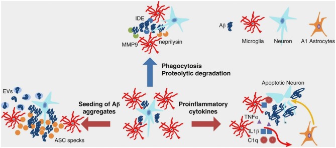
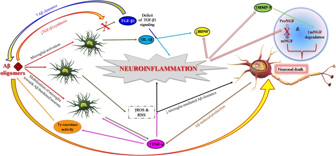
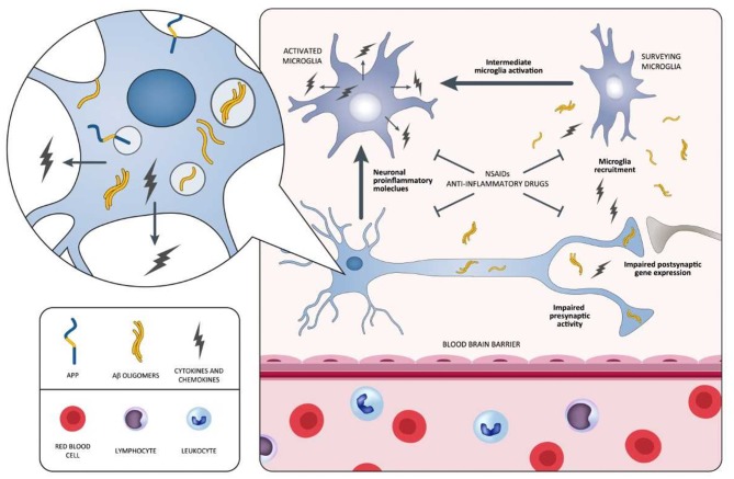
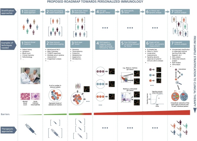
Verification
Claims the paper makes
The claims and statistics of this paper haven’t been checked yet.
Run the check — free, no account needed. It reads the open-access text and checks the sources it cites.
Claims other papers make about this one
Each is checked against this paper’s own text: does it say what the citing paper says it does?
The source backs part of the claim, not all of it — or backs it for a different population, condition or comparison.
In the citing paper
“mitochondrial dysfunction—with reactive oxygen species (ROS) accumulation and neuroinflammation driving microglial activation—plays a central role in AD pathogenesis [56]”
From Theranostic Nanoplatforms for Alzheimer's Disease: A Critical Analysis of Conceptual Contradictions (2026) · cites this paper as [56]
Experimental — accuracy not yet measured
Self-check
Percentages, sums and reported differences re-computed from the paper’s own numbers.
The self-check needs the paper’s full text.
Citations
Cites
1 retracted
RETRACTED AND REPLACED: Nerve growth factor metabolic dysfunction in Down’s syndrome brains
Iulita, M. F., Carmo, S. D., Ower, A. K., Fortress, A. M., Aguilar, L. F., Hanna, M., Wısnıewskı, T., Granholm, A., Buhusi, M. C., Busciglio, J. A., et al. (2014) · Brain · doi:10.1093/brain/awt372 · Cited by 94
Self-citationRetractedMcKhann, G. M., Knopman, D. S., Chertkow, H., Hyman, B. T., Jack, C. R., Kawas, C. H., Klunk, W. E., Koroshetz, W. J., Manly, J. J., Mayeux, R., et al. (2011) · Alzheimer's & Dementia · doi:10.1016/j.jalz.2011.03.005 · Cited by 18,657
NIA‐AA Research Framework: Toward a biological definition of Alzheimer's disease
Jack, C. R., Bennett, D. A., Blennow, K., Carrillo, M. C., Dunn, B., Haeberlein, S. B., Holtzman, D. M., Jagust, W., Jessen, F., Karlawish, J., et al. (2018) · Alzheimer's & Dementia · doi:10.1016/j.jalz.2018.02.018 · Cited by 10,537
Albert, M. S., DeKosky, S. T., Dickson, D., Dubois, B., Feldman, H. H., Fox, N. C., Gamst, A., Holtzman, D. M., Jagust, W. J., Petersen, R. C., et al. (2011) · Alzheimer's & Dementia · doi:10.1016/j.jalz.2011.03.008 · Cited by 10,152 · cited 1×
“, amnestic or non-amnestic), which significantly impacts clinical outcome (8, 179, 180), is inadequately specified in most of the studies.”
BackgroundInflammation in atherosclerosis
Libby, P. (2002) · Nature · doi:10.1038/nature01323 · Cited by 8,315
Neurotoxic reactive astrocytes are induced by activated microglia
Liddelow, S. A., Guttenplan, K. A., Clarke, L. E., Bennett, F. C., Bohlen, C. J., Schirmer, L., Bennett, M. L., Münch, A. E., Chung, W.-S., Peterson, T. C., et al. (2017) · Nature · doi:10.1038/nature21029 · Cited by 7,971
Sperling, R. A., Aisen, P. S., Beckett, L. A., Bennett, D. A., Craft, S., Fagan, A. M., Iwatsubo, T., Jack, C. R., Kaye, J., Montine, T. J., et al. (2011) · Alzheimer's & Dementia · doi:10.1016/j.jalz.2011.03.003 · Cited by 7,155
Neuroinflammation in Alzheimer's disease
Heneka, M. T., Carson, M. J., Khoury, J. E., Landreth, G. E., Brosseron, F., Feinstein, D. L., Jacobs, A. H., Wyss-Coray, T., Vitorica, J., Ransohoff, R. M., et al. (2015) · The Lancet Neurology · doi:10.1016/s1474-4422(15)70016-5 · Cited by 6,226 · cited 5×
“The pathophysiological mechanisms of multifactorial and polygenic AD are not limited to the neuronal tissue; they are related to cerebral immunological responses (12).”
BackgroundInfluentialA New Initiative on Precision Medicine
Collins, F. S., & Varmus, H. (2015) · New England Journal of Medicine · doi:10.1056/nejmp1500523 · Cited by 5,202
Astrocytes: Biology and pathology
Sofroniew, M. V., & Vinters, H. V. (2010) · Acta Neuropathologica · doi:10.1007/s00401-009-0619-8 · Cited by 5,196 · cited 1×
“Moreover, remodeling of astrocytes promotes neuroprotection and recovery of injured neural tissue (80, 81).”
Background
Research works printed but not linked: 1
- Presented at the 11thClinical Trials on Alzheimer's Disease (CTAD) Conference October 24–272018
Cited by
Sorted by citations
Shen, X.-N., Niu, L.-D., Wang, Y.-J., Cao, X.-P., Liu, Q., Tan, L., Zhang, C., & Yu, J.-T. (2019) · Journal of Neurology, Neurosurgery & Psychiatry · doi:10.1136/jnnp-2018-319148 · Cited by 388
Economic burden of Alzheimer disease and managed care considerations
Winston Wong, P. (2020) · The American Journal of Managed Care · doi:10.37765/ajmc.2020.88482 · Cited by 436
The Role of NLRP3 Inflammasome in Alzheimer’s Disease and Potential Therapeutic Targets
Liang, T., Zhang, Y., Wu, S., Chen, Q., & Wang, L. (2022) · Frontiers in Pharmacology · doi:10.3389/fphar.2022.845185 · Cited by 226
Role of Inflammatory Processes in Hemorrhagic Stroke
Ohashi, S. N., DeLong, J. H., Kozberg, M. G., Mazur-Hart, D. J., van Veluw, S. J., Alkayed, N. J., & Sansing, L. H. (2023) · Stroke · doi:10.1161/strokeaha.122.037155 · Cited by 224
The role of innate immune genes in Alzheimer's disease
Griciuc, A., & Tanzi, R. E. (2021) · Current Opinion in Neurology · doi:10.1097/wco.0000000000000911 · Cited by 192
“Neuroinflammation is as an innate immunological response of the central nervous system that is characterized by the activation of microglia and astrocytes, which play a central role in Alzheimer’s disease pathogenesis [24].”
BackgroundSahu, M., Gupta, R. V., Ambasta, R. K., & Kumar, P. (2022) · Progress in molecular biology and translational science · doi:10.1016/bs.pmbts.2022.03.002 · Cited by 214
Di Benedetto, G., Burgaletto, C., Bellanca, C. M., Munafò, A., Bernardini, R., & Cantarella, G. (2022) · Cells · doi:10.3390/cells11172728 · Cited by 189
Role of JAK/STAT in the Neuroinflammation and its Association with Neurological Disorders
Jain, M., Singh, M. K., Shyam, H., Mishra, A., Kumar, S., Kumar, A., & Kushwaha, J. K. (2021) · Annals of Neurosciences · doi:10.1177/09727531211070532 · Cited by 196
Versele, R., Sevin, E., Gosselet, F., Fénart, L., & Candela, P. (2022) · International Journal of Molecular Sciences · doi:10.3390/ijms231810235 · Cited by 187
Neuroinflammation: A Potential Risk for Dementia
Ahmad, M. A., Kareem, O., Khushtar, M., Akbar, M., Haque, M. R., Iqubal, A., Haider, M. F., Pottoo, F. H., Abdulla, F. S., Al-Haidar, M. B., et al. (2022) · International Journal of Molecular Sciences · doi:10.3390/ijms23020616 · Cited by 174
Glia Crosstalk in Neuroinflammatory Diseases
Bernaus, A., Blanco, S., & Sevilla, A. (2020) · Frontiers in Cellular Neuroscience · doi:10.3389/fncel.2020.00209 · Cited by 157
The foundation and architecture of precision medicine in neurology and psychiatry
Hampel, H. J., Gao, P., Cummings, J. L., Toschi, N., Thompson, P. M., Hu, Y., Cho, M., & Vergallo, A. (2023) · Trends in Neurosciences · doi:10.1016/j.tins.2022.12.004 · Cited by 163
“The temporal-spatial dynamics of the neuroinflammatory process could be dissected through multi-omics profiling along with neuroimaging and could potentially be targeted by specific and stage-guided immune-modulator drugs, such as TREM2 agonists, to modify disease progression [157,158].”
Self-citationThe Alzheimer’s disease drug development landscape
van Bokhoven, P., de Wilde, A., Vermunt, L., Leferink, P. S., Heetveld, S., Cummings, J., Scheltens, P., & Vijverberg, E. G. B. (2021) · Alzheimer's Research & Therapy · doi:10.1186/s13195-021-00927-z · Cited by 123
“Targeting pathways involved in (neuro)inflammation remains a strategy of interest both in preclinical and clinical development [11].”
BackgroundThe Future of Precision Medicine in the Cure of Alzheimer’s Disease
Arafah, A., Khatoon, S., Rasool, I., Khan, A., Rather, M. A., Abujabal, K. A., Faqih, Y. A. H., Rashid, H., Rashid, S. M., Bilal Ahmad, S., et al. (2023) · Biomedicines · doi:10.3390/biomedicines11020335 · Cited by 133
Astrocyte biomarkers GFAP and YKL‐40 mediate early Alzheimer's disease progression
Pelkmans, W., Shekari, M., Brugulat‐Serrat, A., Sánchez‐Benavides, G., Minguillón, C., Fauria, K., Molinuevo, J. L., Grau‐Rivera, O., González Escalante, A., Kollmorgen, G., et al. (2024) · Alzheimer's & Dementia · doi:10.1002/alz.13450 · Cited by 122
Exploring ER stress response in cellular aging and neuroinflammation in Alzheimer's disease
Uddin, M. S., Yu, W. S., & Lim, L. W. (2021) · Ageing Research Reviews · doi:10.1016/j.arr.2021.101417 · Cited by 123
Dhiman, K., Blennow, K., Zetterberg, H., Martins, R. N., & Gupta, V. B. (2019) · Cellular and Molecular Life Sciences · doi:10.1007/s00018-019-03040-5 · Cited by 117
The role of the immune system in Alzheimer’s disease
Wu, K., Zhang, Y.-R., Huang, Y., Dong, Q., Tan, L., & Yu, J. (2021) · Ageing Research Reviews · doi:10.1016/j.arr.2021.101409 · Cited by 119
Wang, W., Wang, Y., Wang, Y., Xie, G., Liu, S., Li, Z., Wang, P., Liu, J., & Li, L. (2024) · Phytomedicine · doi:10.1016/j.phymed.2024.155518 · Cited by 107
Inflammatory Cascade in Alzheimer’s Disease Pathogenesis: A Review of Experimental Findings
de Oliveira, J., Kucharska, E., Garcez, M. L., Rodrigues, M. S., Quevedo, J., Moreno‐González, I., & Budni, J. (2021) · Cells · doi:10.3390/cells10102581 · Cited by 110
Alzheimer's disease pathophysiology in the Retina
Gaire, B. P., Koronyo, Y., Fuchs, D.-T. S., Shi, H., Rentsendorj, A., Danziger, R., Vit, J., Mirzaei, N., Doustar, J., Sheyn, J., et al. (2024) · Progress in Retinal and Eye Research · doi:10.1016/j.preteyeres.2024.101273 · Cited by 107
Self-citationmicroRNA-Based Biomarkers in Alzheimer’s Disease (AD)
Zhao, Y., Jaber, V. R., Alexandrov, P. N., Vergallo, A., Lista, S., Hampel, H. J., & Lukiw, W. J. (2020) · Frontiers in Neuroscience · doi:10.3389/fnins.2020.585432 · Cited by 99
Self-citationAlzheimer’s Disease and Inflammaging
Kosyreva, A., Sentyabreva, A., Tsvetkov, I., & Makarova, O. (2022) · Brain Sciences · doi:10.3390/brainsci12091237 · Cited by 98
Neuroinflammation, Its Role in Alzheimer's Disease and Therapeutic Strategies
Király, M., Foss, J. F., & Giordano, T. (2023) · The Journal of Prevention of Alzheimer s Disease · doi:10.14283/jpad.2023.109 · Cited by 111
“Details on the role of microglia in AD, alternative pathways that contribute to their activation to various degrees in between the M1 and M2 states (including NF-κB, mTOR, MAPK, proinflammatory mediators, interleukins, anti-inflammatory cytokines, complement proteins, chemokines, caspases, prostanoids, neuroprotection…”
BackgroundBlood–brain barrier leakage and perivascular inflammation in cerebral amyloid angiopathy
Kozberg, M. G., Yi, I., Freeze, W. M., Auger, C. A., Scherlek, A. A., Greenberg, S. M., & van Veluw, S. J. (2022) · Brain Communications · doi:10.1093/braincomms/fcac245 · Cited by 94
Targeting Cytokine-Mediated Inflammation in Brain Disorders: Developing New Treatment Strategies
Mallick, R., Basak, S., Chowdhury, P., Bhowmik, P., Das, R. K., Banerjee, A., Paul, S., Pathak, S., & Duttaroy, A. K. (2025) · Pharmaceuticals · doi:10.3390/ph18010104 · Cited by 108
“Personalized techniques can enhance therapeutic results by targeting interventions to patients most likely to benefit from cytokine regulation [94–96].”
The immune system on the TRAIL of Alzheimer’s disease
Burgaletto, C., Munafò, A., Benedetto, G. D., Francisci, C. D., Caraci, F., Mauro, R. D., Bucolo, C., Bernardini, R., & Cantarella, G. (2020) · Journal of Neuroinflammation · doi:10.1186/s12974-020-01968-1 · Cited by 93
Self-citationNeuroinflammation as a Potential Therapeutic Target in Alzheimer’s Disease
Liu, P., Wang, Y., Sun, Y., & Peng, G. (2022) · Clinical Interventions in Aging · doi:10.2147/cia.s357558 · Cited by 94
“rivastigmine) and non-competitive N-methyl-D-aspartate antagonists (memantine); these compounds can only temporarily alleviate symptoms already apparent in the dementia stage.(6) Although more than 99% of AD drug trials do not meet efficacy endpoints, the first Phase 3 clinical trial of sodium oligomannate (GV-971) wa…”
BackgroundRen, F., Wei, J., Chen, Q., Hu, M., Yu, L., Mi, J., Zhou, X., Qin, D., Wu, J., & Wu, A. (2025) · Acta Pharmaceutica Sinica B · doi:10.1016/j.apsb.2025.07.030 · Cited by 97
Torrisi, S. A., Geraci, F., Tropea, M. R., Grasso, M., Caruso, G., Fidilio, A., Musso, N., Sanfilippo, G., Tascedda, F., Palmeri, A., et al. (2019) · Frontiers in Pharmacology · doi:10.3389/fphar.2019.00693 · Cited by 90
Self-citationWang, S., Ma, J., Zeng, Y., Zhou, G., Wang, Y., Zhou, W., Sun, X., & Wu, M. (2021) · Drug Design Development and Therapy · doi:10.2147/dddt.s310686 · Cited by 85
Song, J. (2023) · Biomedicine & Pharmacotherapy · doi:10.1016/j.biopha.2023.114647 · Cited by 88
Tesco, G., & Lomoio, S. (2022) · Seminars in Immunology · doi:10.1016/j.smim.2022.101628 · Cited by 102
Lista, S., Imbimbo, B. P., Grasso, M., Fidilio, A., Emanuele, E., Minoretti, P., López-Ortiz, S., Martín-Hernández, J., Gabelle, A., Caruso, G., et al. (2024) · Journal of Neuroinflammation · doi:10.1186/s12974-024-03163-y · Cited by 81
“Therefore, identifying TREM2 carriers can be valuable in improving patient stratification for clinical trials and supporting the development of personalized therapeutic approaches [33].”
BackgroundSelf-citationHeneka, M. T., Gauthier, S. G., Chandekar, S. A., Hahn-Pedersen, J. H., Bentsen, M. A., & Zetterberg, H. (2025) · Molecular Psychiatry · doi:10.1038/s41380-025-02939-9 · Cited by 97
Anti-amyloid-β protein agents for the treatment of Alzheimer’s disease: an update on emerging drugs
Lozupone, M., Solfrizzi, V., D’Urso, F., Di Gioia, I., Sardone, R., Dibello, V., Stallone, R., Liguori, A., Ciritella, C., Daniele, A., et al. (2020) · Expert Opinion on Emerging Drugs · doi:10.1080/14728214.2020.1808621 · Cited by 77
The Potential Role of Cytokines and Growth Factors in the Pathogenesis of Alzheimer’s Disease
Ogunmokun, G., Dewanjee, S., Chakraborty, P., Valupadas, C., Chaudhary, A., Kolli, V., Anand, U., Vallamkondu, J., Goel, P., Paluru, H. P. R., et al. (2021) · Cells · doi:10.3390/cells10102790 · Cited by 85
Do, H. T. T., & Cho, J. (2020) · International Journal of Molecular Sciences · doi:10.3390/ijms21176211 · Cited by 75
Targeting Sigma Receptors for the Treatment of Neurodegenerative and Neurodevelopmental Disorders
Malar, D. S., Thitilertdecha, P., Ruckvongacheep, K. S., Brimson, S., Tencomnao, T., & Brimson, J. M. (2023) · CNS Drugs · doi:10.1007/s40263-023-01007-6 · Cited by 77
“The key components of the disease have been reported to be the accumulation of amyloid precursor protein (APP) fragment amyloidbeta (Aβ) from its overproduction or reduced clearance [35], and the downstream process in hyperphosphorylation of the protein tau, leading to cell death, neurotransmitter deficits [36], reduc…”
BackgroundTargeting NLRP3-Mediated Neuroinflammation in Alzheimer’s Disease Treatment
Barczuk, J., Siwecka, N., Lusa, W., Rozpędek‐Kamińska, W., Kucharska, E., & Majsterek, I. (2022) · International Journal of Molecular Sciences · doi:10.3390/ijms23168979 · Cited by 76
CD33 isoforms in microglia and Alzheimer's disease: Friend and foe
Eskandari‐Sedighi, G., Jung, J., & Macauley, M. S. (2022) · Molecular Aspects of Medicine · doi:10.1016/j.mam.2022.101111 · Cited by 71
Brain Structural and Functional Changes in Cognitive Impairment Due to Alzheimer’s Disease
Ávila‐Villanueva, M., Dolado, A. M., Gómez‐Ramírez, J., & Fernández‐Blázquez, M. A. (2022) · Frontiers in Psychology · doi:10.3389/fpsyg.2022.886619 · Cited by 72
Current and evolving treatment strategies for the Alzheimer disease continuum
Marasco, R. (2020) · The American Journal of Managed Care · doi:10.37765/ajmc.2020.88481 · Cited by 67
Neuroinflammation PET imaging of the translocator protein (TSPO) in Alzheimer's disease: An update
Gouilly, D., Saint‐Aubert, L., Ribeiro, M. J., Salabert, A., Tauber, C., Péran, P., Arlicot, N., Pariente, J., & Payoux, P. (2022) · European Journal of Neuroscience · doi:10.1111/ejn.15613 · Cited by 70
“These results have driven the development of biomarkers to improve our understanding of AD neuroimmune changes and to monitor the engagement of inflammatory treatments (Gyengesi & Münch, 2020; Hampel et al., 2020).”
BackgroundTargeting Microglia-Synapse Interactions in Alzheimer’s Disease
Piccioni, G., Mango, D., Saidi, A., Corbo, M., & Nisticò, R. (2021) · International Journal of Molecular Sciences · doi:10.3390/ijms22052342 · Cited by 64
Self-citationThe road to precision medicine: Eliminating the “One Size Fits All” approach in Alzheimer’s disease
Behl, T., Kaur, I., Sehgal, A., Singh, S., Albarrati, A., Albratty, M., Najmi, A., Meraya, A. M., & BUNGAU, S. G. (2022) · Biomedicine & Pharmacotherapy · doi:10.1016/j.biopha.2022.113337 · Cited by 67
Gertje, E. C., Janelidze, S., van Westen, D., Cullen, N. C., Stomrud, E., Palmqvist, S., Hansson, O., & Mattsson, N. (2023) · Neurology · doi:10.1212/wnl.0000000000207113 · Cited by 62
Locus Coeruleus Modulates Neuroinflammation in Parkinsonism and Dementia
Giorgi, F. S., Biagioni, F., Galgani, A., Pavese, N., Lazzeri, G., & Fornai, F. (2020) · International Journal of Molecular Sciences · doi:10.3390/ijms21228630 · Cited by 63
Fernando, K., & Wijayasinghe, Y. S. (2021) · Frontiers in Cellular Neuroscience · doi:10.3389/fncel.2021.746631 · Cited by 57
Rapamycin Responds to Alzheimer’s Disease: A Potential Translational Therapy
Hou, S.-J., Zhang, S., Li, Y., & Xu, S.-Y. (2023) · Clinical Interventions in Aging · doi:10.2147/cia.s429440 · Cited by 58
“Hyperphosphorylated tau protein aggregates form paired helical filaments in NFTs as one of the key hallmarks of AD.”
InfluentialMenopause hormone therapy significantly alters pathophysiological biomarkers of Alzheimer's disease
Depypere, H. T., Vergallo, A., Lemercier, P., Lista, S., Benedet, A. L., Ashton, N. J., Cavedo, E., Zetterberg, H., Blennow, K., Vanmechelen, E., et al. (2022) · Alzheimer s & Dementia · doi:10.1002/alz.12759 · Cited by 61
Self-citationZhang, W., Hua, H., Guo, Y., Cheng, Y., Pi, F., Yao, W., Xie, Y., & He, Q. (2020) · Journal of Agricultural and Food Chemistry · doi:10.1021/acs.jafc.0c01892 · Cited by 55
Role of TREM2 in Alzheimer’s Disease: A Long Road Ahead
Kulkarni, B., Kumar, D., Martins, N. A., & Sellamuthu, S. (2021) · Molecular Neurobiology · doi:10.1007/s12035-021-02477-9 · Cited by 53
Wyatt-Johnson, S. K., & Brutkiewicz, R. R. (2020) · Frontiers in Aging Neuroscience · doi:10.3389/fnagi.2020.592359 · Cited by 50
Hampel, H. J., Goetzl, E. J., Kapogiannis, D., Lista, S., & Vergallo, A. (2019) · Frontiers in Pharmacology · doi:10.3389/fphar.2019.00310 · Cited by 47
Self-citationBarber, K., Mendonca, P., & Soliman, K. F. A. (2023) · Brain Sciences · doi:10.3390/brainsci13010145 · Cited by 51
“tify the target group and provide the process, contributed to the disappointing results [36].”
BackgroundInfluentialZhang, L., Wang, X., Yu, W., Ying, J., Fang, P., Zheng, Q., Feng, X., Hu, J., Xiao, F., Chen, S., et al. (2022) · Frontiers in Immunology · doi:10.3389/fimmu.2022.836494 · Cited by 46
Alzheimer’s Disease: From Immune Homeostasis to Neuroinflammatory Condition
Cariddi, L. P., Mauri, M. F., Cosentino, M., Versino, M., & Marino, F. (2022) · International Journal of Molecular Sciences · doi:10.3390/ijms232113008 · Cited by 45
Calmodulin binding proteins and neuroinflammation in multiple neurodegenerative diseases
O’Day, D. H., & Huber, R. J. (2022) · BMC Neuroscience · doi:10.1186/s12868-022-00695-y · Cited by 45
“Here, we examined proteins identified by others that are critical to neuroinflammation and other neurodegenerative events involved not only in AD but also in HD, LBD, PD, FTD, MS and other neurodegenerative diseases [18, 19, 22, 50].”
BackgroundBrain targeting based nanocarriers loaded with resveratrol in Alzheimer's disease: A review
Jalili, C., Kiani, A., Gholami, M., Bahrehmand, F., Fakhri, S., Kakehbaraei, S., & Kakebaraei, S. (2023) · IET Nanobiotechnology · doi:10.1049/nbt2.12127 · Cited by 43
“Neuroinflammation has a crucial impress in the induction of AD [84].”
BackgroundYu, W.-Y., Sun, T.-H., Hsu, K.-C., Wang, C., Chien, S.-Y., Tsai, C., & Yang, Y. (2024) · Computers in Biology and Medicine · doi:10.1016/j.compbiomed.2024.108621 · Cited by 40
Roles of TREM2 in the Pathological Mechanism and the Therapeutic Strategies of Alzheimer's Disease
Lin, M., Yu, J., Zhang, W., Lao, F., & Huang, H. (2024) · The Journal of Prevention of Alzheimer s Disease · doi:10.14283/jpad.2024.164 · Cited by 46
Alzheimer Disease Occurs More Frequently In Patients With Inflammatory Bowel Disease
Aggarwal, M., Alkhayyat, M. M., Saleh, M. A., Sarmini, M. T., Singh, A., Garg, R., Garg, P., Mansoor, E., Padival, R., & Cohen, B. L. (2022) · Journal of Clinical Gastroenterology · doi:10.1097/mcg.0000000000001714 · Cited by 41
Hao, Y., Su, C., Liu, X., Sui, H., Shi, Y., & Zhao, L. (2022) · Biomaterials Advances · doi:10.1016/j.bioadv.2022.212770 · Cited by 39
CNS Redox Homeostasis and Dysfunction in Neurodegenerative Diseases
Goldsteins, G., Hakosalo, V., Jaronen, M., Keuters, M. H., Lehtonen, Š., & Koıstınaho, J. (2022) · Antioxidants · doi:10.3390/antiox11020405 · Cited by 42
Associations between saliva and plasma cytokines in cognitively normal, older adults
Parkin, G. M., Kim, S., Mikhail, A. P., Malhas, R., McMillan, L. C., Hollearn, M. K., Granger, D. A., Mapstone, M. E., Yassa, M. A., & Thomas, E. A. (2022) · Aging Clinical and Experimental Research · doi:10.1007/s40520-022-02292-9 · Cited by 42
“We also measured levels of YKL-40, a pro-inflammatory glycoprotein, which has been considered as a pleiotropic cytokine given its secretion by macrophages and involvement in a wide range of inflammatory responses [26, 27] neuroinflammation and astroglial activation [28, 29].”
BackgroundThrombin, a Key Driver of Pathological Inflammation in the Brain
Iannucci, J., & Grammas, P. (2023) · Cells · doi:10.3390/cells12091222 · Cited by 40
“Therefore, targeting inflammation is a rational approach to AD prevention and therapy [141].”
BackgroundPET Agents in Dementia: An Overview
van Waarde, A., Marcolini, S., Deyn, P. P. D., & Dierckx, R. (2021) · Seminars in Nuclear Medicine · doi:10.1053/j.semnuclmed.2020.12.008 · Cited by 39
Knorr, U., Simonsen, A. H., Jensen, C. S., Zetterberg, H., Blennow, K., Akhøj, M., Forman, J. L., Hasselbalch, S. G., & Kessing, L. V. (2021) · Journal of Affective Disorders · doi:10.1016/j.jad.2021.10.074 · Cited by 40
Kirubakaran, D. (2025) · Digital Chinese Medicine · doi:10.1016/j.dcmed.2025.05.002 · Cited by 39
The chaperone Clusterin in neurodegeneration−friend or foe?
Yuste‐Checa, P., Bracher, A., & Hartl, F. U. (2022) · BioEssays · doi:10.1002/bies.202100287 · Cited by 37
“response, providing support for the critical role of neuroinflammation in AD pathogenesis.[131] An upregulation of inflammation-related”
BackgroundTherapeutic Targeting Strategies for Early- to Late-Staged Alzheimer’s Disease
Kang, Y. J., Diep, Y. N., Tran, M., & Cho, H. (2020) · International Journal of Molecular Sciences · doi:10.3390/ijms21249591 · Cited by 36
Astrocyte subtype-specific approach to Alzheimer's disease treatment
Sarkar, S., & Biswas, S. C. (2021) · Neurochemistry International · doi:10.1016/j.neuint.2021.104956 · Cited by 35
Carthamus tinctorius L.: A natural neuroprotective source for anti-Alzheimer's disease drugs
Liang, Y., & Wang, L. (2022) · Journal of Ethnopharmacology · doi:10.1016/j.jep.2022.115656 · Cited by 36
A small-molecule TLR4 antagonist reduced neuroinflammation in female E4FAD mice
Balu, D., Valencia-Olvera, A. C., Nguyen, A., Patnam, M., York, J., Peri, F., Neumann, F., LaDu, M. J., & Tai, L. M. (2023) · Alzheimer's Research & Therapy · doi:10.1186/s13195-023-01330-6 · Cited by 27
Frye, B. M., Craft, S., Latimer, C. S., Keene, C. D., Montine, T. J., Register, T. C., Orr, M. E., Kavanagh, K., Macauley, S. L., & Shively, C. A. (2021) · American Journal of Primatology · doi:10.1002/ajp.23260 · Cited by 34
“, 2011), which are hypothesized to contribute to AD-like pathophysiologic processes (Hampel et al., 2020; Hashemiaghdam & Mroczek, 2020).”
BackgroundBoosting mitochondrial health to counteract neurodegeneration
Burtscher, J., Romani, M., Bernardo, G., Popa, T., Ziviani, E., Hummel, F. C., Sorrentino, V., & Millet, G. P. (2022) · Progress in Neurobiology · doi:10.1016/j.pneurobio.2022.102289 · Cited by 36
Microglial autophagy in Alzheimer’s disease and Parkinson’s disease
Wang, Z., Wang, Q., Li, S., Li, X.-J., Yang, W., & He, D. (2023) · Frontiers in Aging Neuroscience · doi:10.3389/fnagi.2022.1065183 · Cited by 35
Discontinued disease-modifying therapies for Alzheimer’s disease: status and future perspectives
Imbimbo, B. P., Lozupone, M., Watling, M., & Panza, F. (2020) · Expert Opinion on Investigational Drugs · doi:10.1080/13543784.2020.1795127 · Cited by 31
Self-citationYan, Y., Aierken, A., Wang, C., Song, D. F., Ni, J., Wang, Z., Quan, Z., & Qing, H. (2021) · Neuroscience & Biobehavioral Reviews · doi:10.1016/j.neubiorev.2021.11.009 · Cited by 32
A dynamical systems approach for multiscale synthesis of Alzheimer’s pathogenesis
Rollo, J. L., Crawford, J. W., & Hardy, J. (2023) · Neuron · doi:10.1016/j.neuron.2023.04.018 · Cited by 31
“A systems approach will therefore lead to more effective personalised plans in the treatment of the neuroinflammatory mechanisms in AD 69 studying the disease’s progression over time with a dynamical systems approach, will better inform these therapeutic strategies.”
DYRK1A Inhibitors and Perspectives for the Treatment of Alzheimer's Disease
de Souza, M. M., Cenci, A. R., Teixeira, K. F., Machado, V., Schülerb, M. C. G. M., Gonçalves, A. E., Dalmagro, A. P., Cazarin, C. A., Ferreira, L. L. G., de Oliveira, A. S., et al. (2022) · Current Medicinal Chemistry · doi:10.2174/0929867329666220620162018 · Cited by 30
Therapeutics of Alzheimer’s Disease: Recent Developments
Burns, S., Selman, A., Sehar, U., Rawat, P., Reddy, A. P., & Reddy, P. H. (2022) · Antioxidants · doi:10.3390/antiox11122402 · Cited by 36
“Dominant-negative TNF inhibitors, such as XPro®1595 [67] and XENP 345 [68], are considered effective therapeutics for targeting TNF. AL002, a humanized monoclonal IgG1 antibody, is an anti-human TREM2 antibody under phase 2 study; phase 1 study data suggested it to be safe and tolerable by healthy adults [78].”
BackgroundInfluentialNavigating the dementia landscape: Biomarkers and emerging therapies
Maheshwari, S., Singh, A., Ansari, V. A., Mahmood, T., Wasim, R., Akhtar, J., & Verma, A. (2024) · Ageing Research Reviews · doi:10.1016/j.arr.2024.102193 · Cited by 30
Antonioni, A., Martorana, A., Santarnecchi, E., Hampel, H. J., & Koch, G. (2025) · Alzheimer s & Dementia · doi:10.1002/alz.70337 · Cited by 31
Self-citationA Therapeutic Strategy for Alzheimer's Disease Focused on Immune-inflammatory Modulation
Kim, S. H., Noh, M., Kim, H.-J., Oh, K., Park, J., Lee, S.-G., Moon, Y., Kim, Y.-E., Bae, J. S., Jin, H. K., et al. (2019) · Dementia and Neurocognitive Disorders · doi:10.12779/dnd.2019.18.2.33 · Cited by 30
Rasouli, H., Nayeri, F. D., & Khodarahmi, R. (2022) · Frontiers in Nutrition · doi:10.3389/fnut.2022.981984 · Cited by 33
Association of Plasma YKL-40 With MRI, CSF, and Cognitive Markers of Brain Health and Dementia
Pase, M. P., Himali, J. J., Puerta, R., Beiser, A., Gonzales, M. M., Satizábal, C. L., Yang, Q., Aparicio, H. J., Kojis, D. J., DeCarli, C. S., et al. (2024) · Neurology · doi:10.1212/wnl.0000000000208075 · Cited by 29
Targeting Abnormal Tau Phosphorylation for Alzheimer’s Therapeutics
Singh, A., Ansari, V. A., Mahmood, T., Hasan, S. S., Wasim, R., Maheshwari, S., Akhtar, J., Sheikh, S., & Vishwakarma, V. K. (2024) · Hormone and Metabolic Research · doi:10.1055/a-2238-1384 · Cited by 29
“Research has demonstrat - ed that AD patients have a substantial accumulation of tau protein in brain tissue compared to healthy control groups [10–15].”
BackgroundZhang, L., Liu, Y., Wang, X., Wang, D., Wu, H., Chen, H., Chen, J., & Liu, Y. (2022) · Physiology & Behavior · doi:10.1016/j.physbeh.2022.113820 · Cited by 27
Takata, K., Kimura, H., Yanagisawa, D., Harada, K., Nishimura, K., Kitamura, Y., Shimohama, S., & Tooyama, I. (2022) · Molecules · doi:10.3390/molecules27092780 · Cited by 29
“Neuroinflammation is a central pathogenic process in a variety of neurodegenerative diseases [72] and is one of the most important factors in the etiology of AD [73].”
BackgroundWątroba, M., & Szukiewicz, D. (2022) · Frontiers in Physiology · doi:10.3389/fphys.2022.962769 · Cited by 29
Potential of food-derived bioactive peptides in alleviation and prevention of Alzheimer's disease
Zhao, L., Li, D., Qi, X., Guan, K., Chen, H., Wang, R., & Ma, Y. (2022) · Food & Function · doi:10.1039/d2fo02278h · Cited by 29
New Insights into Microglial Mechanisms of Memory Impairment in Alzheimer’s Disease
Li, N., Deng, M., Hu, G., Li, N., Yuan, H., & Zhou, Y. (2022) · Biomolecules · doi:10.3390/biom12111722 · Cited by 30
Zou, W., Liu, J., Jiao, Y., Sun, Y., Chen, N., Zhou, L., Guan, X., Wang, J., Wei, W., Han, C., et al. (2022) · Neural Regeneration Research · doi:10.4103/1673-5374.339002 · Cited by 30
“Studies have consistently demonstrated that disease-associated astrocytes (DAAs) respond to inflammatory damage by promoting release of inflammatory factors, which accelerate activation of astrocytes and microglia (Chávez et al., 2019; Wang et al., 2019; Hampel et al., 2020).”
BackgroundA Dichotomous Role for FABP7 in Sleep and Alzheimer’s Disease Pathogenesis: A Hypothesis
Needham, H., Torpey, G., Flores, C. C., Davis, C. J., Vanderheyden, W. M., & Gerstner, J. R. (2022) · Frontiers in Neuroscience · doi:10.3389/fnins.2022.798994 · Cited by 29
“Despite its likely beneficial role, overexpression of TREM2 is insufficient to ward off AD progression.”
BackgroundInfluentialYang, Y., Seok, M., Kim, Y. E., Choi, Y., Song, J., Sulistio, Y. A., Kim, S., Chang, M., Oh, S., Nam, M., et al. (2022) · Molecular Psychiatry · doi:10.1038/s41380-022-01693-6 · Cited by 30
“[2, 3]).”
Alzheimer’s Disease Treatment: The Search for a Breakthrough
Reiss, A. B., Muhieddine, D., Jacob, B., Mesbah, M., Pinkhasov, A., Gomolin, I. H., Stecker, M. M., Wısnıewskı, T., & Leon, J. D. (2023) · Medicina · doi:10.3390/medicina59061084 · Cited by 31
Li, Q., Rubin, L., Silva, M., Li, S., Yang, C., Lazarovici, P., & Zheng, W. (2022) · Oxidative Medicine and Cellular Longevity · doi:10.1155/2022/3777021 · Cited by 28
“Nevertheless, these drugs provide only a temporary symptomatic relief, failing to improve the patients ’ cognitive function or to provide a de fi nitive cure [7].”
Beyond the amyloid cascade: An update of Alzheimer's disease pathophysiology
Gouilly, D., Rafiq, M., Nogueira, L., Salabert, A., Payoux, P., Péran, P., & Pariente, J. (2023) · Revue Neurologique · doi:10.1016/j.neurol.2022.12.006 · Cited by 31
Metadata
- Authors
- Hampel, H., Caraci, F., Cuello, A. C., Caruso, G., Nisticò, R., Corbo, M., Baldacci, F., Toschi, N., Garaci, F., Chiesa, P. A., Verdooner, S. R., Akman-Anderson, L., Hernández, F., Ávila, J., Emanuele, E., Valenzuela, P. L., Lucía, A., Watling, M., Imbimbo, B. P., Vergallo, A., & Lista, S.
- Affiliations
- Sorbonne Université (FR)
- University of Catania (IT)
- University of Oxford (GB)
- I.R.C.C.S. Oasi Maria SS (IT)
- University of Rome Tor Vergata (IT)
- University of Pisa (IT)
- Centre National de la Recherche Scientifique (FR)
- Consejo Superior de Investigaciones Científicas (ES)
- Universidad de Alcalá (ES)
- Research Institute Hospital 12 de Octubre (ES)
- Chiesi (Italy) (IT)
- Corresponding author
- Hampel, Harald
- ORCID
- Venue
- Frontiers in Immunology
- Publisher
- Frontiers Media SA
- Published
- 2020-03-31
- Volume
- 11
- Pages
- 456-456
- ISSN
- 1664-3224 (ISSN-L)
- PMC
- PMC7137904
- OpenAlex
- W3014540555
- Cited by
- 360
- References
- 297
- Citations by year
- 2026: 43 · 2025: 53 · 2024: 53 · 2023: 66 · 2022: 69 · 2021: 49 · 2020: 22
- FWCI
- 5.13
- Type
- journal article
- Language
- en
- Open access
- gold
- doi.org
- Licence
- Locations
- Frontiers in Immunology (publishedVersion, cc-by)
- PubMed Central (publishedVersion, cc-by)
- PubMed (publishedVersion)
- HAL (submittedVersion, cc-by)
- Cineca Institutional Research Information System (Tor Vergata University) (submittedVersion, cc-by)
- Biblos-e Archivo (Universidad Autónoma de Madrid) (submittedVersion, cc-by)
- ABACUS. Repositorio de Producción Científica (Universidad Europea) (submittedVersion, cc-by-nc-nd)
- CINECA IRIS Institutial research information system (University of Pisa) (submittedVersion, other-oa)
- DIGITAL.CSIC (Spanish National Research Council (CSIC)) (submittedVersion, cc-by)
- LA Referencia (Red Federada de Repositorios Institucionales de Publicaciones Científicas) (submittedVersion, other-oa)
- DOAJ (DOAJ: Directory of Open Access Journals) (submittedVersion, cc-by-sa)
- Hispana (submittedVersion)
- Funders
- AXA Research Fund
- Eisai
- Sorbonne Université
- Fondation pour la Recherche sur Alzheimer
- Topics
- Life Sciences › Neuroscience › Neurology › Neuroinflammation and Neurodegeneration Mechanisms
- Life Sciences › Neuroscience › Cellular and Molecular Neuroscience › Alzheimer's disease research and treatments
- Health Sciences › Medicine › Psychiatry and Mental health › Dementia and Cognitive Impairment Research
- Keywords
- Alzheimer's disease, neuroinflammation, TREM2, microglia, astrocytes, amyloid-beta, tau protein, synaptotoxicity, neurodegeneration, genetic risk variants, cytokines, TNF-α, IL-1β, TGF-beta, biomarkers, positron emission tomography, nonsteroidal anti-inflammatory drugs, disease-modifying therapy, precision medicine
- MeSH terms
- Cell Self Renewal, Alzheimer Disease, Animals, Humans, Signal Transduction, Cytokines, Neurogenic Inflammation, Proteomics, Systems Biology, Lipid Metabolism, Genome-Wide Association Study, Precision Medicine
About these checks
Registry, retraction, Cites and statistics results are computed, not judged, with one exception: a printed reference that no index lists is read once by a model before it counts as a citation error. Claims are extracted by a model and kept only when their quote appears verbatim in the text; each cited source is then checked, with a model’s help, for whether it says what the claim relies on. Those verdicts can be wrong — read the claim’s quote and the cited source before relying on one.
Statistics and arithmetic are recomputed, not judged.
Confidence bands are shown only for fields calibrated on our annotated test set. None is calibrated yet, so no band is shown.
What counts as a claim ›How we measure confidence ›How we check this ›
Are you an author, or do you see a problem with this page? Request a review
