

The DOI matches its Crossref record; 252 of the 255 references it cites were found in a registry or an index.
Excitatory neuronal connectivity in the barrel cortex
Open accessAbstract
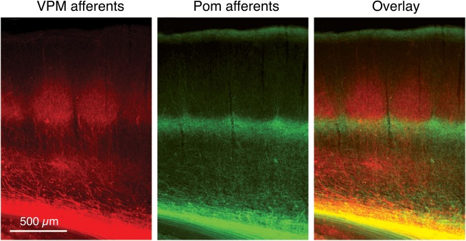
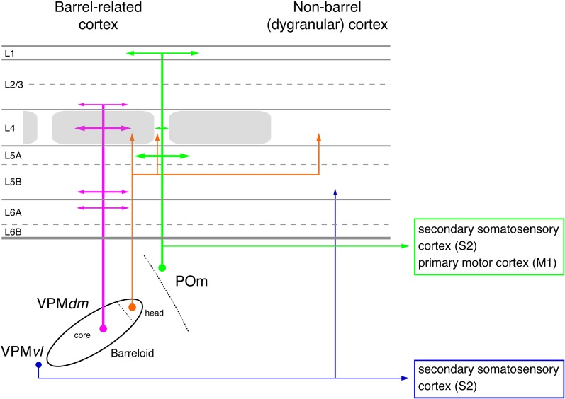
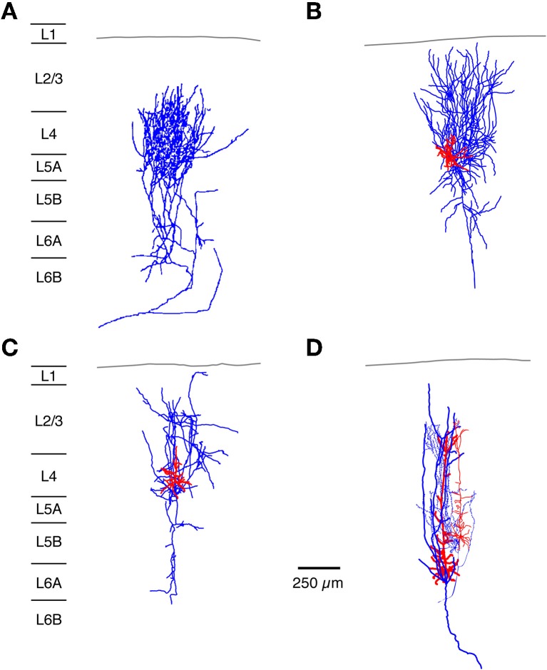
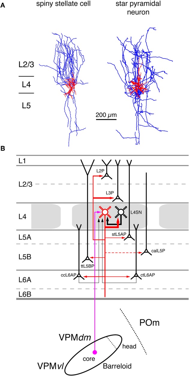
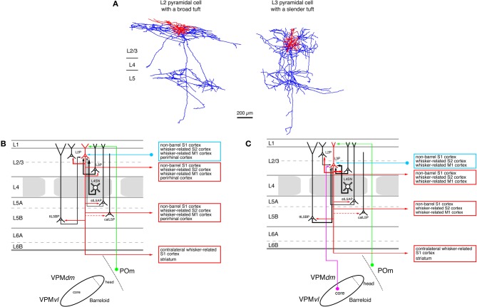
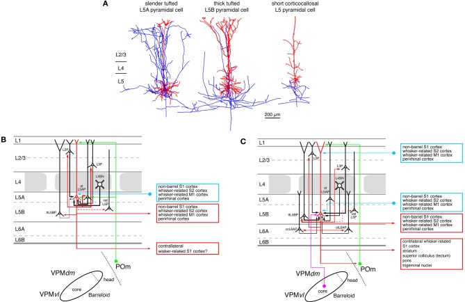
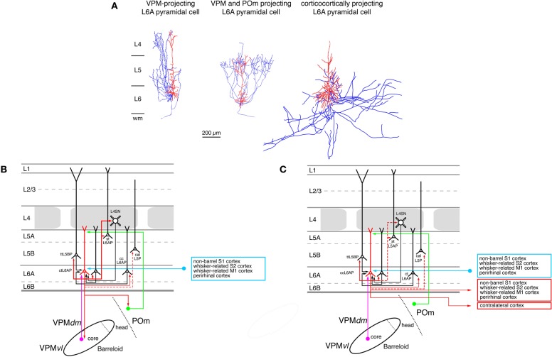
Neocortical areas are believed to be organized into vertical modules, the cortical columns, and the horizontal layers 1-6. In the somatosensory barrel cortex these columns are defined by the readily discernible barrel structure in layer 4. Information processing in the neocortex occurs along vertical and horizontal axes, thereby linking individual barrel-related columns via axons running through the different cortical layers of the barrel cortex. Long-range signaling occurs within the neocortical layers but also through axons projecting through the white matter to other neocortical areas and subcortical brain regions. Because of the ease of identification of barrel-related columns, the rodent barrel cortex has become a prototypical system to study the interactions between different neuronal connections within a sensory cortical area and between this area and other cortical as well subcortical regions. Such interactions will be discussed specifically for the feed-forward and feedback loops between the somatosensory and the somatomotor cortices as well as the different thalamic nuclei. In addition, recent advances concerning the morphological characteristics of excitatory neurons and their impact on the synaptic connectivity patterns and signaling properties of neuronal microcircuits in the whisker-related somatosensory cortex will be reviewed. In this context, their relationship between the structural properties of barrel-related columns and their function as a module in vertical synaptic signaling in the whisker-related cortical areas will be discussed.
Overview
The claims and statistics of this paper haven’t been checked yet.
Run the check — free, no account needed. It reads the open-access text and checks the sources it cites.
Integrity signals
Registry
Cites
Statistics
Figures
Verification
Claims the paper makes
The claims and statistics of this paper haven’t been checked yet.
Run the check — free, no account needed. It reads the open-access text and checks the sources it cites.
Self-check
Percentages, sums and reported differences re-computed from the paper’s own numbers.
The self-check needs the paper’s full text.
Citations
Cites
Neuronal Oscillations in Cortical Networks
Buzsáki, G., & Draguhn, A. (2004) · Science · doi:10.1126/science.1099745 · Cited by 6,576
Receptive fields of single neurones in the cat's striate cortex
Hubel, D. H., & Wiesel, T. N. (1959) · The Journal of Physiology · doi:10.1113/jphysiol.1959.sp006308 · Cited by 4,653
Thalamocortical oscillations in the sleeping and aroused brain
Steriade, M., McCormick, D. A., & Sejnowski, T. J. (1993) · Science · doi:10.1126/science.8235588 · Cited by 3,564
MODALITY AND TOPOGRAPHIC PROPERTIES OF SINGLE NEURONS OF CAT'S SOMATIC SENSORY CORTEX
Mountcastle, V. B. (1957) · Journal of Neurophysiology · doi:10.1152/jn.1957.20.4.408 · Cited by 2,411
The columnar organization of the neocortex
Mountcastle, V. B. (1997) · Brain · doi:10.1093/brain/120.4.701 · Cited by 2,314
The structural organization of layer IV in the somatosensory region (S I) of mouse cerebral cortex
Woolsey, T. A., & Van der Loos, H. (1970) · Brain Research · doi:10.1016/0006-8993(70)90079-x · Cited by 2,254
Highly nonrandom features of synaptic connectivity in local cortical circuits
Song, S., Sjöström, P. J., Reigl, M., Nelson, S., & Chklovskii, D. B. (2005) · PLoS Biology · doi:10.1371/journal.pbio.0030068 · Cited by 1,512
Petilla terminology: nomenclature of features of GABAergic interneurons of the cerebral cortex
The Petilla Interneuron Nomenclature Group (PING) (2008) · Nature Reviews Neuroscience · doi:10.1038/nrn2402 · Cited by 1,491
A new cellular mechanism for coupling inputs arriving at different cortical layers
Larkum, M. E., Zhu, J. J., & Sakmann, B. (1999) · Nature · doi:10.1038/18686 · Cited by 1,180
The high-conductance state of neocortical neurons in vivo
Alain Destexhe, A., Rudolph, M., & Paré, D. (2003) · Nature reviews. Neuroscience · doi:10.1038/nrn1198 · Cited by 1,062
Research works printed but not linked: 5
- Alloway K. D. Mutic J. J. Hoffer Z. S. Hoover J. E. (2000). Overlapping corticostriatal projections from the rodent vibrissal representations in primary and secondary somatosensory cortex. J. Comp. Neurol. 428, 51– 67 11185904
- Douglas R. Markram H. Martin K. (2004). Neocortex, in The Synaptic Organization of the Brain, 5th Edn ed Shepherd G. M. (New York, NY: Oxford University Press), 499– 558
- Fox K. D. (2008). Barrel Cortex, 1st Edn Cambridge, UK: Cambridge University Press
- Jones E. G. (1984). Laminar distribution of cortical efferent cells, in Cerebral Cortex, Vol. 1, Cellular Components of the Cerebral Cortex, eds Peters A. Jones E. G. (New York, NY: Plenum Press), 521– 533
- Tömböl T. (1984). Layer VI cells, in Cerebral Cortex, eds Peters A. Jones E. G. (New York, London: Plenum Press), 479– 519
Cited by
Sorted by citations
The neocortical circuit: themes and variations
Harris, K. D., & Shepherd, G. M. G. (2015) · Nature Neuroscience · doi:10.1038/nn.3917 · Cited by 1,215
Dysregulation of brain and choroid plexus cell types in severe COVID-19
Yang, A. C., Kern, F., Losada, P. M., Agam, M. R., Maat, C. A., Schmartz, G. P., Fehlmann, T., Stein, J. A., Schaum, N., Lee, D. P., et al. (2021) · Nature · doi:10.1038/s41586-021-03710-0 · Cited by 620
Deep Cortical Layers Are Activated Directly by Thalamus
Constantinople, C. M., & Bruno, R. M. (2013) · Science · doi:10.1126/science.1236425 · Cited by 484
Feldmeyer, D., Brecht, M., Helmchen, F., Petersen, C. C. H., Poulet, J. F. A., Staiger, J. F., Luhmann, H. J., & Schwarz, C. (2013) · Progress in Neurobiology · doi:10.1016/j.pneurobio.2012.11.002 · Cited by 354
“revealed that the major excitatory pathways of the canonical microcircuitry defined by Douglas and Martin are present, as recently reviewed in detail (Feldmeyer, 2012; Ahissar and Staiger, 2010).”
BackgroundInfluentialSelf-citationA Cellular Resolution Map of Barrel Cortex Activity during Tactile Behavior
Peron, S., Freeman, J., Iyer, V., Guo, C., & Svoboda, K. (2015) · Neuron · doi:10.1016/j.neuron.2015.03.027 · Cited by 334
Evolution of the human brain: when bigger is better
Hofman, M. A. (2014) · Frontiers in Neuroanatomy · doi:10.3389/fnana.2014.00015 · Cited by 303
Higher-Order Thalamocortical Inputs Gate Synaptic Long-Term Potentiation via Disinhibition
Williams, L. E., & Holtmaat, A. J. D. G. (2018) · Neuron · doi:10.1016/j.neuron.2018.10.049 · Cited by 282
Sensorimotor processing in the rodent barrel cortex
Petersen, C. C. H. (2019) · Nature reviews. Neuroscience · doi:10.1038/s41583-019-0200-y · Cited by 283
Sensory-evoked LTP driven by dendritic plateau potentials in vivo
Gambino, F., Pagès, S., Kehayas, V., Baptista, D., Tatti, R., Carleton, A., & Holtmaat, A. (2014) · Nature · doi:10.1038/nature13664 · Cited by 253
Control of synaptic plasticity in deep cortical networks
Roelfsema, P. R., & Holtmaat, A. J. D. G. (2018) · Nature reviews. Neuroscience · doi:10.1038/nrn.2018.6 · Cited by 254
A cortex-like canonical circuit in the avian forebrain
Stacho, M., Herold, C., Rook, N., Wagner, H., Axer, M., Amunts, K., & Güntürkün, O. (2020) · Science · doi:10.1126/science.abc5534 · Cited by 250
Dendritic channelopathies contribute to neocortical and sensory hyperexcitability in Fmr1−/y mice
Zhang, Y., Bonnan, A., Bony, G., Férézou, I., Pietropaolo, S., Ginger, M. R., Sans, N., Rossier, J., Oostra, B. A., LeMasson, G., et al. (2014) · Nature Neuroscience · doi:10.1038/nn.3864 · Cited by 239
Reinhard, S. M., Razak, K., & Ethell, I. M. (2015) · Frontiers in Cellular Neuroscience · doi:10.3389/fncel.2015.00280 · Cited by 219
Beltramo, R., D’Urso, G., Maschio, M. D., Farisello, P., Bovetti, S., Clovis, Y. M., Lassi, G., Tucci, V., Tonelli, D. D. P., & Fellin, T. (2013) · Nature Neuroscience · doi:10.1038/nn.3306 · Cited by 238
The Sparseness of Mixed Selectivity Neurons Controls the Generalization–Discrimination Trade-Off
Barak, O., Rigotti, M., & Fusi, S. (2013) · Journal of Neuroscience · doi:10.1523/jneurosci.2753-12.2013 · Cited by 199
Ji, X.-Y., Zingg, B., Mesik, L., Xiao, Z., Zhang, L. I., & Tao, H. W. (2016) · Cerebral Cortex · doi:10.1093/cercor/bhv099 · Cited by 176
Baker, A. L., Kalmbach, B. E., Morishima, M., Kim, J., Juavinett, A., Li, N., & Dembrow, N. C. (2018) · Journal of Neuroscience · doi:10.1523/jneurosci.0150-18.2018 · Cited by 205
Type-I-interferon-responsive microglia shape cortical development and behavior
Escoubas, C. C., Dorman, L. C., Nguyen, P. T., Lagares-Linares, C., Nakajo, H., Anderson, S. R., Barron, J. J., Wade, S. D., Cuevas, B., Vainchtein, I. D., et al. (2024) · Cell · doi:10.1016/j.cell.2024.02.020 · Cited by 179
Early postnatal development of pyramidal neurons across layers of the mouse medial prefrontal cortex
Kroon, T., van Hugte, E., van Linge, L., Mansvelder, H. D., & Meredith, R. M. (2019) · Scientific Reports · doi:10.1038/s41598-019-41661-9 · Cited by 161
Connectivity of mouse somatosensory and prefrontal cortex examined with trans-synaptic tracing
DeNardo, L. A., Berns, D. S., DeLoach, K. E., & Luo, L. (2015) · Nature Neuroscience · doi:10.1038/nn.4131 · Cited by 180
van Aerde, K. I., & Feldmeyer, D. (2013) · Cerebral Cortex · doi:10.1093/cercor/bht278 · Cited by 180
Self-citationLayer 6 Corticothalamic Neurons Activate a Cortical Output Layer, Layer 5a
Kim, J., Matney, C. J., Blankenship, A. G., Hestrin, S., & Brown, S. P. (2014) · Journal of Neuroscience · doi:10.1523/jneurosci.1325-14.2014 · Cited by 170
Iascone, D. M., Li, Y., Sümbül, U., Doron, M., Chen, H., Andreu, V., Goudy, F., Blockus, H., Abbott, L. F., Segev, I., et al. (2020) · Neuron · doi:10.1016/j.neuron.2020.02.015 · Cited by 177
Neuronal circuits in barrel cortex for whisker sensory perception
Staiger, J. F., & Petersen, C. C. H. (2021) · Physiological Reviews · doi:10.1152/physrev.00019.2019 · Cited by 165
Coupling the State and Contents of Consciousness
Aru, J., Suzuki, M., Rutiku, R., Larkum, M. E., & Bachmann, T. (2019) · Frontiers in Systems Neuroscience · doi:10.3389/fnsys.2019.00043 · Cited by 164
Bloem, B., Poorthuis, R. B., & Mansvelder, H. D. (2014) · Frontiers in Neural Circuits · doi:10.3389/fncir.2014.00017 · Cited by 163
Valiullina, F., Akhmetshina, D., Nasretdinov, A., Mukhtarov, M. R., Valeeva, G. R., Khazipov, R., & Rozov, A. V. (2016) · Frontiers in Neural Circuits · doi:10.3389/fncir.2016.00001 · Cited by 147
Complementary networks of cortical somatostatin interneurons enforce layer specific control
Naka, A., Veit, J., Shababo, B., Chance, R. K., Risso, D., Stafford, D., Snyder, B., Egladyous, A., Chu, D., Sridharan, S., et al. (2019) · eLife · doi:10.7554/elife.43696 · Cited by 117
A genuine layer 4 in motor cortex with prototypical synaptic circuit connectivity
Yamawaki, N., Borges, K., Suter, B. A., Harris, K. D., & Shepherd, G. M. G. (2014) · eLife · doi:10.7554/elife.05422 · Cited by 141
Inhibitory Interneurons and their Circuit Motifs in the Many Layers of the Barrel Cortex
Feldmeyer, D., Qi, G., Emmenegger, V., & Staiger, J. F. (2017) · Neuroscience · doi:10.1016/j.neuroscience.2017.05.027 · Cited by 140
Self-citationRecurrent synapses and circuits in the CA3 region of the hippocampus: an associative network
Duigou, C. L., Simonnet, J., Teleńczuk, M. T., Fricker, D., & Miles, R. (2014) · Frontiers in Cellular Neuroscience · doi:10.3389/fncel.2013.00262 · Cited by 145
Lozovaya, N., Gataullina, S., Tsintsadze, T. S., Tsintsadze, V., Pallesi‐Pocachard, E., Minlebaev, M., Goriounova, N. A., Buhler, E., Watrin, F., Shityakov, S., et al. (2014) · Nature Communications · doi:10.1038/ncomms5563 · Cited by 123
Deitcher, Y., Eyal, G., Kanari, L., Verhoog, M. B., Kahou, G. A. A., Mansvelder, H. D., de Kock, C. P. J., & Segev, I. (2017) · Cerebral Cortex · doi:10.1093/cercor/bhx226 · Cited by 117
Differential Structure of Hippocampal CA1 Pyramidal Neurons in the Human and Mouse
Benavides‐Piccione, R., Regalado-Reyes, M., Fernaud-Espinosa, I., Kastanauskaite, A., Tapia‐González, S., León‐Espinosa, G., Rojo, C., Insausti, R., Segev, I., & DeFelipe, J. (2019) · Cerebral Cortex · doi:10.1093/cercor/bhz122 · Cited by 115
Development and plasticity of the corpus callosum
de León-Reyes, N. S., Bragg-Gonzalo, L., & Nieto, M. (2020) · Development · doi:10.1242/dev.189738 · Cited by 122
Cracking the Function of Layers in the Sensory Cortex
Adesnik, H., & Naka, A. (2018) · Neuron · doi:10.1016/j.neuron.2018.10.032 · Cited by 121
A thalamic bridge from sensory perception to cognition
Wolff, M., Morceau, S., Folkard, R., Martín-Cortecero, J., & Groh, A. (2020) · Neuroscience & Biobehavioral Reviews · doi:10.1016/j.neubiorev.2020.11.013 · Cited by 114
Whisker-Mediated Touch System in Rodents: From Neuron to Behavior
Adibi, M. (2019) · Frontiers in Systems Neuroscience · doi:10.3389/fnsys.2019.00040 · Cited by 110
A direct translaminar inhibitory circuit tunes cortical output
Pluta, S. R., Naka, A., Veit, J., Telian, G. I., Yao, L., Hakim, R., Taylor, D., & Adesnik, H. (2015) · Nature Neuroscience · doi:10.1038/nn.4123 · Cited by 104
Modularity in the Organization of Mouse Primary Visual Cortex
Ji, W., Gămănuț, R., Bista, P., D’Souza, R. D., Wang, Q., & Burkhalter, A. (2015) · Neuron · doi:10.1016/j.neuron.2015.07.004 · Cited by 95
Reyes‐Puerta, V., Sun, J., Kim, S., Kilb, W., & Luhmann, H. J. (2014) · Cerebral Cortex · doi:10.1093/cercor/bhu007 · Cited by 95
Microcircuits for spatial coding in the medial entorhinal cortex
Tukker, J. J., Beed, P. S., Brecht, M., Kempter, R., Moser, E. I., & Schmitz, D. (2021) · Physiological Reviews · doi:10.1152/physrev.00042.2020 · Cited by 95
Bio‐Inspired 3D Artificial Neuromorphic Circuits
Liu, X., Wang, F., Su, J., Zhou, Y., & Ramakrishna, S. (2022) · Advanced Functional Materials · doi:10.1002/adfm.202113050 · Cited by 82
Schoonover, C. E., Tapia, J.-C., Schilling, V. C., Wimmer, V. C., Blazeski, R., Zhang, W., Mason, C. A., & Bruno, R. M. (2014) · Journal of Neuroscience · doi:10.1523/jneurosci.0305-14.2014 · Cited by 79
A Canonical Circuit for Generating Phase-Amplitude Coupling
Onslow, A. C. E., Jones, M. W., & Bogacz, R. (2014) · PLoS ONE · doi:10.1371/journal.pone.0102591 · Cited by 82
Pyramidal Cell Subtypes and Their Synaptic Connections in Layer 5 of Rat Frontal Cortex
Kawaguchi, Y. (2017) · Cerebral Cortex · doi:10.1093/cercor/bhx252 · Cited by 85
Frandolig, J. E., Matney, C. J., Lee, K., Kim, J., Chevée, M., Kim, S.-J., Bickert, A. A., & Brown, S. P. (2019) · Cell Reports · doi:10.1016/j.celrep.2019.08.048 · Cited by 78
Szegedi, V., Paizs, M., Csakvari, E., Molnar, G., Barzo, P., Tamas, G., & Lamsa, K. (2016) · PLOS Biology · doi:10.1371/journal.pbio.2000237 · Cited by 75
Cortical fosGFP Expression Reveals Broad Receptive Field Excitatory Neurons Targeted by POm
Jouhanneau, J.-S., Ferrarese, L., Estebanez, L., Audette, N. J., Brecht, M., Barth, A. L., & Poulet, J. F. A. (2014) · Neuron · doi:10.1016/j.neuron.2014.10.014 · Cited by 76
Harding-Forrester, S., & Feldman, D. E. (2018) · Handbook of clinical neurology · doi:10.1016/b978-0-444-63622-5.00004-8 · Cited by 77
Projection-Specific Visual Feature Encoding by Layer 5 Cortical Subnetworks
Lür, G., Vinck, M. A., Tang, L., Cardin, J. A., & Higley, M. J. (2016) · Cell Reports · doi:10.1016/j.celrep.2016.02.050 · Cited by 71
The Functioning of a Cortex without Layers
Guy, J., & Staiger, J. F. (2017) · Frontiers in Neuroanatomy · doi:10.3389/fnana.2017.00054 · Cited by 75
Stasi, A. M. D., Farisello, P., Marcon, I., Cavallari, S., Forli, A., Vecchia, D., Losi, G., Mantegazza, M., Panzeri, S., Carmignoto, G., et al. (2016) · Cerebral Cortex · doi:10.1093/cercor/bhw002 · Cited by 69
Koelbl, C., Helmstaedter, M., Lübke, J., & Feldmeyer, D. (2015) · Cerebral Cortex · doi:10.1093/cercor/bht263 · Cited by 68
Self-citationDecreasing-Rate Pruning Optimizes the Construction of Efficient and Robust Distributed Networks
Navlakha, S., Barth, A. L., & Bar‐Joseph, Z. (2015) · PLoS Computational Biology · doi:10.1371/journal.pcbi.1004347 · Cited by 73
Neural coding: A single neuron’s perspective
Azarfar, A., Calcini, N., Huang, C., Zeldenrust, F., & Celikel, T. (2018) · Neuroscience & Biobehavioral Reviews · doi:10.1016/j.neubiorev.2018.09.007 · Cited by 70
Khazipov, R., Minlebaev, M., & Valeeva, G. R. (2013) · Neuroscience · doi:10.1016/j.neuroscience.2013.07.019 · Cited by 68
A Non-canonical Feedback Circuit for Rapid Interactions between Somatosensory Cortices
Minamisawa, G., Kwon, S. E., Chevée, M., Brown, S. P., & O’Connor, D. H. (2018) · Cell Reports · doi:10.1016/j.celrep.2018.04.115 · Cited by 67
Circuit organization of the excitatory sensorimotor loop through hand/forelimb S1 and M1
Yamawaki, N., Tapies, M. G. R., Stults, A. M., Smith, G. A., & Shepherd, G. M. (2021) · eLife · doi:10.7554/elife.66836 · Cited by 60
Tjia, M., Yu, X., Jammu, L. S., Lu, J., & Zuo, Y. (2017) · Frontiers in Neural Circuits · doi:10.3389/fncir.2017.00043 · Cited by 73
Juczewski, K., von Richthofen, H. J., Bagni, C., Celikel, T., Fisone, G., & Krieger, P. (2016) · Neurobiology of Disease · doi:10.1016/j.nbd.2016.09.007 · Cited by 59
Staiger, J. F., Bojak, I., Miceli, S., & Schubert, D. (2014) · Brain Structure and Function · doi:10.1007/s00429-014-0726-8 · Cited by 60
Circuits and components of delta wave regulation
Uygun, D. S., & Basheer, R. (2022) · Brain Research Bulletin · doi:10.1016/j.brainresbull.2022.06.006 · Cited by 60
Cadwell, C. R., Scala, F., Fahey, P. G., Kobak, D., Mulherkar, S. A., Sinz, F. H., Papadopoulos, S., Tan, Z. H., Johnsson, P., Hartmanis, L., et al. (2020) · eLife · doi:10.7554/elife.52951 · Cited by 56
Jarre, G., Altwegg‐Boussac, T., Williams, M. S., Studer, F., Chipaux, M., Olivier David, A., Charpier, S., Depaulis, A., Mahon, S., & Guillemain, I. (2017) · Cerebral Cortex · doi:10.1093/cercor/bhx174 · Cited by 57
Antonini, A., Sattin, A., Moroni, M., Bovetti, S., Moretti, C., Succol, F., Forli, A., Vecchia, D., Rajamanickam, V. P., Bertoncini, A., et al. (2020) · eLife · doi:10.7554/elife.58882 · Cited by 56
Johnstone, V., Yan, E. B., Alwis, D. S., & Rajan, R. (2013) · PLoS ONE · doi:10.1371/journal.pone.0063454 · Cited by 55
Cell Type-Specific Effects of Adenosine on Cortical Neurons
van Aerde, K. I., Qi, G., & Feldmeyer, D. (2013) · Cerebral Cortex · doi:10.1093/cercor/bht274 · Cited by 53
Self-citationThe neurogenesis of P1 and N1: A concurrent EEG/LFP study
Bruyns‐Haylett, M., Luo, J., Kennerley, A. J., Harris, S., Boorman, L. W., Milne, E., Vautrelle, N., Hayashi, Y., Whalley, B. J., Jones, M., et al. (2016) · NeuroImage · doi:10.1016/j.neuroimage.2016.09.034 · Cited by 55
Gao, L., Liu, S., Wang, Y., Wu, Q., Gou, L., & Yan, J. (2023) · Nature Neuroscience · doi:10.1038/s41593-023-01339-y · Cited by 53
Ultra high-resolution fMRI and electrophysiology of the rat primary somatosensory cortex
Shih, Y. I., Chen, Y., Lai, H., Kao, Y. J., Shyu, B., & Duong, T. Q. (2013) · NeuroImage · doi:10.1016/j.neuroimage.2013.01.062 · Cited by 50
Rajkovich, K. E., Loerwald, K. W., Hale, C. F., Hess, C. T., Gibson, J. R., & Huber, K. M. (2016) · Neuron · doi:10.1016/j.neuron.2016.11.022 · Cited by 48
Miceli, S., Negwer, M., van Eijs, F., Kalkhoven, C., van Lierop, I., Homberg, J., & Schubert, D. (2013) · Frontiers in Cellular Neuroscience · doi:10.3389/fncel.2013.00088 · Cited by 49
Physiology and Impact of Horizontal Connections in Rat Neocortex
Schnepel, P., Kumar, A., Zohar, M., Aertsen, A., & Boucsein, C. (2014) · Cerebral Cortex · doi:10.1093/cercor/bhu265 · Cited by 52
Qi, G., van Aerde, K. I., Abel, T. G., & Feldmeyer, D. (2016) · Cerebral Cortex · doi:10.1093/cercor/bhw243 · Cited by 47
Self-citationWang, Y., Ye, M., Kuang, X., Li, Y., & Hu, S. (2018) · IBRO Reports · doi:10.1016/j.ibror.2018.10.001 · Cited by 50
Rate and Temporal Coding Convey Multisensory Information in Primary Sensory Cortices
Bieler, M., Sieben, K., Cichon, N., Schildt, S., Röder, B., & Hanganu‐Opatz, I. L. (2017) · eNeuro · doi:10.1523/eneuro.0037-17.2017 · Cited by 47
Li, M.-Y., Miao, W., Wu, Q.-Z., He, S., Yan, G., Yang, Y., Liu, J., Taketo, M. M., & Yu, X. (2017) · Neuron · doi:10.1016/j.neuron.2017.05.022 · Cited by 46
Transcriptional and Post-Transcriptional Mechanisms of the Development of Neocortical Lamination
Popovitchenko, T., & Rašin, M. (2017) · Frontiers in Neuroanatomy · doi:10.3389/fnana.2017.00102 · Cited by 45
Rodent somatosensory thalamocortical circuitry: Neurons, synapses, and connectivity
O’Reilly, C., Iavarone, E., Yi, J., & Hill, S. (2021) · Neuroscience & Biobehavioral Reviews · doi:10.1016/j.neubiorev.2021.03.015 · Cited by 43
Apicella, A. J., & Marchionni, I. (2022) · Frontiers in Cellular Neuroscience · doi:10.3389/fncel.2022.811484 · Cited by 45
Specialized Somatosensory–Motor Integration Functions in Musicians
Hirano, M., Kimoto, Y., & Furuya, S. (2019) · Cerebral Cortex · doi:10.1093/cercor/bhz154 · Cited by 44
Farinella, M., Ruedt, D. T., Gleeson, P., Lanore, F., & Silver, R. A. (2014) · PLoS Computational Biology · doi:10.1371/journal.pcbi.1003590 · Cited by 50
Yetman, M. J., Washburn, E. K., Hyun, J. H., Osakada, F., Hayano, Y., Zeng, H., Callaway, E. M., Kwon, H., & Taniguchi, H. (2019) · Nature Neuroscience · doi:10.1038/s41593-018-0322-y · Cited by 39
NPY+-, but not PV+-GABAergic neurons mediated long-range inhibition from infra- to prelimbic cortex
Saffari, R., Teng, Z., Zhang, M., Kravchenko, M., Hohoff, C., Ambrée, O., & Zhang, W. (2016) · Translational Psychiatry · doi:10.1038/tp.2016.7 · Cited by 39
Superficial Layers Suppress the Deep Layers to Fine-tune Cortical Coding
Pluta, S. R., Telian, G. I., Naka, A., & Adesnik, H. (2019) · Journal of Neuroscience · doi:10.1523/jneurosci.1459-18.2018 · Cited by 39
Terlau, J., Yang, J., Khastkhodaei, Z., Seidenbecher, T., Luhmann, H. J., Pape, H., & Lüttjohann, A. (2020) · The Journal of Physiology · doi:10.1113/jp279483 · Cited by 39
Critical aspects of neurodevelopment
Chakraborty, R., Kumar, M., & Clement, J. P. (2021) · Neurobiology of Learning and Memory · doi:10.1016/j.nlm.2021.107415 · Cited by 41
Ebner, F. F., & Kaas, J. H. (2014) · Elsevier eBooks · doi:10.1016/b978-0-12-374245-2.00024-3 · Cited by 38
Developmental genetic programs and activity-dependent mechanisms instruct neocortical area mapping
Simi, A., & Studer, M. (2018) · Current Opinion in Neurobiology · doi:10.1016/j.conb.2018.06.007 · Cited by 43
Mapping GPR88-Venus illuminates a novel role for GPR88 in sensory processing
Ehrlich, A. T., Semache, M., Bailly, J., Wojcik, S., Arefin, T. M., Colley, C., Gouill, C. L., Gross, F., Lukasheva, V., Hogue, M., et al. (2017) · Brain Structure and Function · doi:10.1007/s00429-017-1547-3 · Cited by 39
Luo, H., Hasegawa, K., Liu, M., & Song, W. (2017) · Frontiers in Neuroanatomy · doi:10.3389/fnana.2017.00115 · Cited by 38
Rabies virus glycoprotein variants display different patterns in rabies monosynaptic tracing
Mori, T., & Morimoto, K. (2014) · Frontiers in Neuroanatomy · doi:10.3389/fnana.2013.00047 · Cited by 35
Layer 1 of somatosensory cortex: an important site for input to a tiny cortical compartment
Ledderose, J. M. T., Zolnik, T. A., Toumazou, M., Trimbuch, T., Rosenmund, C., Eickholt, B. J., Jaeger, D., Larkum, M. E., & Sachdev, R. N. S. (2023) · Cerebral Cortex · doi:10.1093/cercor/bhad371 · Cited by 39
Distinct Neocortical Progenitor Lineages Fine-tune Neuronal Diversity in a Layer-specific Manner.
Guillamon-Vivancos, T., Tyler, W. A., Medalla, M., Chang, W. W.-E., Okamoto, M., Haydar, T. F., & Luebke, J. I. (2019) · Cerebral Cortex · doi:10.1093/cercor/bhy019 · Cited by 34
Qi, G., & Feldmeyer, D. (2015) · Cerebral Cortex · doi:10.1093/cercor/bhu334 · Cited by 36
Self-citationChang, P., Lorenzo Fabrizi, D., Olhede, S. C., & Fitzgerald, M. (2016) · Cerebral Cortex · doi:10.1093/cercor/bhw330 · Cited by 32
Dufour, A., Rollenhagen, A., Sätzler, K., & Lübke, J. H. R. (2015) · Cerebral Cortex · doi:10.1093/cercor/bhv270 · Cited by 39
Dipolar extracellular potentials generated by axonal projections
McColgan, T., Liu, J., Kuokkanen, P. T., Carr, C. E., Wagner, H., & Kempter, R. (2017) · eLife · doi:10.7554/elife.26106 · Cited by 29
Studies of cortical connectivity using optical circuit mapping methods
Anastasiades, P. G., Marques–Smith, A., & Butt, S. J. B. (2017) · The Journal of Physiology · doi:10.1113/jp273463 · Cited by 32
Metadata
- Authors
- Feldmeyer, D.
- Affiliations
- Forschungszentrum Jülich (DE)
- Corresponding author
- Feldmeyer, Dirk
- ORCID
- Feldmeyer, Dirk
- Venue
- Frontiers in Neuroanatomy
- Publisher
- Frontiers Media SA
- Published
- 2012
- Volume
- 6
- Pages
- 24-24
- ISSN
- 1662-5129 (ISSN-L)
- PMC
- PMC3394394
- OpenAlex
- W2156728993
- Cited by
- 298
- References
- 255
- Citations by year
- 2026: 16 · 2025: 15 · 2024: 15 · 2023: 19 · 2022: 16 · 2021: 31 · 2020: 26 · 2019: 25 · 2018: 25 · 2017: 23 · 2016: 24 · 2015: 23 · 2014: 25 · 2013: 14 · 2012: 1
- FWCI
- 10.31
- Type
- journal article
- Language
- en
- Open access
- gold
- doi.org
- Licence
- cc-by
- Locations
- Frontiers in Neuroanatomy (publishedVersion, cc-by)
- Europe PMC (PubMed Central) (publishedVersion)
- PubMed Central (publishedVersion)
- PubMed (publishedVersion)
- DOAJ (DOAJ: Directory of Open Access Journals) (submittedVersion, cc-by-sa)
- JuSER (Forschungszentrum Jülich) (submittedVersion, other-oa)
- RWTH Publications (RWTH Aachen) (submittedVersion, other-oa)
- RWTH Publications (RWTH Aachen)
- Funders
- Deutsche Forschungsgemeinschaft
- Topics
- Life Sciences › Neuroscience › Cognitive Neuroscience › Neural dynamics and brain function
- Life Sciences › Neuroscience › Cellular and Molecular Neuroscience › Neuroscience and Neuropharmacology Research
- Life Sciences › Neuroscience › Cognitive Neuroscience › Tactile and Sensory Interactions
- Keywords
- barrel cortex, excitatory neurons, cortical columns, somatosensory cortex, somatomotor cortex, thalamic nuclei, neuronal microcircuits, neuronal morphology, synaptic connectivity
About these checks
Registry, retraction, Cites and statistics results are computed, not judged, with one exception: a printed reference that no index lists is read once by a model before it counts as a citation error. Claims are extracted by a model and kept only when their quote appears verbatim in the text; each cited source is then checked, with a model’s help, for whether it says what the claim relies on. Those verdicts can be wrong — read the claim’s quote and the cited source before relying on one.
Statistics and arithmetic are recomputed, not judged.
Confidence bands are shown only for fields calibrated on our annotated test set. None is calibrated yet, so no band is shown.
What counts as a claim ›How we measure confidence ›How we check this ›
Are you an author, or do you see a problem with this page? Request a review
