

The DOI matches its Crossref record; 12 of the 12 references it cites were found in a registry or an index.
Recovery sleep attenuates impairments in working memory following total sleep deprivation
Abstract
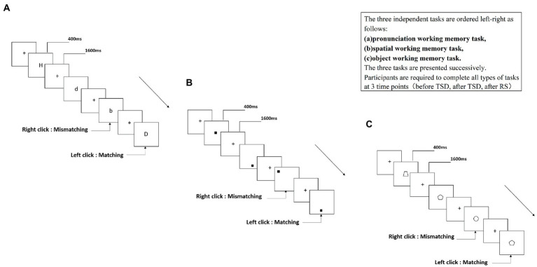
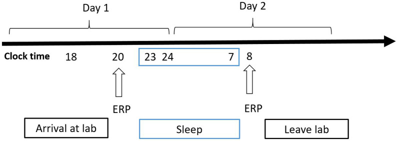
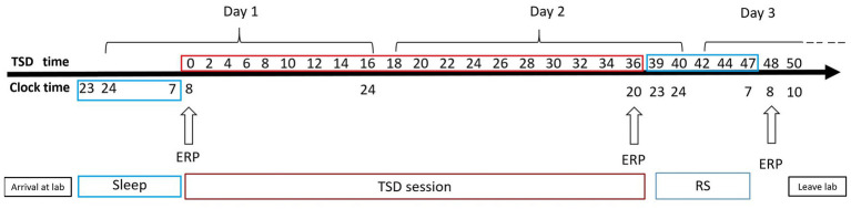
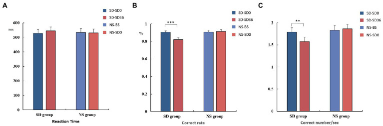
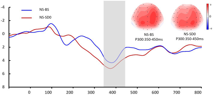
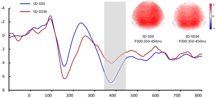
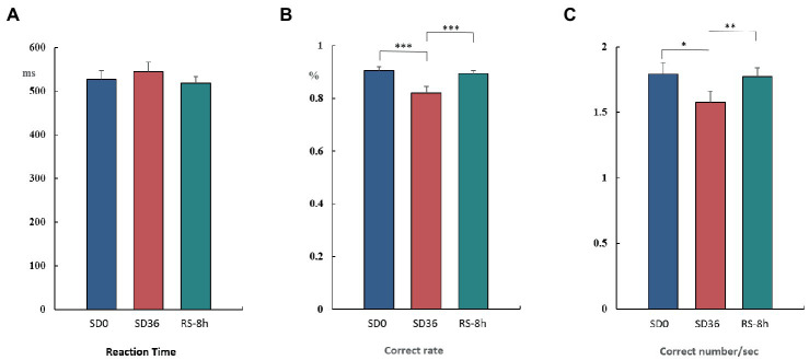
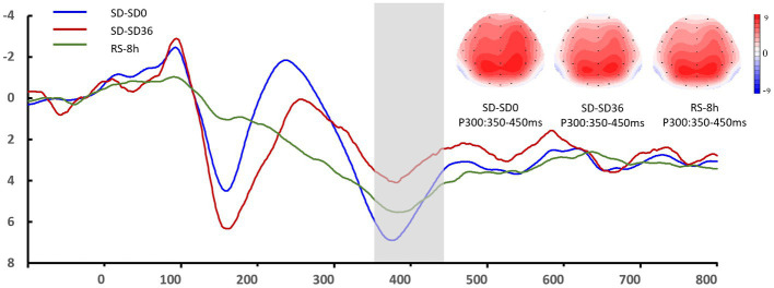
Introduction: The detrimental effects of sleep deprivation (SD) on cognitive function and quality of life are well known, and sleep disturbances are a major physical and mental health issue worldwide. Working memory plays an important role in many complex cognitive processes. Therefore, it is necessary to identify strategies that can effectively counteract the negative effects of SD on working memory. Methods: In the present study, we utilized event-related potentials (ERPs) to investigate the restorative effects of 8 h of recovery sleep (RS) on working memory impairments induced by total sleep deprivation for 36 h. We analyzed ERP data from 42 healthy male participants who were randomly assigned to two groups. The nocturnal sleep (NS) group completed a 2-back working memory task before and after normal sleep for 8 h. The sleep deprivation (SD) group completed a 2-back working memory task before and after 36 h of total sleep deprivation (TSD) and after 8 h of RS. Electroencephalographic data were recorded during each task. Results: The N2 and P3 components—which are related to working memory—exhibited low-amplitude and slow-wave characteristics after 36 h of TSD. Additionally, we observed a significant decrease in N2 latency after 8 h of RS. RS also induced significant increases in the amplitude of the P3 component and in the behavioral indicators. Discussion: Overall, 8 h of RS attenuated the decrease in working memory performance caused by 36 h of TSD. However, the effects of RS appear to be limited.
Overview
The claims and statistics of this paper haven’t been checked yet.
Run the check — free, no account needed. It reads the open-access text and checks the sources it cites.
Integrity signals
Registry
Cites
Statistics
Figures
Verification
Claims the paper makes
The claims and statistics of this paper haven’t been checked yet.
Run the check — free, no account needed. It reads the open-access text and checks the sources it cites.
Claims other papers make about this one
Each is checked against this paper’s own text: does it say what the citing paper says it does?
In the citing paper
“Meanwhile, acute SD has been shown to significantly impair various cognitive capabilities, including working memory, cognitive control, and emotional regulation (10–13).”
From Sleep deprivation alters utilization of negative feedback in risky decision-making (2024) · cites this paper as [12]
Self-check
Percentages, sums and reported differences re-computed from the paper’s own numbers.
The self-check needs the paper’s full text.
Citations
Cites
The episodic buffer: a new component of working memory?
Baddeley, A. (2000) · Trends in Cognitive Sciences · doi:10.1016/s1364-6613(00)01538-2 · Cited by 7,359 · cited 1×
“As such, it acts as a bridge between short- and long-term memory (Baddeley, 2000) and is involved in operating, processing, and executing various control processes.”
BackgroundSleep-dependent memory consolidation
Stickgold, R. (2005) · Nature · doi:10.1038/nature04286 · Cited by 1,857
Sleep-Dependent Learning and Memory Consolidation
Walker, M. P., & Stickgold, R. (2004) · Neuron · doi:10.1016/j.neuron.2004.08.031 · Cited by 1,113 · cited 2×
“Although proper sleep has been shown to exert bene cial effects on memory, cognitive function, work performance, and immune-related parameters [1, 2, 3, 4], the incidence of sleep problems continues to increase.”
BackgroundThe neurocognitive consequences of sleep restriction: A meta-analytic review
Lowe, C. J., Safati, A., & Hall, P. A. (2017) · Neuroscience & Biobehavioral Reviews · doi:10.1016/j.neubiorev.2017.07.010 · Cited by 520 · cited 1×
“The deterioration of sustained attention seems to be a longlasting negative effect of SD (de Bruin et al., 2017; Lowe et al., 2017), and is likely caused by decreased arousal of the central nervous system (CNS) (Schneider and Fisk, 1984; Cote et al.”
BackgroundLo, J. C., Ong, J. L., Leong, R. L. F., Gooley, J. J., & Chee, M. W. L. (2016) · Sleep · doi:10.5665/sleep.5552 · Cited by 446
Smallwood, J., Brown, K., Baird, B., & Schooler, J. W. (2012) · Brain Research · doi:10.1016/j.brainres.2011.03.072 · Cited by 379
Lo, J. C., Groeger, J. A., Santhi, N., Arbon, E. L., Lazar, A. S., Hasan, S., von Schantz, M., Archer, S. N., & Dijk, D.-J. (2012) · PLoS ONE · doi:10.1371/journal.pone.0045987 · Cited by 342
Huster, R. J., Westerhausen, R., Pantev, C., & Konrad, C. (2010) · Human Brain Mapping · doi:10.1002/hbm.20933 · Cited by 189
Simulation of daytime vigilance by the additive interaction of a homeostatic and a circadian process
Achermann, P., & Borbély, A. A. (1994) · Biological Cybernetics · doi:10.1007/bf00197314 · Cited by 180
Meta-Analysis of the Relationship Between Total Sleep Deprivation and Performance
Koslowsky, M., & Babkoff, H. (1992) · Chronobiology International · doi:10.3109/07420529209064524 · Cited by 158
Research works printed but not linked: 60
- Alhola, 2007, Sleep deprivation: impact on cognitive performance, Neuropsychiatr. Dis. Treat., 3, 553
- Almklov, 2015, The effects of sleep deprivation on brain functioning in older adults, Behav. Sleep Med., 13, 324, 10.1080/15402002.2014.905474 · DOI
- Anderson, 2006, Sleepiness enhances distraction during a monotonous task, Sleep, 29, 573, 10.1093/sleep/29.4.573 · DOI
- Anderson, 2011, Sleep deprivation lowers inhibition and enhances impulsivity to negative stimuli, Behav. Brain Res., 217, 463, 10.1016/j.bbr.2010.09.020 · DOI
- Baddeley, 2010, Working memory, Curr. Biol., 20, R136, 10.1016/j.cub.2009.12.014 · DOI
- Banks, 2010, Neurobehavioral dynamics following chronic sleep restriction: dose-response effects of one night for recovery, Sleep, 33, 1013, 10.1093/sleep/33.8.1013 · DOI · Paper page
- Biggs, 2007, Perception of simulated driving performance after sleep restriction and caffeine, J. Psychosom. Res., 63, 573, 10.1016/j.jpsychores.2007.06.017 · DOI
- Boardman, 2018, The ability to self-monitor cognitive performance during 60 h total sleep deprivation and following 2 nights recovery sleep, J. Sleep Res., 27, e12633, 10.1111/jsr.12633 · DOI
- Borbély, 1982, A two process model of sleep regulation, Hum. Neurobiol., 1, 195
- Buysse, 1989, The Pittsburgh sleep quality index: a new instrument for psychiatric practice and research, Psychiatry Res., 28, 193, 10.1016/0165-1781(89)90047-4 · DOI · Paper page
- Carskadon, 1985, Sleep loss in elderly volunteers, Sleep, 8, 207, 10.1093/sleep/8.3.207 · DOI
- Chai, 2020, Two nights of recovery sleep restores hippocampal connectivity but not episodic memory after total sleep deprivation, Sci. Rep., 10, 8774, 10.1038/s41598-020-65086-x · DOI
- Choo, 2005, Dissociation of cortical regions modulated by both working memory load and sleep deprivation and by sleep deprivation alone, NeuroImage, 25, 579, 10.1016/j.neuroimage.2004.11.029 · DOI · Paper page
- Chua, 2014, Sustained attention performance during sleep deprivation associates with instability in behavior and physiologic measures at baseline, Sleep, 37, 27, 10.5665/sleep.3302 · DOI
- Cote, 2009, CNS arousal and neurobehavioral performance in a short-term sleep restriction paradigm, J. Sleep Res., 18, 291, 10.1111/j.1365-2869.2008.00733.x · DOI
- Daan, 1984, Timing of human sleep: recovery process gated by a circadian pacemaker, Am. J. Phys., 246, R161, 10.1152/ajpregu.1984.246.2.R161 · DOI
- de Bruijn, 2020, Enhanced error-related brain activations for mistakes that harm others: ERP evidence from a novel social performance-monitoring paradigm, NeuroImage, 204, 116238, 10.1016/j.neuroimage.2019.116238 · DOI
- de Bruin, 2017, Effects of sleep manipulation on cognitive functioning of adolescents: a systematic review, Sleep Med. Rev., 32, 45, 10.1016/j.smrv.2016.02.006 · DOI · Paper page
- Dehaene, 2011, Experimental and theoretical approaches to conscious processing, Neuron, 70, 200, 10.1016/j.neuron.2011.03.018 · DOI · Paper page
- Dinges, 1986, Differential effects of prior wakefulness and circadian phase on nap sleep, Electroencephalogr. Clin. Neurophysiol., 64, 224, 10.1016/0013-4694(86)90170-7 · DOI
- Djonlagic, 2009, Sleep enhances category learning, Learn. Mem., 16, 751, 10.1101/lm.1634509 · DOI · Paper page
- Donchin, 1991, The Use of Event-Related Brain Potentials in the Study of Memory: Is P300 a Measure of event distinctiveness?, 58
- Doty, 2017, Limited efficacy of caffeine and recovery costs during and following 5 days of chronic sleep restriction, Sleep, 40, zsx171, 10.1093/sleep/zsx171 · DOI
- Drummond, 2006, Effects of two nights sleep deprivation and two nights recovery sleep on response inhibition, J. Sleep Res., 15, 261, 10.1111/j.1365-2869.2006.00535.x · DOI
- Folstein, 2008, Influence of cognitive control and mismatch on the N2 component of the ERP: a review, Psychophysiology, 45, 152, 10.1111/j.1469-8986.2007.00602.x · DOI · Paper page
- Gerhardsson, 2019, Effect of sleep deprivation on emotional working memory, J. Sleep Res., 28, e12744, 10.1111/jsr.12744 · DOI
- Gosselin, 2005, Total sleep deprivation and novelty processing: implications for frontal lobe functioning, Clin. Neurophysiol., 116, 211, 10.1016/j.clinph.2004.07.033 · DOI
- Gumenyuk, 2014, Sleep loss, circadian mismatch, and abnormalities in reorienting of attention in night workers with shift work disorder, Sleep, 37, 545, 10.5665/sleep.3494 · DOI
- Hennecke, 2019, Reestablishment of individual sleep structure during a single 14-h recovery sleep episode after 58 h of wakefulness, J. Sleep Res., 28, e12641, 10.1111/jsr.12641 · DOI
- Hilditch, 2017, A review of short naps and sleep inertia: do naps of 30 min or less really avoid sleep inertia and slow-wave sleep?, Sleep Med., 32, 176, 10.1016/j.sleep.2016.12.016 · DOI
- Honn, 2020, New insights into the cognitive effects of sleep deprivation by decomposition of a cognitive throughput task, Sleep, 43, zsz319, 10.1093/sleep/zsz319 · DOI
- Jiang, 2018, 0218 Effects of sleep deprivation and recovery sleep on human brain network organization, Sleep, 41, A85, 10.1093/sleep/zsy061.217 · DOI
- Jin, 2015, Recovery sleep reverses impaired response inhibition due to sleep restriction: evidence from a visual event related potentials study, PLoS One, 10, e0142361, 10.1371/journal.pone.0142361 · DOI
- Kiesel, 2008, Measurement of ERP latency differences: a comparison of single-participant and jackknife-based scoring methods, Psychophysiology, 45, 250, 10.1111/j.1469-8986.2007.00618.x · DOI
- Killgore, 2006, Impaired decision making following 49 h of sleep deprivation, J. Sleep Res., 15, 7, 10.1111/j.1365-2869.2006.00487.x · DOI · Paper page
- Kusztor, 2019, Sleep deprivation differentially affects subcomponents of cognitive control, Sleep, 42, zsz016, 10.1093/sleep/zsz016 · DOI
- Maddirala, 2016, Motion artifact removal from single channel electroencephalogram signals using singular spectrum analysis, Biomed. Signal Process. Control., 30, 79, 10.1016/j.bspc.2016.06.017 · DOI
- Malik, 2018, Sleep-wake classification via quantifying heart rate variability by convolutional neural network, Physiol. Meas., 39, 085004, 10.1088/1361-6579/aad5a9 · DOI
- Miccoli, 2008, Comparing sleep-loss sleepiness and sleep inertia: lapses make the difference, Chronobiol. Int., 25, 725, 10.1080/07420520802397228 · DOI
- Montplaisir, 1981, Depression and biological rhythm: therapeutic effects of sleep deprivation, Union Med. Can., 110, 272
- Nilsson, 2005, Less effective executive functioning after one night’s sleep deprivation, J. Sleep Res., 14, 1, 10.1111/j.1365-2869.2005.00442.x · DOI
- Panjwani, 2010, Electrophysiological correlates of cognition improve with nap during sleep deprivation, Eur. J. Appl. Physiol., 108, 549, 10.1007/s00421-009-1222-3 · DOI
- Peng, 2020, Effect of sleep deprivation on the working memory-related N2-P3 components of the event-related potential waveform, Front. Neurosci., 14, 469, 10.3389/fnins.2020.00469 · DOI
- Polich, 2007, Updating P300: an integrative theory of P3a and P3b, Clin. Neurophysiol., 118, 2128, 10.1016/j.clinph.2007.04.019 · DOI · Paper page
- Raud, 2017, The temporal dynamics of response inhibition and their modulation by cognitive control, Brain Topogr., 30, 486, 10.1007/s10548-017-0566-y · DOI
- Roehrs, 2012, Pain sensitivity and recovery from mild chronic sleep loss, Sleep, 35, 1667, 10.5665/sleep.2240 · DOI
- Rosenthal, 1991, Alerting effects of caffeine after normal and restricted sleep, Neuropsychopharmacology, 4, 103
- Ruggiero, 2014, Effects of napping on sleepiness and sleep-related performance deficits in night-shift workers: a systematic review, Biol. Res. Nurs., 16, 134, 10.1177/1099800413476571 · DOI
- Rupp, 2009, Banking sleep: realization of benefits during subsequent sleep restriction and recovery, Sleep, 32, 311, 10.1093/sleep/32.3.311 · DOI · Paper page
- Saletin, 2016, Human hippocampal structure: a novel biomarker predicting mnemonic vulnerability to, and recovery from, sleep deprivation, J. Neurosci., 36, 2355, 10.1523/JNEUROSCI.3466-15.2016 · DOI
- Schacht, 2008, The appraisal of facial beauty is rapid but not mandatory, Cogn. Affect. Behav. Neurosci., 8, 132, 10.3758/cabn.8.2.132 · DOI
- Schneider, 1984, Automatic category search and its transfer, J. Exp. Psychol. Learn. Mem. Cogn., 10, 1, 10.1037//0278-7393.10.1.1 · DOI
- Skurvydas, 2020, One night of sleep deprivation impairs executive function but does not affect psychomotor or motor performance, Biol. Sport, 37, 7, 10.5114/biolsport.2020.89936 · DOI
- Spano, 2019, Sleep deprivation by exposure to novel objects increases synapse density and axon-spine Interface in the hippocampal CA1 region of adolescent mice, J. Neurosci., 39, 6613, 10.1523/JNEUROSCI.0380-19.2019 · DOI
- Stroemel-Scheder, 2020, The effects of recovery sleep on pain perception: a systematic review, Neurosci. Biobehav. Rev., 113, 408, 10.1016/j.neubiorev.2020.03.028 · DOI
- Studte, 2015, Nap sleep preserves associative but not item memory performance, Neurobiol. Learn. Mem., 120, 84, 10.1016/j.nlm.2015.02.012 · DOI
- Verleger, 2005, Evidence for an integrative role of P3b in linking reaction to perception, J. Psychophysiol., 19, 165, 10.1027/0269-8803.19.3.165 · DOI · Paper page
- Wen, 2013, Top-down regulation of default mode activity in spatial visual attention, J. Neurosci., 33, 6444, 10.1523/JNEUROSCI.4939-12.2013 · DOI · Paper page
- Zhang, 2014, A visual ERP study of impulse inhibition following a zaleplon-induced nap after sleep deprivation, PLoS One, 9, e95653, 10.1371/journal.pone.0095653 · DOI
- Zhang, 2019, Decreased information replacement of working memory after sleep deprivation: evidence from an event-related potential study, Front. Neurosci., 13, 408, 10.3389/fnins.2019.00408 · DOI
Cited by
Sorted by citations
Sleep deprivation alters utilization of negative feedback in risky decision-making
Xu, W., Wang, L., Yang, L., Zhu, Y., & Chen, P. (2024) · Frontiers in Psychiatry · doi:10.3389/fpsyt.2024.1307408 · Cited by 2
Metadata
- Authors
- Peng, Z., Hou, Y., Xu, L., Wang, H., Wu, S., Song, T., Shao, Y., & Yang, Y.
- Venue
- Frontiers in Neuroscience
- Publisher
- Frontiers Media SA
- Published
- 2023-04-18
- Volume
- 17
- ISSN
- 1662-453X
- PMC
- PMC10151529
- Cited by
- 13
- References
- 12
About these checks
Registry, retraction, Cites and statistics results are computed, not judged, with one exception: a printed reference that no index lists is read once by a model before it counts as a citation error. Claims are extracted by a model and kept only when their quote appears verbatim in the text; each cited source is then checked, with a model’s help, for whether it says what the claim relies on. Those verdicts can be wrong — read the claim’s quote and the cited source before relying on one.
Statistics and arithmetic are recomputed, not judged.
Confidence bands are shown only for fields calibrated on our annotated test set. None is calibrated yet, so no band is shown.
What counts as a claim ›How we measure confidence ›How we check this ›
Are you an author, or do you see a problem with this page? Request a review
How to cite
Cite CoChat for the checks on this page, and the paper for its own findings.
- This page
- CoChat. (2026). Citation check: Recovery sleep attenuates impairments in working memory following total sleep deprivation. https://app.cochat.ai/papers/10.3389/fnins.2023.1056788
- The paper
- Peng, Z., Hou, Y., Xu, L., Wang, H., Wu, S., Song, T., Shao, Y., & Yang, Y. (2023). Recovery sleep attenuates impairments in working memory following total sleep deprivation. Frontiers in Neuroscience. https://doi.org/10.3389/fnins.2023.1056788
