

The DOI matches its Crossref record; 13 of the 13 references it cites were found in a registry or an index.
Bacterial Biofilm in Chronic Wounds and Possible Therapeutic Approaches
Abstract
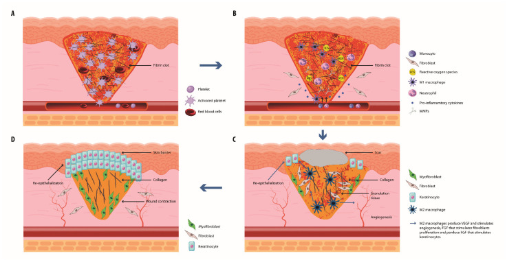
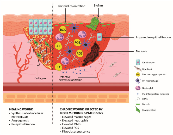
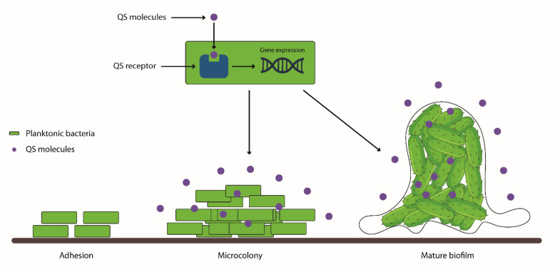
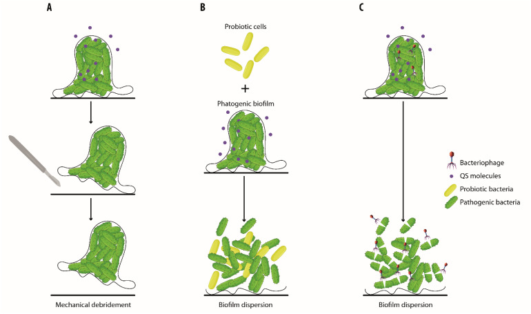
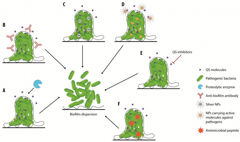
Wound repair and skin regeneration is a very complex orchestrated process that is generally composed of four phases: hemostasis, inflammation, proliferation, and remodeling. Each phase involves the activation of different cells and the production of various cytokines, chemokines, and other inflammatory mediators affecting the immune response. The microbial skin composition plays an important role in wound healing. Indeed, skin commensals are essential in the maintenance of the epidermal barrier function, regulation of the host immune response, and protection from invading pathogenic microorganisms. Chronic wounds are common and are considered a major public health problem due to their difficult-to-treat features and their frequent association with challenging chronic infections. These infections can be very tough to manage due to the ability of some bacteria to produce multicellular structures encapsulated into a matrix called biofilms. The bacterial species contained in the biofilm are often different, as is their capability to influence the healing of chronic wounds. Biofilms are, in fact, often tolerant and resistant to antibiotics and antiseptics, leading to the failure of treatment. For these reasons, biofilms impede appropriate treatment and, consequently, prolong the wound healing period. Hence, there is an urgent necessity to deepen the knowledge of the pathophysiology of delayed wound healing and to develop more effective therapeutic approaches able to restore tissue damage. This work covers the wound-healing process and the pathogenesis of chronic wounds infected by biofilm-forming pathogens. An overview of the strategies to counteract biofilm formation or to destroy existing biofilms is also provided.
Overview
The claims and statistics of this paper haven’t been checked yet.
Run the check — free, no account needed. It reads the open-access text and checks the sources it cites.
Integrity signals
Registry
Cites
Statistics
Figures
Verification
Claims the paper makes
The claims and statistics of this paper haven’t been checked yet.
Run the check — free, no account needed. It reads the open-access text and checks the sources it cites.
Claims other papers make about this one
Each is checked against this paper’s own text: does it say what the citing paper says it does?
In the citing paper
“Chronic wounds with biofilms are more likely to develop recalcitrant severe infections, leading to complications such as amputation and tissue necrosis.3”
From SEMTWIST Quantification of Biofilm Infection in Human Chronic Wound Using Scanning Electron Microscopy and Machine Learning (2025) · cites this paper as [3]
In the citing paper
“The extent to which the BFI burden negatively impacts wound healing outcomes depends on the balance between BFI threat and countering defenses, including host factors and therapeutic intervention.1,3,14,15”
From SEMTWIST Quantification of Biofilm Infection in Human Chronic Wound Using Scanning Electron Microscopy and Machine Learning (2025) · cites this paper as [3]
Self-check
Percentages, sums and reported differences re-computed from the paper’s own numbers.
The self-check needs the paper’s full text.
Citations
Cites
Principles of nanoparticle design for overcoming biological barriers to drug delivery
Blanco, E., Shen, H., & Ferrari, M. (2015) · Nature Biotechnology · doi:10.1038/nbt.3330 · Cited by 7,002 · cited 1×
“Nanoparticles (NPs) can cross biological barriers and, therefore, can efficiently release the antimicrobial agent directly within biofilms [117,118].”
BackgroundByrd, A. L., Belkaid, Y., & Segre, J. A. (2018) · Nature Reviews Microbiology · doi:10.1038/nrmicro.2017.157 · Cited by 2,806
Transition from inflammation to proliferation: a critical step during wound healing
Landén, N. X., Li, D., & Ståhle, M. (2016) · Cellular and Molecular Life Sciences · doi:10.1007/s00018-016-2268-0 · Cited by 1,980
Epidermal homeostasis: a balancing act of stem cells in the skin
Blanpain, C., & Fuchs, E. (2009) · Nature Reviews Molecular Cell Biology · doi:10.1038/nrm2636 · Cited by 1,270
Nakatsuji, T., Chen, T. H., Narala, S., Chun, K. A., Two, A. M., Yun, T., Shafiq, F., Kotol, P. F., Bouslimani, A., Melnik, A. V., et al. (2017) · Science Translational Medicine · doi:10.1126/scitranslmed.aah4680 · Cited by 1,095
Biofilms and Inflammation in Chronic Wounds
Zhao, G., Usui, M. L., Lippman, S. I., James, G. A., Stewart, P. S., Fleckman, P., & Olerud, J. E. (2013) · Advances in Wound Care · doi:10.1089/wound.2012.0381 · Cited by 448 · cited 3×
“Chronic wounds represent an ideal environment for biofilm formation as necrotic tissue and debris allow for bacterial attachment, and wounds are more susceptible to infection due to an impaired host immune response [69,70].”
BackgroundPastar, I., Nusbaum, A. G., Gil, J., Patel, S. B., Chen, J., Valdes, J., Stojadinovic, O., Plano, L. R., Tomic-Canic, M., & Davis, S. C. (2013) · PLoS ONE · doi:10.1371/journal.pone.0056846 · Cited by 391
The Neuropathic Diabetic Foot Ulcer Microbiome Is Associated With Clinical Factors
Gardner, S. E., Hillis, S. L., Heilmann, K., Segre, J. A., & Grice, E. A. (2013) · Diabetes · doi:10.2337/db12-0771 · Cited by 320
Pakshir, P., Noskovicova, N., Lodyga, M., Son, D. O., Schuster, R., Goodwin, A., Karvonen, H., & Hinz, B. (2020) · Journal of Cell Science · doi:10.1242/jcs.227900 · Cited by 293 · cited 1×
“In this phase, ECM components are modified to form a stronger and more organized matrix [28].”
BackgroundVan De Water, L., Varney, S., & Tomasek, J. J. (2013) · Advances in Wound Care · doi:10.1089/wound.2012.0393 · Cited by 251
Research works printed but not linked: 123
- Yousef, H., Alhajj, M., and Sharma, S. (2022, November 14). Anatomy, Skin (Integument), Epidermis, Available online: https://www.ncbi.nlm.nih.gov/books/NBK470464/.
- Blair, 2020, Skin structure-function relationships and the wound healing response to intrinsic aging, Adv. Wound Care, 9, 127, 10.1089/wound.2019.1021 · DOI
- Williams, 2021, Diverse cellular players orchestrate regeneration after wounding, Exp. Dermatol., 30, 605, 10.1111/exd.14248 · DOI
- Wilkinson, H.N., and Hardman, M.J. (2020). Wound healing: Cellular mechanisms and pathological outcomes. Open Biol., 10. · DOI
- Frykberg, 2015, Challenges in the treatment of chronic wounds, Adv. Wound Care, 4, 560, 10.1089/wound.2015.0635 · DOI
- Cavallo, I., Lesnoni La Parola, I., Sivori, F., Toma, L., Koudriavtseva, T., Sperduti, I., Kovacs, D., D’Agosto, G., Trento, E., and Cameli, N. (2022). homocysteine and inflammatory cytokines in the clinical assessment of infection in venous leg ulcers. Antibiotics, 11. · DOI
- Rodrigues, 2019, Wound healing: A cellular perspective, Physiol. Rev., 99, 665, 10.1152/physrev.00067.2017 · DOI
- Opneja, 2019, Contribution of platelets, the coagulation and fibrinolytic systems to cutaneous wound healing, Thromb. Res., 179, 56, 10.1016/j.thromres.2019.05.001 · DOI
- Kim, 2019, Macrophages in wound healing, activation and plasticity, Immunol. Cell Biol., 97, 258, 10.1111/imcb.12236 · DOI
- Demidova-Rice, T.N., Wolf, L., Deckenback, J., Hamblin, M.R., and Herman, I.M. (2012). Human platelet-rich plasma- and extracellular matrix-derived peptides promote impaired cutaneous wound healing in vivo. PLoS ONE, 7. · DOI
- Jiang, 2020, Cytokinocytes, the diverse contribution of keratinocytes to immune responses in skin, JCI Insight, 5, e142067, 10.1172/jci.insight.142067 · DOI
- Nguyen, A.V., and Soulika, A.M. (2019). The dynamics of the skin’s immune system. Int. J. Mol. Sci., 20. · DOI
- Munoz, L.D., Sweeney, M.J., and Jameson, J.M. (2020). Skin resident γδ T cell function and regulation in wound repair. Int. J. Mol. Sci., 21. · DOI
- Morgun, E.I., and Vorotelyak, E.A. (2020). Epidermal stem cells in hair follicle cycling and skin regeneration, a view from the perspective of inflammation. Front. Cell Dev. Biol., 8. · DOI
- Dunnill, 2017, Reactive oxygen species (ROS) and wound healing, the functional role of ROS and emerging ROS-modulating technologies for augmentation of the healing process, Int. Wound J., 14, 89, 10.1111/iwj.12557 · DOI
- Larouche, 2018, Immune regulation of skin wound healing, mechanisms and novel therapeutic targets, Adv. Wound Care, 7, 209, 10.1089/wound.2017.0761 · DOI
- Zhao, R., Liang, H., Clarke, E., Jackson, C., and Xue, M. (2016). Inflammation in chronic wounds. Int. J. Mol. Sci., 17. · DOI
- Cheng, 2016, Vimentin coordinates fibroblast proliferation and keratinocyte differentiation in wound healing via TGF-β-Slug signaling, Proc. Natl. Acad. Sci. USA, 113, E4320, 10.1073/pnas.1519197113 · DOI
- Schmidt, 2013, Intradermal adipocytes mediate fibroblast recruitment during skin wound healing, Development, 140, 1517, 10.1242/dev.087593 · DOI · Paper page
- Hao, 2023, Review: Research progress of adipose-derived stem cells in the treatment of chronic wounds, Front. Chem., 11, 1094693, 10.3389/fchem.2023.1094693 · DOI
- Diller, R.B., and Tabor, A.J. (2022). The role of the extracellular matrix (ECM) in wound healing: A review. Biomimetics, 7. · DOI
- Burgess, 2020, Skin microbiota and its interplay with wound healing, Am. J. Clin. Dermatol., 21, 36, 10.1007/s40257-020-00536-w · DOI
- Brun, 2016, Phenotypic and functional changes in dermal primary fibroblasts isolated from intrinsically aged human skin, Exp. Dermatol., 25, 113, 10.1111/exd.12874 · DOI
- Vallée, A., and Lecarpentier, Y. (2019). TGF-β in fibrosis by acting as a conductor for contractile properties of myofibroblasts. Cell Biosci., 9. · DOI
- Sorg, 2017, Skin wound healing: An update on the current knowledge and concepts, Eur. Surg. Res., 58, 81, 10.1159/000454919 · DOI
- Wang, 2017, The human microbiota in health and disease, Engineering, 3, 71, 10.1016/J.ENG.2017.01.008 · DOI
- Hasan, 2019, Factors affecting the composition of the gut microbiota, and its modulation, PeerJ, 7, e7502, 10.7717/peerj.7502 · DOI
- Hannigan, 2015, The human skin double-stranded DNA virome: Topographical and temporal diversity, genetic enrichment, and dynamic associations with the host microbiome, mBio, 6, e01578-15, 10.1128/mBio.01578-15 · DOI
- Grice, 2011, The skin microbiome, Nat. Rev. Microbiol., 9, 244, 10.1038/nrmicro2537 · DOI · Paper page
- Paharik, 2017, Coagulase-negative staphylococcal strain prevents Staphylococcus aureus colonization and skin infection by blocking quorum sensing, Cell Host Microbe, 22, 746, 10.1016/j.chom.2017.11.001 · DOI
- Huan, Y., Kong, Q., Mou, H., and Yi, H. (2020). Antimicrobial peptides, classification, design, application and research progress in multiple Fields. Front. Microbiol., 11. · DOI
- Swaney, 2021, Living in your skin: Microbes, molecules, and mechanisms, Infect. Immun., 89, e00695-e20, 10.1128/IAI.00695-20 · DOI
- Sakaguchi, 2008, Regulatory T cells and immune tolerance, Cell, 133, 775, 10.1016/j.cell.2008.05.009 · DOI
- Belkhodja, 2020, The commensal skin microbiota triggers type I IFN-dependent innate repair responses in injured skin, Nat. Immunol., 21, 1034, 10.1038/s41590-020-0721-6 · DOI
- Zheng, 2022, Commensal Staphylococcus epidermidis contributes to skin barrier homeostasis by generating protective ceramides, Cell Host Microbe, 30, 301, 10.1016/j.chom.2022.01.004 · DOI
- Alves, 2021, Update on the role of antiseptics in the management of chronic wounds with critical colonisation and/or biofilm, Int. Wound J., 18, 342, 10.1111/iwj.13537 · DOI
- Kadam, S., Nadkarni, S., Lele, J., Sakhalkar, S., Mokashi, P., and Kaushik, K.S. (2019). Bioengineered platforms for chronic wound infection studies: How can we make them more human-relevant?. Front. Bioeng. Biotechnol., 7. · DOI
- Escandon, 2011, High mortality in patients with chronic wounds, Wound Repair Regen., 19, 526, 10.1111/j.1524-475X.2011.00699.x · DOI
- Raziyeva, K., Kim, Y., Zharkinbekov, Z., Kassymbek, K., Jimi, S., and Saparov, A. (2021). immunology of acute and chronic wound healing. Biomolecules, 11. · DOI
- Caley, 2015, Metalloproteinases and wound healing, Adv. Wound Care, 4, 225, 10.1089/wound.2014.0581 · DOI
- Schilrreff, P., and Alexiev, U. (2022). Chronic inflammation in non-healing skin wounds and promising natural bioactive compounds treatment. Int. J. Mol. Sci., 23. · DOI
- Parnham, 2018, The influence of matrix metalloproteases and biofilm on chronic wound healing: A discussion, Br. J. Community Nurs., 23, S22, 10.12968/bjcn.2018.23.Sup3.S22 · DOI
- Sun, 2020, Challenges and new therapeutic approaches in the management of chronic wounds, Curr. Drug Targets, 21, 1264, 10.2174/1389450121666200623131200 · DOI
- Zeeuwen, P.L., Boekhorst, J., van den Bogaard, E.H., de Koning, H.D., van de Kerkhof, P.M., Saulnier, D.M., van Swam, I.I., van Hijum, S.A., Kleerebezem, M., and Schalkwijk, J. (2012). Microbiome dynamics of human epidermis following skin barrier disruption. Genome Biol., 13. · DOI · Paper page
- Misic, 2014, The wound microbiome, modern approaches to examining the role of microorganisms in impaired chronic wound healing, Adv. Wound Care, 3, 502, 10.1089/wound.2012.0397 · DOI · Paper page
- Williams, 2021, A systematic review and critical appraisal of metagenomic and culture studies in hidradenitis suppurativa, Exp. Dermatol., 30, 1388, 10.1111/exd.14141 · DOI
- Verbanic, S., Shen, Y., Lee, J., Deacon, J.M., and Chen, I.A. (2020). Microbial predictors of healing and short-term effect of debridement on the microbiome of chronic wounds. NPJ Biofilms Microbiomes, 6. · DOI
- Price, 2011, Macroscale spatial variation in chronic wound microbiota, a cross-sectional study, Wound Repair Regen., 19, 80, 10.1111/j.1524-475X.2010.00628.x · DOI
- Wolcott, R.D., Gontcharova, V., Sun, Y., and Dowd, S.E. (2009). Evaluation of the bacterial diversity among and within individual venous leg ulcers using bacterial tag-encoded FLX and titanium amplicon pyrosequencing and metagenomic approaches. BMC Microbiol., 27. · DOI
- Wolcott, 2016, Analysis of the chronic wound microbiota of 2,963 patients by 16S rDNA pyrosequencing, Wound Repair Regen., 24, 163, 10.1111/wrr.12370 · DOI
- Negut, I., Grumezescu, V., and Grumezescu, A.M. (2018). Treatment strategies for infected wounds. Molecules, 23. · DOI
- Duell, 2012, Recent insights into microbial triggers of interleukin-10 production in the host and the impact on infectious disease pathogenesis, FEMS Immunol. Med. Microbiol., 64, 295, 10.1111/j.1574-695X.2012.00931.x · DOI
- Di Domenico, E.G., Marchesi, F., Cavallo, I., Toma, L., Sivori, F., Papa, E., Spadea, A., Cafarella, G., Terrenato, I., and Prignano, G. (2021). The impact of bacterial biofilms on end-organ disease and mortality in patients with hematologic malignancies developing a bloodstream infection. Microbiol. Spectr., 9. · DOI
- Flemming, H.C., Wingender, J., Szewzyk, U., Steinberg, P., Rice, S.A., and Kjelleberg, S. (2016). Biofilms: An emergent form of bacterial life. Nat. Rev. Microbiol., 14. · DOI · Paper page
- Percival, 2015, Biofilms and wounds: An identification algorithm and potential treatment options, Adv. Wound Care, 4, 389, 10.1089/wound.2014.0574 · DOI
- Prakash, 2003, Biofilms: A Survival Strategy of Bacteria, Curr. Sci., 85, 1299
- Kostakioti, 2013, Bacterial biofilms, development, dispersal, and therapeutic strategies in the dawn of the postantibiotic era, Cold Spring Harb. Perspect. Med., 3, a010306, 10.1101/cshperspect.a010306 · DOI
- Siddiqui, 2010, Chronic wound infection, facts and controversies, Clin. Dermatol., 28, 519, 10.1016/j.clindermatol.2010.03.009 · DOI
- Sanchez, C.J., Mende, K., Beckius, M.L., Akers, K.S., Romano, D.R., Wenke, J.C., and Murray, C.K. (2013). Biofilm formation by clinical isolates and the implications in chronic infections. BMC Infect. Dis., 13. · DOI
- Harika, 2020, Detection of biofilm production and its impact on antibiotic resistance profile of bacterial isolates from chronic wound infections, J. Glob. Infect. Dis., 12, 129, 10.4103/jgid.jgid_150_19 · DOI
- Kalan, 2019, Strain- and species-level variation in the microbiome of diabetic wounds is associated with clinical outcomes and therapeutic efficacy, Cell Host Microbe, 25, 641, 10.1016/j.chom.2019.03.006 · DOI
- Fedtke, 2004, Bacterial evasion of innate host defenses—The Staphylococcus aureus lesson, Int. J. Med. Microbiol., 294, 189, 10.1016/j.ijmm.2004.06.016 · DOI
- Jensen, 2007, Rapid necrotic killing of polymorphonuclear leukocytes is caused by quorum-sensing-controlled production of rhamnolipid by Pseudomonas aeruginosa, Microbiology, 153, 1329, 10.1099/mic.0.2006/003863-0 · DOI
- Fazli, 2009, Nonrandom distribution of Pseudomonas aeruginosa and Staphylococcus aureus in chronic wounds, J. Clin. Microbiol., 47, 4084, 10.1128/JCM.01395-09 · DOI
- Di Domenico, E.G., Farulla, I., Prignano, G., Gallo, M.T., Vespaziani, M., Cavallo, I., Sperduti, I., Pontone, M., Bordignon, V., and Cilli, L. (2017). Biofilm is a major virulence determinant in bacterial colonization of chronic skin ulcers independently from the multidrug resistant phenotype. Int. J. Mol. Sci., 18. · DOI
- Ryder, V.J., Chopra, I., and O’Neill, A.J. (2012). Increased mutability of Staphylococci in biofilms as a consequence of oxidative stress. PLoS ONE, 7. · DOI
- Omar, A., Wright, J.B., Schultz, G., Burrell, R., and Nadworny, P. (2017). Microbial biofilms and chronic wounds. Microorganisms, 5. · DOI
- Li, 2023, Biofilms in wound healing: A bibliometric and visualised study, Int. Wound J., 20, 313, 10.1111/iwj.13878 · DOI
- Versey, 2021, Biofilm-innate immune interface: Contribution to chronic wound formation, Front. Immunol., 12, 648554, 10.3389/fimmu.2021.648554 · DOI · Paper page
- Shahrour, 2019, AMPs as anti-biofilm agents for human therapy and prophylaxis, Adv. Exp. Med. Biol., 1117, 257, 10.1007/978-981-13-3588-4_14 · DOI
- Wu, 2019, Biofilms in chronic wounds, pathogenesis and diagnosis, Trends Biotechnol., 37, 505, 10.1016/j.tibtech.2018.10.011 · DOI
- Caldwell, 2020, Bacteria and antibiotics in wound healing, Surg. Clin. N. Am., 100, 757, 10.1016/j.suc.2020.05.007 · DOI
- Falcone, 2021, Challenges in the management of chronic wound infections, J. Glob. Antimicrob. Resist., 26, 140, 10.1016/j.jgar.2021.05.010 · DOI · Paper page
- Falabella, 2006, Debridement and wound bed preparation, Dermatol. Ther., 19, 317, 10.1111/j.1529-8019.2006.00090.x · DOI
- Thomas, 2021, The role of debridement in wound bed preparation in chronic wound: A narrative review, Ann. Med. Surg., 71, 102876, 10.1016/j.amsu.2021.102876 · DOI
- Shi, 2011, pH in the bacteria-contaminated wound and its impact on clostridium histolyticum collagenase activity, implications for the use of collagenase wound debridement agents, J. Wound Ostomy Continence Nurs., 38, 514, 10.1097/WON.0b013e31822ad034 · DOI
- Madhok, 2013, New techniques for wound debridement, Int. Wound J., 10, 247, 10.1111/iwj.12045 · DOI
- Liu, 2017, Combined debridement in chronic wounds, A literature review, Chin. Nurs. Res., 4, 5, 10.1016/j.cnre.2017.03.003 · DOI
- Goswami, 2023, Biofilm and wound healing, from bench to bedside, Eur. J. Med. Res., 28, 157, 10.1186/s40001-023-01121-7 · DOI · Paper page
- Sen, 2021, Biofilm management in wound care, Plast. Reconstr. Surg., 148, 275e, 10.1097/PRS.0000000000008142 · DOI · Paper page
- Lipsky, 2016, Antimicrobial stewardship in wound care: A Position Paper from the British Society for Antimicrobial Chemotherapy and European Wound Management Association, J. Antimicrob. Chemother., 71, 3026, 10.1093/jac/dkw287 · DOI
- Yang, 2017, A surfactant-based wound dressing can reduce bacterial biofilms in a porcine skin explant model, Int. Wound J., 14, 408, 10.1111/iwj.12619 · DOI
- Phillips, 2015, Antimicrobial dressing efficacy against mature Pseudomonas aeruginosa biofilm on porcine skin explants, Int. Wound J., 12, 469, 10.1111/iwj.12142 · DOI
- Malone, 2019, Effect on total microbial load and community composition with two vs six-week topical Cadexomer Iodine for treating chronic biofilm infections in diabetic foot ulcers, Int. Wound J., 16, 1477, 10.1111/iwj.13219 · DOI
- Malone, 2017, Effect of cadexomer iodine on the microbial load and diversity of chronic non-healing diabetic foot ulcers complicated by biofilm in vivo, J. Antimicrob. Chemother., 72, 2093, 10.1093/jac/dkx099 · DOI
- Zaver, V., and Kankanalu, P. (2022, September 09). Negative Pressure Wound Therapy, Available online: https://www.ncbi.nlm.nih.gov/books/NBK576388/.
- Tang, 2023, Role of wound microbiome, strategies of microbiota delivery system and clinical management, Adv. Drug Deliv. Rev., 192, 114671, 10.1016/j.addr.2022.114671 · DOI
- Moysidis, 2022, The 3-D configuration of excisional skin wound healing after topical probiotic application, Injury, 53, 1385, 10.1016/j.injury.2022.02.006 · DOI
- Paluch, 2020, Prevention of biofilm formation by quorum quenching, Appl. Microbiol. Biotechnol., 104, 1871, 10.1007/s00253-020-10349-w · DOI
- Knackstedt, 2020, The role of topical probiotics on wound healing, A review of animal and human studies, Int. Wound J., 17, 1687, 10.1111/iwj.13451 · DOI
- Salman, M.K., Abuqwider, J., and Mauriello, G. (2023). Anti-quorum sensing activity of probiotics: The mechanism and role in food and gut health. Microorganisms, 11. · DOI
- Al-Shamiri, M.M., Wang, J., Zhang, S., Li, P., Odhiambo, W.O., Chen, Y., Han, B., Yang, E., Xun, M., and Han, L. (2023). Probiotic Lactobacillus species and their biosurfactants eliminate Acinetobacter baumannii biofilm in various manners. Microbiol. Spectr., 11. · DOI
- Walencka, 2008, The influence of Lactobacillus acidophilus-derived surfactants on staphylococcal adhesion and biofilm formation, Folia Microbiol., 53, 61, 10.1007/s12223-008-0009-y · DOI
- Wilson, R.M., Walker, J.M., and Yin, K. (2021). Different concentrations of Lactobacillus acidophilus cell free filtrate have differing anti-biofilm and immunomodulatory effects. Front. Cell. Infect. Microbiol., 11. · DOI
- Gan, 2002, Lactobacillus fermentum RC-14 inhibits Staphylococcus aureus infection of surgical implants in rats, J. Infect. Dis., 185, 1369, 10.1086/340126 · DOI
- Rybalchenko, 2015, Inhibitory effects of Lactobacillus fermentum on microbial growth and biofilm formation, Arch. Microbiol., 197, 1027, 10.1007/s00203-015-1140-1 · DOI
- Peral, 2009, Bacteriotherapy with Lactobacillus plantarum in burns, Int. Wound J., 6, 73, 10.1111/j.1742-481X.2008.00577.x · DOI
- Venosi, 2019, Infected chronic ischemic wound topically treated with a multi-strain probiotic formulation, a novel tailored treatment strategy, J. Transl. Med., 17, 364, 10.1186/s12967-019-2111-0 · DOI
- Starkey, M., Lepine, F., Maura, D., Bandyopadhaya, A., Lesic, B., He, J., Kitao, T., Righi, V., Milot, S., and Tzika, A. (2014). Identification of anti-virulence compounds that disrupt quorum-sensing regulated acute and persistent pathogenicity. PLoS Pathog., 10. · DOI
- Guendouze, A., Plener, L., Bzdrenga, J., Jacquet, P., Rémy, B., Elias, M., Lavigne, J.P., Daudé, D., and Chabrière, E. (2017). Effect of quorum quenching lactonase in clinical isolates of Pseudomonas aeruginosa and comparison with quorum sensing inhibitors. Front. Microbiol., 8. · DOI
- Mion, S., Rémy, B., Plener, L., Brégeon, F., Chabrière, E., and Daudé, D. (2019). Quorum quenching lactonase strengthens bacteriophage and antibiotic arsenal against Pseudomonas aeruginosa clinical isolates. Front. Microbiol., 10. · DOI
- Fernandes, 2023, Curcumin and 10-undecenoic acid as natural quorum sensing inhibitors of LuxS/AI-2 of Bacillus subtilis and LasI/LasR of Pseudomonas aeruginosa, Food Res. Int., 165, 112519, 10.1016/j.foodres.2023.112519 · DOI
- Bruna, T., Maldonado-Bravo, F., Jara, P., and Caro, N. (2021). Silver nanoparticles and their antibacterial applications. Int. J. Mol. Sci., 22. · DOI
- Kumari, A., Raina, N., Wahi, A., Goh, K.W., Sharma, P., Nagpal, R., Jain, A., Ming, L.C., and Gupta, M. (2022). Wound-healing effects of curcumin and its nanoformulations: A comprehensive review. Pharmaceutics, 14. · DOI
- Fahimirad, 2021, Wound healing performance of PCL/chitosan based electrospun nanofiber electrosprayed with curcumin loaded chitosan nanoparticles, Carbohydr. Polym., 259, 117640, 10.1016/j.carbpol.2021.117640 · DOI
- Geng, 2021, An innovative role for luteolin as a natural quorum sensing inhibitor in Pseudomonas aeruginosa, Life Sci., 274, 119325, 10.1016/j.lfs.2021.119325 · DOI
- Sun, 2022, Luteolin inhibits the biofilm formation and cytotoxicity of methicillin-resistant Staphylococcus aureus via decreasing bacterial toxin synthesis, Evid. Based Complement. Altern. Med., 2022, 4476339
- Gurunathan, 2014, Enhanced antibacterial and anti-biofilm activities of silver nanoparticles against Gram-negative and Gram-positive bacteria, Nanoscale Res. Lett., 9, 373, 10.1186/1556-276X-9-373 · DOI
- Chegini, Z., Khoshbayan, A., Taati Moghadam, M., Farahani, I., Jazireian, P., and Shariati, A. (2020). Bacteriophage therapy against Pseudomonas aeruginosa biofilms, a review. Ann. Clin. Microbiol. Antimicrob., 19. · DOI
- Gupta, 2019, Bacteriophage therapy of chronic nonhealing wound, clinical study, Int. J. Low. Extrem. Wounds, 18, 171, 10.1177/1534734619835115 · DOI
- Jault, 2019, Efficacy and tolerability of a cocktail of bacteriophages to treat burn wounds infected by Pseudomonas aeruginosa (PhagoBurn), A randomised, controlled, double-blind phase 1/2 trial, Lancet Infect. Dis., 19, 35, 10.1016/S1473-3099(18)30482-1 · DOI
- Ramezanalizadeh, 2022, Vaccination with a combination of planktonic and biofilm virulence factors confers protection against carbapenem-resistant Acinetobacter baumannii strains, Sci. Rep., 12, 19909, 10.1038/s41598-022-24163-z · DOI
- Ramezanalizadeh, 2021, Protective response against Acinetobacter baumannii with ferric iron receptors HemTR-BauA in a murine sepsis model, Future Microbiol., 16, 159, 10.2217/fmb-2020-0133 · DOI
- Ahmadi, 2021, Anti-flagellin IgY antibodies protect against Pseudomonas aeruginosa infection in both acute pneumonia and burn wound murine models in a non-type-specific mode, Mol. Immunol., 136, 118, 10.1016/j.molimm.2021.06.002 · DOI
- Ahmadi, 2024, Polyclonal anti-whole cell IgY passive immunotherapy shields against P. aeruginosa-induced acute pneumonia and burn wound infections in murine models, Sci. Rep., 14, 405, 10.1038/s41598-023-50859-x · DOI
- Raafat, 2019, Fighting Staphylococcus aureus biofilms with monoclonal antibodies, Trends Microbiol., 27, 303, 10.1016/j.tim.2018.12.009 · DOI
- Zhang, 2016, Antimicrobial peptides, Curr. Biol., 26, R14, 10.1016/j.cub.2015.11.017 · DOI
- Mansour, 2014, Host defense peptides, front-line immunomodulators, Trends Immunol., 35, 443, 10.1016/j.it.2014.07.004 · DOI
- Lundquist, 2011, S100A8/A9 deficiency in nonhealing venous leg ulcers uncovered by multiplexed antibody microarray profiling, Br. J. Dermatol., 165, 292, 10.1111/j.1365-2133.2011.10384.x · DOI
- Holstein, 2016, S100A8/A9 is an important host defence mediator in neuropathic foot ulcers in patients with type 2 diabetes mellitus, Arch. Dermatol. Res., 308, 347, 10.1007/s00403-016-1646-7 · DOI
- Thapa, 2020, Topical antimicrobial peptide formulations for wound healing: Current developments and future prospects, Acta Biomater., 103, 52, 10.1016/j.actbio.2019.12.025 · DOI
- Makabenta, 2023, Antimicrobial polymer-loaded hydrogels for the topical treatment of multidrug-resistant wound biofilm infections, J. Control. Release, 362, 513, 10.1016/j.jconrel.2023.09.002 · DOI
- Grassi, L., Maisetta, G., Esin, S., and Batoni, G. (2017). Combination strategies to enhance the efficacy of antimicrobial peptides against bacterial biofilms. Front. Microbiol., 8. · DOI
Cited by
Sorted by citations
Singh, S., Muniz De Oliveira, F., Wang, C., Kumar, M., Xuan, Y., DeMazumder, D., Sen, C. K., & Roy, S. (2025) · Advances in Wound Care · doi:10.1089/wound.2024.0291 · Cited by 6
Metadata
- Authors
- Cavallo, I., Sivori, F., Mastrofrancesco, A., Abril, E., Pontone, M., Di Domenico, E. G., & Pimpinelli, F.
- Venue
- Biology
- Publisher
- MDPI AG
- Published
- 2024-02-09
- Volume
- 13
- Issue
- 2
- Pages
- 109
- ISSN
- 2079-7737
- PMC
- PMC10886835
- Cited by
- 213
- References
- 13
- Language
- en
- Funders
- Italian Ministry of Health (Ricerca Corrente)
About these checks
Registry, retraction, Cites and statistics results are computed, not judged, with one exception: a printed reference that no index lists is read once by a model before it counts as a citation error. Claims are extracted by a model and kept only when their quote appears verbatim in the text; each cited source is then checked, with a model’s help, for whether it says what the claim relies on. Those verdicts can be wrong — read the claim’s quote and the cited source before relying on one.
Statistics and arithmetic are recomputed, not judged.
Confidence bands are shown only for fields calibrated on our annotated test set. None is calibrated yet, so no band is shown.
What counts as a claim ›How we measure confidence ›How we check this ›
Are you an author, or do you see a problem with this page? Request a review
