

The DOI matches its Crossref record; 35 of the 35 references it cites were found in a registry or an index.
Biofilm-Innate Immune Interface: Contribution to Chronic Wound Formation
Abstract
Delayed wound healing can cause significant issues for immobile and ageing individuals as well as those living with co-morbid conditions such as diabetes, cardiovascular disease, and cancer. These delays increase a patient’s risk for infection and, in severe cases, can result in the formation of chronic, non-healing ulcers (e.g., diabetic foot ulcers, surgical site infections, pressure ulcers and venous leg ulcers). Chronic wounds are very difficult and expensive to treat and there is an urgent need to develop more effective therapeutics that restore healing processes. Sustained innate immune activation and inflammation are common features observed across most chronic wound types. However, the factors driving this activation remain incompletely understood. Emerging evidence suggests that the composition and structure of the wound microbiome may play a central role in driving this dysregulated activation but the cellular and molecular mechanisms underlying these processes require further investigation. In this review, we will discuss the current literature on: 1) how bacterial populations and biofilms contribute to chronic wound formation, 2) the role of bacteria and biofilms in driving dysfunctional innate immune responses in chronic wounds, and 3) therapeutics currently available (or underdevelopment) that target bacteria-innate immune interactions to improve healing. We will also discuss potential issues in studying the complexity of immune-biofilm interactions in chronic wounds and explore future areas of investigation for the field.
Overview
The claims and statistics of this paper haven’t been checked yet.
Run the check — free, no account needed. It reads the open-access text and checks the sources it cites.
Integrity signals
Registry
Cites
Statistics
Figures
Verification
Claims the paper makes
The claims and statistics of this paper haven’t been checked yet.
Run the check — free, no account needed. It reads the open-access text and checks the sources it cites.
Claims other papers make about this one
Each is checked against this paper’s own text: does it say what the citing paper says it does?
In the citing paper
“Biofilms create a persistent inflammatory state that disrupts normal wound healing processes by impairing reepithelialization and angiogenesis.2”
From SEMTWIST Quantification of Biofilm Infection in Human Chronic Wound Using Scanning Electron Microscopy and Machine Learning (2025) · cites this paper as [2]
Self-check
Percentages, sums and reported differences re-computed from the paper’s own numbers.
The self-check needs the paper’s full text.
Citations
Cites
Exploring the full spectrum of macrophage activation
Mosser, D. M., & Edwards, J. P. (2008) · Nature Reviews Immunology · doi:10.1038/nri2448 · Cited by 9,144
Factors affecting wound healing
Guo, S., & DiPietro, L. A. (2010) · Journal of Dental Research · doi:10.1177/0022034509359125 · Cited by 5,792
Protective and pathogenic functions of macrophage subsets
Murray, P. J., & Wynn, T. A. (2011) · Nature Reviews Immunology · doi:10.1038/nri3073 · Cited by 5,269
Byrd, A. L., Belkaid, Y., & Segre, J. A. (2018) · Nature Reviews Microbiology · doi:10.1038/nrmicro.2017.157 · Cited by 2,806
Methicillin-Resistant S. aureus Infections among Patients in the Emergency Department
Moran, G. J., Krishnadasan, A., Gorwitz, R. J., Fosheim, G. E., McDougal, L. K., Carey, R. B., & Talan, D. A. (2006) · New England Journal of Medicine · doi:10.1056/nejmoa055356 · Cited by 2,285
Transition from inflammation to proliferation: a critical step during wound healing
Landén, N. X., Li, D., & Ståhle, M. (2016) · Cellular and Molecular Life Sciences · doi:10.1007/s00018-016-2268-0 · Cited by 1,980 · cited 9×
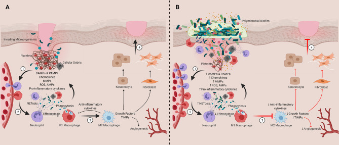
“The innate immune system plays a critical role in modulating these processes as well as the transition from inflammation to proliferation phases, which includes a transition of inflammatory macrophages (M1) into anti-inflammatory wound healing cells (M2; Step 3) (86, 87).”
BackgroundInfluentialTopographic diversity of fungal and bacterial communities in human skin
NIH Intramural Sequencing Center Comparative Sequencing Program, Findley, K., Oh, J., Yang, J., Conlan, S., Deming, C., Meyer, J. A., Schoenfeld, D., Nomicos, E., Park, M., et al. (2013) · Nature · doi:10.1038/nature12171 · Cited by 1,206
Sindrilaru, A., Peters, T., Wieschalka, S., Baican, C., Baican, A., Peter, H., Hainzl, A., Schatz, S., Qi, Y., Schlecht, A., et al. (2011) · Journal of Clinical Investigation · doi:10.1172/jci44490 · Cited by 1,100
Edwards, R., & Harding, K. G. (2004) · Current Opinion in Infectious Diseases · doi:10.1097/00001432-200404000-00004 · Cited by 1,099
Phagocyte partnership during the onset and resolution of inflammation
Soehnlein, O., & Lindbom, L. (2010) · Nature Reviews Immunology · doi:10.1038/nri2779 · Cited by 1,054
Research works printed but not linked: 225
- Morton, 2016, Wound healing and treating wounds, J Am Acad Dermatol, 74, 589, 10.1016/j.jaad.2015.08.068 · DOI · Paper page
- Järbrink, 2017, The humanistic and economic burden of chronic wounds: a protocol for a systematic review, Systematic Rev, 6, 15, 10.1186/s13643-016-0400-8 · DOI
- Tricco, 2015, A systematic review of cost-effectiveness analyses of complex wound interventions reveals optimal treatments for specific wound types, BMC Med, 13, 90, 10.1186/s12916-015-0326-3 · DOI
- Percival, 2010, Biofilms and bacterial imbalances in chronic wounds: anti-Koch, Int Wound J, 7, 10.1111/j.1742-481X.2010.00668.x · DOI
- Wolcott, 2016, Analysis of the chronic wound microbiota of 2,963 patients by 16S rDNA pyrosequencing, Wound Repair Regeneration, 24, 10.1111/wrr.12370 · DOI
- Metcalf, 2013, Biofilm delays wound healing: A review of the evidence, Burns Trauma, 1, 5, 10.4103/2321-3868.113329 · DOI
- Lopez, 2010, Biofilms, Cold Spring Harbor Perspect Biol, 2, a000398, 10.1101/cshperspect.a000398 · DOI
- Malone, 2017, The prevalence of biofilms in chronic wounds: a systematic review and meta-analysis of published data, J Wound Care, 26, 10.12968/jowc.2017.26.1.20 · DOI
- Kirketerp-Møller, 2020, The zone model: A conceptual model for understanding the microenvironment of chronic wound infection, Wound Repair Regeneration, 28, 10.1111/wrr.12841 · DOI
- Frieri, 2017, Antibiotic resistance, J Infect Public Health, 10, 10.1016/j.jiph.2016.08.007 · DOI
- Bjarnsholt, 2013, Applying insights from biofilm biology to drug development — can a new approach be developed, Nat Rev Drug Discovery, 12, 791, 10.1038/nrd4000 · DOI
- Høiby, 2015, ESCMID∗ guideline for the diagnosis and treatment of biofilm infections 2014, Clin Microbiol Infect, 21, S1, 10.1016/j.cmi.2014.10.024 · DOI · Paper page
- Hall, 2017, Molecular mechanisms of biofilm-based antibiotic resistance and tolerance in pathogenic bacteria, FEMS Microbiol Rev, 41, 276, 10.1093/femsre/fux010 · DOI
- Høiby, 2010, Antibiotic resistance of bacterial biofilms, Int J Antimicrobial Agents, 35, 10.1016/j.ijantimicag.2009.12.011 · DOI
- Percival, 2012, A review of the scientific evidence for biofilms in wounds, Wound Repair Regeneration, 20, 10.1111/j.1524-475X.2012.00836.x · DOI
- Flemming, 2016, Biofilms: an emergent form of bacterial life, Nat Rev Microbiol, 14, 10.1038/nrmicro.2016.94 · DOI · Paper page
- Wu, 2019, Biofilms in Chronic Wounds: Pathogenesis and Diagnosis, Trends Biotechnol, 37, 10.1016/j.tibtech.2018.10.011 · DOI
- Lebeaux, 2013, From in vitro to in vivo Models of Bacterial Biofilm-Related Infections, Pathogens, 2, 288, 10.3390/pathogens2020288 · DOI
- Martin, 2010, Molecular Microbiology: New Dimensions for Cutaneous Biology and Wound Healing, J Invest Dermatol, 130, 38, 10.1038/jid.2009.221 · DOI
- Ursell, 2012, Defining the human microbiome, Nutr Rev, 70, 10.1111/j.1753-4887.2012.00493.x · DOI · Paper page
- Ursell, 2014, The Intestinal Metabolome: An Intersection Between Microbiota and Host, Gastroenterology, 146, 10.1053/j.gastro.2014.03.001 · DOI
- Wang, 2017, The Human Microbiota in Health and Disease, Engineering, 3, 71, 10.1016/J.ENG.2017.01.008 · DOI
- Sender, 2016, Are We Really Vastly Outnumbered? Revisiting the Ratio of Bacterial to Host Cells in Humans, Cell, 164, 10.1016/j.cell.2016.01.013 · DOI · Paper page
- Hasan, 2019, Factors affecting the composition of the gut microbiota, and its modulation, PeerJ, 7, e7502, 10.7717/peerj.7502 · DOI
- Grice, 2011, The skin microbiome, Nat Rev Microbiol, 9, 10.1038/nrmicro2537 · DOI · Paper page
- Grice, 2009, Topographical and Temporal Diversity of the Human Skin Microbiome, Science, 324, 10.1126/science.1171700 · DOI · Paper page
- Daeschlein, 2013, Antimicrobial and antiseptic strategies in wound management, Int Wound J, 10, 9, 10.1111/iwj.12175 · DOI
- Bowler, 2001, Wound Microbiology and Associated Approaches to Wound Management, Clin Microbiol Rev, 14, 10.1128/CMR.14.2.244-269.2001 · DOI · Paper page
- Orsted, 2017, Best practice recommendations for the prevention and management of wounds, 74
- Negut, 2018, Treatment Strategies for Infected Wounds, Molecules, 23, 10.3390/molecules23092392 · DOI
- Kingsley, 2003, The wound infection continuum and its application to clinical practice, Ostomy Manage, 49, 1
- Gardner, 2008, Wound bioburden and infection-related complications in diabetic foot ulcers, Biol Res Nurs, 10, 44, 10.1177/1099800408319056 · DOI
- Baviera, 2014, Microbiota in healthy skin and in atopic eczema, BioMed Res Int, 2014, 10.1155/2014/436921 · DOI
- Grice, 2008, A diversity profile of the human skin microbiota, Genome Res, 18, 10.1101/gr.075549.107 · DOI
- Tuttle, 2015, Association Between Microbial Bioburden and Healing Outcomes in Venous Leg Ulcers: A Review of the Evidence, Adv Wound Care, 4, 1, 10.1089/wound.2014.0535 · DOI
- Smith, 2016, One step closer to understanding the role of bacteria in diabetic foot ulcers: characterising the microbiome of ulcers, BMC Microbiol, 16, 54, 10.1186/s12866-016-0665-z · DOI
- Wolcott, 2016, Analysis of the chronic wound microbiota of 2,963 patients by 16S rDNA pyrosequencing, Wound Repair Regeneration, 24, 10.1111/wrr.12370 · DOI
- Malone, 2017, Next Generation DNA Sequencing of Tissues from Infected Diabetic Foot Ulcers, EBioMedicine, 21, 10.1016/j.ebiom.2017.06.026 · DOI
- Kalan, 2019, Strain- and Species-Level Variation in the Microbiome of Diabetic Wounds Is Associated with Clinical Outcomes and Therapeutic Efficacy, Cell Host Microbe, 25, 10.1016/j.chom.2019.03.006 · DOI
- Kpeli, 2019, “Secondary infection of Buruli ulcer lesions,”, 10.1007/978-3-030-11114-4_13 · DOI
- Meisel, 2016, Skin Microbiome Surveys Are Strongly Influenced by Experimental Design, J Invest Dermatol, 136, 10.1016/j.jid.2016.01.016 · DOI
- Kong, 2017, Performing Skin Microbiome Research: A Method to the Madness, J Invest Dermatol, 137, 10.1016/j.jid.2016.10.033 · DOI
- Travis, 2020, The microbiome of diabetic foot ulcers: a comparison of swab and tissue biopsy wound sampling techniques using 16S rRNA gene sequencing, BMC Microbiol, 20, 163, 10.1186/s12866-020-01843-2 · DOI
- Wolcott, 2008, A rapid molecular method for characterising bacterial bioburden in chronic wounds, J Wound Care, 17, 10.12968/jowc.2008.17.12.31769 · DOI
- Jneid, 2017, The diabetic foot microbiota: A review, Hum Microbiome J, 1, 10.1016/j.humic.2017.09.002 · DOI
- Tomic-Canic, 2020, Skin Microbiota and its Interplay with Wound Healing, Am J Clin Dermatol, 21, 36, 10.1007/s40257-020-00536-w · DOI
- Jahns, 2012, An increased incidence of Propionibacterium acnes biofilms in acne vulgaris: a case-control study, Br J Dermatol, 167, 10.1111/j.1365-2133.2012.10897.x · DOI
- Lazarus, 1994, Definitions and Guidelines for Assessment of Wounds and Evaluation of Healing, Arch Dermatol, 130, 489, 10.1001/archderm.1994.01690040093015 · DOI
- Pereira, 2017, Microbiota of Chronic Diabetic Wounds: Ecology, Impact, and Potential for Innovative Treatment Strategies, Front Microbiol, 8, 10.3389/fmicb.2017.01791 · DOI
- Rahim, 2017, Bacterial Contribution in Chronicity of Wounds, Microbial Ecol, 73, 10.1007/s00248-016-0867-9 · DOI
- Percival, 2018, Role of anaerobes in polymicrobial communities and biofilms complicating diabetic foot ulcers, Int Wound J, 15, 10.1111/iwj.12926 · DOI
- Sadeghpour Heravi, 2019, Bacterial Diversity of Diabetic Foot Ulcers: Current Status and Future Prospectives, J Clin Med, 8, 10.3390/jcm8111935 · DOI
- Dowd, 2008, Survey of bacterial diversity in chronic wounds using Pyrosequencing, DGGE, and full ribosome shotgun sequencing, BMC Microbiol, 8, 10.1186/1471-2180-8-43 · DOI
- Noor, 2016, Culture-Based Screening of Aerobic Microbiome in Diabetic Foot Subjects and Developing Non-healing Ulcers, Front Microbiol, 7, 10.3389/fmicb.2016.01792 · DOI
- Jneid, 2018, Exploring the Microbiota of Diabetic Foot Infections With Culturomics, Front Cell Infect Microbiol, 8, 10.3389/fcimb.2018.00282 · DOI
- Suryaletha, 2018, Metataxonomic approach to decipher the polymicrobial burden in diabetic foot ulcer and its biofilm mode of infection, Int Wound J, 15, 10.1111/iwj.12888 · DOI
- Johani, 2019, Understanding the microbiome of diabetic foot osteomyelitis: insights from molecular and microscopic approaches, Clin Microbiol Infect, 25, 10.1016/j.cmi.2018.04.036 · DOI
- Jnana, 2020, Microbial Community Distribution and Core Microbiome in Successive Wound Grades of Individuals with Diabetic Foot Ulcers, Appl Environ Microbiol, 86, 10.1128/AEM.02608-19 · DOI
- Min, 2020, Association between baseline abundance of Peptoniphilus, a Gram-positive anaerobic coccus, and wound healing outcomes of DFUs, PloS One, 15, e0227006, 10.1371/journal.pone.0227006 · DOI
- Sweere, 2020, The Immune Response to Chronic Pseudomonas aeruginosa Wound Infection in Immunocompetent Mice, Adv Wound Care, 9, 35, 10.1089/wound.2019.1039 · DOI
- Klein, 2018, A porcine model of skin wound infected with a polybacterial biofilm, Biofouling, 34, 10.1080/08927014.2018.1425684 · DOI
- Trøstrup, 2017, Chronic Pseudomonas aeruginosa Biofilm Infection Impairs Murine S100A8/A9 and Neutrophil Effector Cytokines – Implications for Delayed Wound Closure, Pathog Dis, 75, ftx068, 10.1093/femspd/ftx068 · DOI
- Trøstrup, 2018, Pseudomonas aeruginosa biofilm hampers murine central wound healing by suppression of vascular epithelial growth factor, Int Wound J, 15, 10.1111/iwj.12846 · DOI
- Canesso, 2014, Skin Wound Healing Is Accelerated and Scarless in the Absence of Commensal Microbiota, J Immunol, 193, 10.4049/jimmunol.1400625 · DOI
- Seth, 2012, Comparative Analysis of Single-Species and Polybacterial Wound Biofilms Using a Quantitative, In Vivo, Rabbit Ear Model, PloS One, 7, e42897, 10.1371/journal.pone.0042897 · DOI
- Gurjala, 2011, Development of a novel, highly quantitative in vivo model for the study of biofilm-impaired cutaneous wound healing, Wound Repair Regeneration, 19, 10.1111/j.1524-475X.2011.00690.x · DOI
- Kim, 2008, Dynamics of Neutrophil Infiltration during Cutaneous Wound Healing and Infection Using Fluorescence Imaging, J Invest Dermatol, 128, 10.1038/sj.jid.5701223 · DOI
- van der Laan, 2001, Immunohistopathological appearance of three different types of injury in human skin, Inflammation Res, 50, 10.1007/PL00000255 · DOI
- Thomsen, 2010, The bacteriology of chronic venous leg ulcer examined by culture-independent molecular methods, Wound Repair Regeneration, 18, 38, 10.1111/j.1524-475X.2009.00561.x · DOI
- Horton, 2015, The Cutaneous Microbiome in Outpatients Presenting With Acute Skin Abscesses, J Infect Dis, 211, 10.1093/infdis/jiv003 · DOI
- Prieto-Borja, 2016, Sonication of Abdominal Drains: Clinical Implications of Quantitative Cultures for the Diagnosis of Surgical Site Infection, Surg Infect, 17, 10.1089/sur.2015.268 · DOI
- Cuchí, 2020, Relationship between skin and urine colonization and surgical site infection in the proximal femur fracture: a prospective study, Int Orthopaedics, 44, 10.1007/s00264-020-04525-w · DOI
- de Wert, 2020, The cutaneous microbiome in hospitalized patients with pressure ulcers, Sci Rep, 10, 5963, 10.1038/s41598-020-62918-8 · DOI
- Johnson, 2016, Bacterial Etiology and Risk Factors Associated with Cellulitis and Purulent Skin Abscesses in Military Trainees, PloS One, 11, e0165491, 10.1371/journal.pone.0165491 · DOI
- Vierhout, 2019, Inguinal microbiome in patients undergoing an endovascular aneurysm repair: Application of next-generation sequencing of the 16S-23S rRNA regions, Med Hypotheses, 132, 10.1016/j.mehy.2019.109358 · DOI
- Eming, 2014, Wound repair and regeneration: Mechanisms, signaling, and translation, Sci Trans Med, 6, 10.1126/scitranslmed.3009337 · DOI · Paper page
- Larouche, 2018, Immune Regulation of Skin Wound Healing: Mechanisms and Novel Therapeutic Targets, Adv Wound Care, 7, 10.1089/wound.2017.0761 · DOI
- Cañedo-Dorantes, 2019, Skin Acute Wound Healing: A Comprehensive Review, Int J Inflammation, 2019, 1, 10.1155/2019/3706315 · DOI
- Nguyen, 2019, The Dynamics of the Skin’s Immune System, Int J Mol Sci, 20, 10.3390/ijms20081811 · DOI
- Rodrigues, 2019, Wound Healing: A Cellular Perspective, Physiol Rev, 99, 665, 10.1152/physrev.00067.2017 · DOI
- Martins-Green, 2013, Chemokines and Their Receptors Are Key Players in the Orchestra That Regulates Wound Healing, Adv Wound Care, 2, 10.1089/wound.2012.0380 · DOI
- de Oliveira, 2016, Neutrophil migration in infection and wound repair: going forward in reverse, Nat Rev Immunol, 16, 10.1038/nri.2016.49 · DOI · Paper page
- Theilgaard-Mönch, 2004, The Transcriptional Activation Program of Human Neutrophils in Skin Lesions Supports Their Important Role in Wound Healing, J Immunol, 172, 10.4049/jimmunol.172.12.7684 · DOI
- Kruger, 2015, Neutrophils: Between Host Defence, Immune Modulation, and Tissue Injury, PloS Pathog, 11, e1004651, 10.1371/journal.ppat.1004651 · DOI
- Krzyszczyk, 2018, The Role of Macrophages in Acute and Chronic Wound Healing and Interventions to Promote Pro-wound Healing Phenotypes, Front Physiol, 9, 10.3389/fphys.2018.00419 · DOI
- Rodero, 2014, In Vivo Imaging Reveals a Pioneer Wave of Monocyte Recruitment into Mouse Skin Wounds, PloS One, 9, e108212, 10.1371/journal.pone.0108212 · DOI
- Ishida, 2008, Chemokine receptor CX3CR1 mediates skin wound healing by promoting macrophage and fibroblast accumulation and function, J Immunol, 180, 10.4049/jimmunol.180.1.569 · DOI · Paper page
- Ferrante, 2012, Regulation of Macrophage Polarization and Wound Healing, Adv Wound Care, 1, 10.1089/wound.2011.0307 · DOI
- Sorokin, 2010, The impact of the extracellular matrix on inflammation, Nat Rev Immunol, 10, 10.1038/nri2852 · DOI
- Daley, 2010, The phenotype of murine wound macrophages, J Leukocyte Biol, 87, 59, 10.1189/jlb.0409236 · DOI · Paper page
- Elliott, 2017, Efferocytosis Signaling in the Regulation of Macrophage Inflammatory Responses, J Immunol, 198, 10.4049/jimmunol.1601520 · DOI · Paper page
- Zhao, 2016, Inflammation in Chronic Wounds, Int J Mol Sci, 17, 10.3390/ijms17122085 · DOI
- Sindrilaru, 2013, Disclosure of the Culprits: Macrophages—Versatile Regulators of Wound Healing, Adv Wound Care, 2, 10.1089/wound.2012.0407 · DOI · Paper page
- Wysocki, 1993, Wound Fluid from Chronic Leg Ulcers Contains Elevated Levels of Metalloproteinases MMP-2 and MMP-9, J Invest Dermatol, 101, 10.1111/1523-1747.ep12359590 · DOI · Paper page
- Bullen, 1995, Tissue Inhibitor of Metalloproteinases-1 Is Decreased and Activated Gelatinases Are Increased in Chronic Wounds, J Invest Dermatol, 104, 10.1111/1523-1747.ep12612786 · DOI
- Lobmann, 2002, Expression of matrix-metalloproteinases and their inhibitors in the wounds of diabetic and non-diabetic patients, Diabetologia, 45, 10.1007/s00125-002-0868-8 · DOI
- Diegelmann, 2003, Excessive neutrophils characterize chronic pressure ulcers, Wound Repair Regeneration, 11, 10.1046/j.1524-475X.2003.11617.x · DOI
- Diegelmann, 2004, Wound healing: an overview of acute, fibrotic and delayed healing, Front Biosci: J Virtual Library, 9, 283, 10.2741/1184 · DOI · Paper page
- Herrick, 1997, Up-regulation of elastase in acute wounds of healthy aged humans and chronic venous leg ulcers are associated with matrix degradation, Lab Invest, 77
- Schafer, 2008, Oxidative stress in normal and impaired wound repair, Pharmacol Res, 58, 10.1016/j.phrs.2008.06.004 · DOI · Paper page
- Sen, 2009, Wound healing essentials: Let there be oxygen, Wound Repair Regeneration, 17, 1, 10.1111/j.1524-475X.2008.00436.x · DOI
- Wong, 2015, Diabetes primes neutrophils to undergo NETosis, which impairs wound healing, Nat Med, 21, 10.1038/nm.3887 · DOI · Paper page
- Dunnill, 2017, Reactive oxygen species (ROS) and wound healing: the functional role of ROS and emerging ROS-modulating technologies for augmentation of the healing process, Int Wound J, 14, 89, 10.1111/iwj.12557 · DOI
- Klinkert, 2017, Selective M2 Macrophage Depletion Leads to Prolonged Inflammation in Surgical Wounds, Eur Surg Res, 58, 10.1159/000451078 · DOI
- Trøstrup, 2016, S100A8/A9 is an important host defence mediator in neuropathic foot ulcers in patients with type 2 diabetes mellitus, Arch Dermatol Res, 308, 10.1007/s00403-016-1646-7 · DOI
- Zhao, 2010, Delayed wound healing in diabetic (db/db) mice with Pseudomonas aeruginosa biofilm challenge: a model for the study of chronic wounds, Wound Repair Regeneration, 18, 10.1111/j.1524-475X.2010.00608.x · DOI · Paper page
- Zhao, 2012, Time course study of delayed wound healing in a biofilm-challenged diabetic mouse model, Wound Repair Regeneration, 20, 10.1111/j.1524-475X.2012.00793.x · DOI · Paper page
- Watters, 2013, Pseudomonas aeruginosa biofilms perturb wound resolution and antibiotic tolerance in diabetic mice, Med Microbiol Immunol, 202, 10.1007/s00430-012-0277-7 · DOI
- Brandenburg, 2018, Novel murine model for delayed wound healing using a biological wound dressing with Pseudomonas aeruginosa biofilms, Microbial Pathogen, 122, 10.1016/j.micpath.2018.05.043 · DOI
- Kim, 2019, High Levels of Oxidative Stress and Skin Microbiome are Critical for Initiation and Development of Chronic Wounds in Diabetic Mice, Sci Rep, 9, 19318, 10.1038/s41598-019-55644-3 · DOI
- Bjarnsholt, 2008, Why chronic wounds will not heal: a novel hypothesis, Wound Repair Regeneration, 16, 2, 10.1111/j.1524-475X.2007.00283.x · DOI
- Trøstrup, 2020, Insights into Host–Pathogen Interactions in Biofilm-Infected Wounds Reveal Possibilities for New Treatment Strategies, Antibiotics, 9, 10.3390/antibiotics9070396 · DOI
- Kirker, 2017, In vitro studies evaluating the effects of biofilms on wound-healing cells: a review, APMIS, 125, 10.1111/apm.12678 · DOI
- Ramond, 2019, Pivotal Role of Mitochondria in Macrophage Response to Bacterial Pathogens, Front Immunol, 10, 10.3389/fimmu.2019.02461 · DOI
- Yamada, 2019, Biofilm-Leukocyte Cross-Talk: Impact on Immune Polarization and Immunometabolism, J Innate Immun, 11, 10.1159/000492680 · DOI
- Meisel, 2018, Commensal microbiota modulate gene expression in the skin, Microbiome, 6, 20, 10.1186/s40168-018-0404-9 · DOI · Paper page
- di Domizio, 2020, The commensal skin microbiota triggers type I IFN–dependent innate repair responses in injured skin, Nat Immunol, 21, 10.1038/s41590-020-0721-6 · DOI
- Brandt, 2018, Macrophage-derived LTB4 promotes abscess formation and clearance of Staphylococcus aureus skin infection in mice, PloS Pathog, 14, e1007244, 10.1371/journal.ppat.1007244 · DOI
- Brandt, 2018, Excessive localized leukotriene B4 levels dictate poor skin host defense in diabetic mice, JCI Insight, 3, e120220, 10.1172/jci.insight.120220 · DOI
- Na, 2018, Lack of Receptor for Advanced Glycation End Products Leads to Less Severe Staphylococcal Skin Infection but More Skin Abscesses and Prolonged Wound Healing, J Infect Dis, 218, 791, 10.1093/infdis/jiy007 · DOI
- Thurlow, 2018, Peroxisome Proliferator-Activated Receptor γ Is Essential for the Resolution of Staphylococcus aureus Skin Infections, Cell Host Microbe, 24, 10.1016/j.chom.2018.07.001 · DOI
- Tanaka, 2017, MiR-142 Is Required for Staphylococcus aureus Clearance at Skin Wound Sites via Small GTPase-Mediated Regulation of the Neutrophil Actin Cytoskeleton, J Invest Dermatol, 137, 10.1016/j.jid.2016.11.018 · DOI
- Feuerstein, 2015, MyD88 in Macrophages Is Critical for Abscess Resolution in Staphylococcal Skin Infection, J Immunol, 194, 10.4049/jimmunol.1402566 · DOI
- Li, 2014, Interleukin-33 Increases Antibacterial Defense by Activation of Inducible Nitric Oxide Synthase in Skin, PloS Pathog, 10, e1003918, 10.1371/journal.ppat.1003918 · DOI
- Cho, 2012, Neutrophil-derived IL-1β Is Sufficient for Abscess Formation in Immunity against Staphylococcus aureus in Mice, PloS Pathog, 8, e1003047, 10.1371/journal.ppat.1003047 · DOI
- Kamen, 2011, Pyk2 Is Required for Neutrophil Degranulation and Host Defense Responses to Bacterial Infection, J Immunol, 186, 10.4049/jimmunol.1002093 · DOI
- Mahoney, 2002, Bacterial Colonization and the Expression of Inducible Nitric Oxide Synthase in Murine Wounds, Am J Pathol, 161, 10.1016/S0002-9440(10)64492-6 · DOI · Paper page
- Chen, 2018, Pseudomonas aeruginosa infection alters the macrophage phenotype switching process during wound healing in diabetic mice, Cell Biol Int, 42, 10.1002/cbin.10955 · DOI
- Nguyen, 2013, Deficient cytokine expression and neutrophil oxidative burst contribute to impaired cutaneous wound healing in diabetic, biofilm-containing chronic wounds, Wound Repair Regeneration, 21, 10.1111/wrr.12109 · DOI
- McInnes, 2014, Contrasting host immuno-inflammatory responses to bacterial challenge within venous and diabetic ulcers, Wound Repair Regeneration, 22, 58, 10.1111/wrr.12133 · DOI
- Fazli, 2011, Quantitative analysis of the cellular inflammatory response against biofilm bacteria in chronic wounds, Wound Repair Regeneration, 19, 10.1111/j.1524-475X.2011.00681.x · DOI · Paper page
- Chan, 2017, Innate Immune Memory Contributes to Host Defense against Recurrent Skin and Skin Structure Infections Caused by Methicillin-Resistant Staphylococcus aureus, Infect Immun, 85, 10.1128/IAI.00876-16 · DOI
- Chan, 2018, Protective immunity in recurrent Staphylococcus aureus infection reflects localized immune signatures and macrophage-conferred memory, Proc Natl Acad Sci, 115, 10.1073/pnas.1808353115 · DOI
- McCaig, 2006, Staphylococcus aureus –associated Skin and Soft Tissue Infections in Ambulatory Care, Emerg Infect Dis, 12, 10.3201/eid1211.060190 · DOI
- Sivamani, 2014, Eicosanoids and Keratinocytes in Wound Healing, Adv Wound Care, 3, 10.1089/wound.2014.0523 · DOI
- Lohwasser, 2006, The Receptor for Advanced Glycation End Products Is Highly Expressed in the Skin and Upregulated by Advanced Glycation End Products and Tumor Necrosis Factor-Alpha, J Invest Dermatol, 126, 10.1038/sj.jid.5700070 · DOI
- Ramasamy, 2012, The diverse ligand repertoire of the receptor for advanced glycation endproducts and pathways to the complications of diabetes, Vasc Pharmacol, 57, 10.1016/j.vph.2012.06.004 · DOI
- Mori, 2018, Identification and functional analysis of inflammation-related miRNAs in skin wound repair, Development Growth Differentiation, 60, 10.1111/dgd.12542 · DOI
- Xu, 2016, NADPH Oxidases Are Essential for Macrophage Differentiation, J Biol Chem, 291, 10.1074/jbc.M116.731216 · DOI
- Guo, 2016, AGEs Induced Autophagy Impairs Cutaneous Wound Healing via Stimulating Macrophage Polarization to M1 in Diabetes, Sci Rep, 6, 10.1038/srep36416 · DOI
- Gould, 2015, Chronic Wound Repair and Healing in Older Adults: Current Status and Future Research, J Am Geriatrics Soc, 63, 10.1111/jgs.13332 · DOI
- Swift, 1999, Impaired wound repair and delayed angiogenesis in aged mice, Lab Investigation J Tech Methods Pathol, 79
- Gosain, 2004, Aging and Wound Healing, World J Surg, 28, 10.1007/s00268-003-7397-6 · DOI
- Gomez, 2005, The aging innate immune system, Curr Opin Immunol, 17, 10.1016/j.coi.2005.07.013 · DOI
- Franceschi, 2006, Inflamm-aging: An Evolutionary Perspective on Immunosenescence, Ann New Y Acad Sci, 908, 10.1111/j.1749-6632.2000.tb06651.x · DOI · Paper page
- Lu, 1999, Chronic exercise increases macrophage-mediated tumor cytolysis in young and old mice, Am J Physiol-Regulatory Integr Comp Physiol, 276, 10.1152/ajpregu.1999.276.2.R482 · DOI
- Fulop, 2004, Signal transduction and functional changes in neutrophils with aging, Aging Cell, 3, 10.1111/j.1474-9728.2004.00110.x · DOI
- Armstrong, 2017, Diabetic Foot Ulcers and Their Recurrence, New Engl J Med, 376, 10.1056/nejmra1615439 · DOI · Paper page
- Baltzis, 2014, Pathogenesis and treatment of impaired wound healing in diabetes mellitus: new insights, Adv Ther, 31, 10.1007/s12325-014-0140-x · DOI
- Pradhan, 2009, Inflammation and neuropeptides: the connection in diabetic wound healing, Expert Rev Mol Med, 11, e2, 10.1017/S1462399409000945 · DOI
- Netea, 2019, Innate and Adaptive Immune Memory: an Evolutionary Continuum in the Host’s Response to Pathogens, Cell Host Microbe, 25, 13, 10.1016/j.chom.2018.12.006 · DOI
- Wolcott, 2010, Biofilm maturity studies indicate sharp debridement opens a time-dependent therapeutic window, J Wound Care, 19, 10.12968/jowc.2010.19.8.77709 · DOI
- Yang, 2017, Effect of Negative Pressure Wound Therapy With Instillation on Bioburden in Chronically Infected Wounds, Wounds: Compendium Clin Res Pract, 29
- Guoqi, 2018, Negative pressure wound therapy reduces the motility of Pseudomonas aeruginosa and enhances wound healing in a rabbit ear biofilm infection model, Antonie Van Leeuwenhoek, 111, 10.1007/s10482-018-1045-5 · DOI
- Li, 2019, Impact of negative-pressure wound therapy on bacterial behaviour and bioburden in a contaminated full-thickness wound, Int Wound J, 16, 10.1111/iwj.13197 · DOI
- Glass, 2014, Systematic review of molecular mechanism of action of negative-pressure wound therapy, Br J Surg, 101, 10.1002/bjs.9636 · DOI
- Borys, 2019, Negative pressure wound therapy use in diabetic foot syndrome—from mechanisms of action to clinical practice, Eur J Clin Invest, 49, e13067, 10.1111/eci.13067 · DOI
- Karam, 2018, al Saeed M. Effect of negative pressure wound therapy on molecular markers in diabetic foot ulcers, Gene, 667, 56, 10.1016/j.gene.2018.05.032 · DOI
- Wang, 2019, Negative pressure wound therapy promoted wound healing by suppressing inflammation via down-regulating MAPK-JNK signaling pathway in diabetic foot patients, Diabetes Res Clin Pract, 150, 10.1016/j.diabres.2019.02.024 · DOI
- Hübner, 2010, Review on the Efficacy, Safety and Clinical Applications of Polihexanide, a Modern Wound Antiseptic, Skin Pharmacol Physiol, 23, 17, 10.1159/000318264 · DOI
- Koburger, 2010, Standardized comparison of antiseptic efficacy of triclosan, PVP-iodine, octenidine dihydrochloride, polyhexanide and chlorhexidine digluconate, J Antimicrobial Chemother, 65, 10.1093/jac/dkq212 · DOI
- Sopata, 2016, Antiseptic with modern wound dressings in the treatment of venous leg ulcers: Clinical and microbiological aspects, J Wound Care, 25, 10.12968/jowc.2016.25.8.419 · DOI
- Bigliardi, 2017, Povidone iodine in wound healing: A review of current concepts and practices, Int J Surg, 44, 10.1016/j.ijsu.2017.06.073 · DOI
- Gold, 2020, Topical stabilized hypochlorous acid: The future gold standard for wound care and scar management in dermatologic and plastic surgery procedures, J Cosmetic Dermatol, 19, 10.1111/jocd.13280 · DOI
- Leaper, 2015, Approach to chronic wound infections, Br J Dermatol, 173, 10.1111/bjd.13677 · DOI
- Wilkinson, 2011, Silver and nanoparticles of silver in wound dressings: A review of efficacy and safety, J Wound Care, 20, 10.12968/jowc.2011.20.11.543 · DOI
- Rodriguez-Arguello, 2018, A Scoping Review of the Use of Silver-impregnated Dressings for the Treatment of Chronic Wounds, Ostomy Wound Manage, 64, 14, 10.25270/owm.2018.3.1431 · DOI
- Schwarzer, 2020, The efficacy of topical agents used in wounds for managing chronic biofilm infections: A systematic review, J Infect, 80, 10.1016/j.jinf.2019.12.017 · DOI
- Nikolić, 2019, The Antiseptic Octenidine Inhibits Langerhans Cell Activation and Modulates Cytokine Expression upon Superficial Wounding with Tape Stripping, J Immunol Res, 2019, 1, 10.1155/2019/5143635 · DOI
- Pavlík, 2019, Dual role of iodine, silver, chlorhexidine and octenidine as antimicrobial and antiprotease agents, PloS One, 14, e0211055, 10.1371/journal.pone.0211055 · DOI
- Tzaneva, 2016, Antibiotic treatment and resistance in chronic wounds of vascular origin, Med Pharm Rep, 89, 10.15386/cjmed-647 · DOI
- Hernandez, 2006, The use of systemic antibiotics in the treatment of chronic wounds, Dermatol Ther, 19, 10.1111/j.1529-8019.2006.00091.x · DOI
- Lipsky, 2009, Topical Antimicrobial Therapy for Treating Chronic Wounds, Clin Infect Dis, 49, 10.1086/644732 · DOI
- CowlingT JonesS Topical Antibiotics for Infected Wounds: A Review of the Clinical Effectiveness and Guidelines2017
- Boyko, 2018, Review of the Current Management of Pressure Ulcers, Adv Wound Care, 7, 57, 10.1089/wound.2016.0697 · DOI
- Ramirez-Acuña, 2019, Diabetic Foot Ulcers: Current Advances in Antimicrobial Therapies and Emerging Treatments, Antibiotics, 8, 10.3390/antibiotics8040193 · DOI
- Lipsky, 2012, Infectious Diseases Society of America Clinical Practice Guideline for the Diagnosis and Treatment of Diabetic Foot Infectionsa, Clin Infect Dis, 10.1093/cid/cis346 · DOI
- Rao, 2011, Treating Osteomyelitis: Antibiotics and Surgery, Plast Reconstruct Surg, 127, 10.1097/PRS.0b013e3182001f0f · DOI
- Shettigar, 2018, Severity of drug resistance and co-existence of Enterococcus faecalis in diabetic foot ulcer infections, Folia Microbiol, 63, 10.1007/s12223-017-0547-2 · DOI
- Chiang, 2013, Extracellular DNA Shields against Aminoglycosides in Pseudomonas aeruginosa Biofilms, Antimicrobial Agents Chemother, 57, 10.1128/AAC.00001-13 · DOI
- Germoni, 2016, The effect of alginate lyase on the gentamicin resistance of Pseudomonas aeruginosa in mucoid biofilms, J Appl Microbiol, 121, 10.1111/jam.13153 · DOI
- Fjell, 2012, Designing antimicrobial peptides: form follows function, Nat Rev Drug Discovery, 11, 37, 10.1038/nrd3591 · DOI
- Luong, 2020, Antimicrobial peptides – Advances in development of therapeutic applications, Life Sci, 260, 10.1016/j.lfs.2020.118407 · DOI
- Mookherjee, 2020, Antimicrobial host defence peptides: functions and clinical potential, Nat Rev Drug Discovery, 19, 10.1038/s41573-019-0058-8 · DOI · Paper page
- Mangoni, 2016, Antimicrobial peptides and wound healing: biological and therapeutic considerations, Exp Dermatol, 25, 10.1111/exd.12929 · DOI
- Strempel, 2014, Potential Application of Antimicrobial Peptides in the Treatment of Bacterial Biofilm Infections, Curr Pharm Design, 21, 67, 10.2174/1381612820666140905124312 · DOI
- Eckert, 2004, S100 proteins in the epidermis, J Invest Dermatol, 123, 23, 10.1111/j.0022-202X.2004.22719.x · DOI
- Schröder, 2006, Antimicrobial skin peptides and proteins, Cell Mol Life Sci, 63, 10.1007/s00018-005-5364-0 · DOI
- Overhage, 2008, Human host defense peptide LL-37 prevents bacterial biofilm formation, Infect Immun, 76, 10.1128/IAI.00318-08 · DOI
- Semple, 2011, Human β-defensin 3 affects the activity of pro-inflammatory pathways associated with MyD88 and TRIF, Eur J Immunol, 41, 10.1002/eji.201141648 · DOI
- Semple, 2010, Human β-defensin 3 has immunosuppressive activity in vitro and in vivo, Eur J Immunol, 40, 10.1002/eji.200940041 · DOI
- Jin, 2010, An Antimicrobial Peptide Regulates Tumor-Associated Macrophage Trafficking via the Chemokine Receptor CCR2, a Model for Tumorigenesis, PloS One, 5, e10993, 10.1371/journal.pone.0010993 · DOI
- Ramos, 2011, Wound healing activity of the human antimicrobial peptide LL37, Peptides, 32, 10.1016/j.peptides.2011.06.005 · DOI
- Duplantier, 2013, The Human Cathelicidin Antimicrobial Peptide LL-37 as a Potential Treatment for Polymicrobial Infected Wounds, Front Immunol, 4, 10.3389/fimmu.2013.00143 · DOI
- Tokumaru, 2005, Induction of Keratinocyte Migration via Transactivation of the Epidermal Growth Factor Receptor by the Antimicrobial Peptide LL-37, J Immunol, 175, 10.4049/jimmunol.175.7.4662 · DOI
- Koczulla, 2003, An angiogenic role for the human peptide antibiotic LL-37/hCAP-18, J Clin Invest, 111, 10.1172/JCI17545 · DOI
- Felgueiras, 2020, Antimicrobial action and clotting time of thin, hydrated poly(vinyl alcohol)/cellulose acetate films functionalized with LL37 for prospective wound-healing applications, J Appl Polymer Sci, 137, 10.1002/app.48626 · DOI
- Fumakia, 2016, Nanoparticles Encapsulated with LL37 and Serpin A1 Promotes Wound Healing and Synergistically Enhances Antibacterial Activity, Mol Pharmaceut, 13, 10.1021/acs.molpharmaceut.6b00099 · DOI
- Bolatchiev, 2020, Effect of antimicrobial peptides HNP-1 and hBD-1 on Staphylococcus aureus strains in vitro and in vivo, Fundam Clin Pharmacol, 34, 10.1111/fcp.12499 · DOI
- Garcia-Orue, 2016, LL37 loaded nanostructured lipid carriers (NLC): A new strategy for the topical treatment of chronic wounds, Eur J Pharmaceut Biopharmaceut, 108, 10.1016/j.ejpb.2016.04.006 · DOI
200 of 225 shown
Cited by
Sorted by citations
Singh, S., Muniz De Oliveira, F., Wang, C., Kumar, M., Xuan, Y., DeMazumder, D., Sen, C. K., & Roy, S. (2025) · Advances in Wound Care · doi:10.1089/wound.2024.0291 · Cited by 6
Metadata
- Authors
- Versey, Z., da Cruz Nizer, W. S., Russell, E., Zigic, S., DeZeeuw, K. G., Marek, J. E., Overhage, J., & Cassol, E.
- Venue
- Frontiers in Immunology
- Publisher
- Frontiers Media SA
- Published
- 2021-04-09
- Volume
- 12
- ISSN
- 1664-3224
- PMC
- PMC8062706
- Cited by
- 253
- References
- 35
About these checks
Registry, retraction, Cites and statistics results are computed, not judged, with one exception: a printed reference that no index lists is read once by a model before it counts as a citation error. Claims are extracted by a model and kept only when their quote appears verbatim in the text; each cited source is then checked, with a model’s help, for whether it says what the claim relies on. Those verdicts can be wrong — read the claim’s quote and the cited source before relying on one.
Statistics and arithmetic are recomputed, not judged.
Confidence bands are shown only for fields calibrated on our annotated test set. None is calibrated yet, so no band is shown.
What counts as a claim ›How we measure confidence ›How we check this ›
Are you an author, or do you see a problem with this page? Request a review
How to cite
Cite CoChat for the checks on this page, and the paper for its own findings.
- This page
- CoChat. (2026). Citation check: Biofilm-Innate Immune Interface: Contribution to Chronic Wound Formation. https://app.cochat.ai/papers/10.3389/fimmu.2021.648554
- The paper
- Versey, Z., da Cruz Nizer, W. S., Russell, E., Zigic, S., DeZeeuw, K. G., Marek, J. E., Overhage, J., & Cassol, E. (2021). Biofilm-Innate Immune Interface: Contribution to Chronic Wound Formation. Frontiers in Immunology. https://doi.org/10.3389/fimmu.2021.648554
